Research Article
A Novel Heterozygous Intronic Mutation in the
FBN1
Gene Contributes to
FBN1
RNA Missplicing
Events in the Marfan Syndrome
Mario Torrado,
1Emilia Maneiro,
2Juan Pablo Trujillo-Quintero,
2Arturo Evangelista,
3Alexander T. Mikhailov
,
1and Lorenzo Monserrat
21Institute of Health Sciences, University of A Coru˜na, A Coru˜na, Spain 2Health in Code, A Coru˜na, Spain
3Cardiology Department, Hospital General Universitari Vall d'Hebron, Barcelona, Spain
Correspondence should be addressed to Alexander T. Mikhailov; [email protected]
Received 26 February 2018; Accepted 29 April 2018; Published 29 May 2018
Academic Editor: Shoichiro Ono
Copyright © 2018 Mario Torrado et al. This is an open access article distributed under the Creative Commons Attribution License, which permits unrestricted use, distribution, and reproduction in any medium, provided the original work is properly cited.
Marfan syndrome (MFS) is an autosomal dominantly inherited connective tissue disorder, mostly caused by mutations in the
fibrillin-1 (FBN1) gene. We, by using targeted next-generation sequence analysis, identified a novel intronicFBN1mutation (the
c.2678-15C>A variant) in a MFS patient with aortic dilatation. The computational predictions showed that the heterozygous
c.2678-15C>A intronic variant might influence the splicing process by differentially affecting canonical versus cryptic splice site utilization
within intron 22 of theFBN1gene. RT-PCR and Western blot analyses, usingFBN1minigenes transfected into HeLa and COS-7
cells, revealed that the c.2678-15C>A variant disrupts normal splicing of intron 22 leading to aberrant 13-nt intron 22 inclusion,
frameshift, and premature termination codon. Collectively, the results strongly suggest that the c.2678-15C>A variant could lead to
haploinsufficiency of the FBN1 functional protein and structural connective tissue fragility in MFS complicated by aorta dilation, a finding that further expands on the genetic basis of aortic pathology.
1. Introduction
Aberrant pre-mRNA splicing is a frequent cause of human genetic disease and, according to some estimates, mutations affecting splicing comprise up to 60% of all disease-causing mutations in human genes [1, 2]. Molecular diagnostics, par-ticularly with implementation of next-generation sequencing (NGS) platforms, continues to identify new causative muta-tions for rare genetic disorders such as Marfan syndrome (MFS) that is often crucial for appropriate patient manage-ment as well as for genetic risk assessmanage-ment in families.
MFS is a heritable connective tissue disorder with clin-ical pathologclin-ical manifestations in the skeletal, ocular, car-diovascular, and other systems [3]. In MFS, the develop-ment of structural cardiovascular alterations such as aortic aneurysms, mitral valve prolapse, dilated cardiomyopathy, and arrhythmia greatly contributes to patient morbidity and
early mortality [4]. In situations of clinical uncertainty or presymptomatic counseling [5], molecular genetic testing and outcome predictions are an integral part of the diagnostic decision process [6–8].
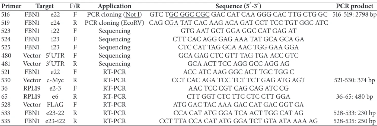
Table 1: Primers used in this study.
Primer Target F/R Application Sequence (5-3) PCR product
516 FBN1 e22 F PCR cloning (Not I) GTC TGC GGC CGC GAC CAT CAA GGG CAC TTG CTG GC 516-519: 2798 bp
519 FBN1 e24 R PCR cloning (EcoRV) CAG CGA TAT CAC AAG ACA GAT CCT TCC TGT GGC ATC
523 FBN1 i22 F Sequencing GTG AAT GCT GGA GGC CAT GAG AT
524 FBN1 i23 F Sequencing CTT CAC AGG GAG AAA TAT GCA GCA GA
525 FBN1 i23 F Sequencing CTC CAT TAG GCA AAC TGG GAA GGA
480 Vector 5UTR F Sequencing GCA GAG CTC GTT TAG TGA ACC GTC
481 Vector 3UTR R Sequencing GCA ACT TCC AGG GCC AGG AG
521 FBN1 e22 F RT-PCR ACC ATC AAG GGC ACT TGC TGG C
530 Vector c-Myc R RT-PCR CCT CAC AGA TCC TCT TCT GAG ATG AGT 521-530: 374 bp
36 RPL19 e2-3 F RT-PCR AAC TCC CGT CAG CAG ATC CG
65 RPL19 e6 R RT-PCR CTT GGT CTC TTC CTC CTT GGA 36-65: 480 bp
528 Vector FLAG F RT-PCR ATG GAC TAC AAA GAC CAT GAC GGT GA
533 FBN1 e23-22 R RT-PCR CCA CAT ATG GGA TCA ACT TGG CAT AG 528-533: 230 bp
535 FBN1 e23-i22 R RT-PCR CCT TTA CCA CAT ATG GGA TCT GTA ATA AAA AG 528-535: 250 bp
patients revealed aberrantFBN1pseudoexon inclusions due to intronic mutations which activate cryptic splice sites [13– 17].
We describe a patient with a diagnosis of MFS where there is no mutation identified in theFBN1coding sequence. Targeted NGS of the genomic DNA from this patient revealed a novel variant located in intron 22 of theFBN1 gene. The results of both in silico and functional studies using minigene splicing assays strongly suggest that this variant leads to a cryptic acceptor splice site activation and subsequent frame shift and premature termination codon (PTC) in exon 23.
2. Materials and Methods
2.1. Patient Information. The index patient was a 32-year-old male with MFS. Clinical findings included aortic root dilata-tion (41 mm aortic sinus, 44 mm or 2.17cm/m2 ascending aorta), ectopia lentis, and typical skeletal manifestations. His father had aortic dilatation and died in his forties due to endocarditis after the implantation of an aortic prosthesis. His mother, his brother, and one paternal aunt were clinically unaffected. Since no relevant mutation was identified in a previous genetic study limited to exons and close flanking intronic regions of the FBN1 gene, the patient requested a new evaluation as he and his couple were considering the possibility ofin vitro fertilization with preimplantation genetic diagnosis. Three nonaffected patient’s relatives as well as 4752 individuals with phenotypes other than MFS were also genetically tested. Written informed consent was obtained from all subjects before the study. The project was approved by each local ethics committee.
2.2. Genomic DNA Extraction. The patient’s and relative’s genomic DNA was extracted from saliva samples collected with the DNA Sample Collection Kit OG-500 (Oragene), on the QIAsymphony SP robot using the QIAsymphony DNA midi Kit (Qiagen). Purified genomic DNA was quantified using Nanodrop 1000 Spectrophotometer (Thermo Scien-tific) and DNA integrity was determined on the 2200 TapeS-tation (Agilent Technologies) following the manufacturer’s recommendation.
2.3. Next-Generation Sequencing. Custom probe RNA baits (120 nucleotides) for NGS were designed using Agilent’s SureDesign online tool to cover all exons and exon/intron boundaries of the humanFBN1gene. In addition, another 34 genes included in the Health in Code panel (see the Genetic Testing Registry at NCBI, Test ID: GTR000530671.1, www.ncbi.nlm.nih.gov/gtr/tests/530671) related to aortic diseases and phenocopies were NGS analyzed. For NGS
library construction, patient genomic DNA (3 𝜇g) was
sheared into 150-200 bp fragments by Covaris E220 fo-cused-ultrasonicator. Preparation and capture of NGS libraries were performed on the Bravo instrument (Ag-ilent Technologies) following the manufacturer’s instruc-tions. The DNA libraries were normalized to 10 nM and then pooled for multiplexed sequencing (HiSeq 1500, Illumina). Filtering of variants was performed using in-house data sets, the database of single-nucleotide polymorphisms (www.ncbi.nlm.nih.gov/projects/SNP/, build 132), Human Gene Mutation Database (HGMD, http://www.hgmd.cf.ac .uk/ac/index.php), and the Health in Code database. In order to confirm theFBN1variants identified by NGS, a standard PCR amplification and bidirectional Sanger sequencing were performed. In silico analysis of identified variants was performed using the splicing prediction module of the Alamut Visual v.2.9.0 software (Interactive Biosoftware), running five independent algorithms for splice signal detection: SpliceSiteFinder-like (SSF), MaxEntScan (MES), NNSPLICE (NNS), GeneSplicer (GSP), and Human Splicing Finder (HSF).
…g t t t t t ccg c t t t t t a t t a c a g A T C C C A T A T G T G G T…
2678 2690
2678-10
2678-20 2685
E23 i22
RS:
…g t t t t t aa g c t t t t t a t t a c a g A T C C C A T A T G T G G T…
2678 2690
2678-10
2678-20 2685
E23 i22
MS:
c.2678-15C>A
c.2678
c.2678-13 c.2678
(a)
0 20 40 60 80 100
0 3 6 9 12 15
0,0 0,2 0,4 0,6 0,8 1,0
0 3 6 9 12 15
0 20 40 60 80 100
SSF
MES
NNS
GSP
HSF
c.2678
score
Algorithm:
RS
MS
RS MS
MS
RS
MS
RS
MS RS
(b)
0 20 40 60 80 100
0 3 6 9 12 15
0,0 0,2 0,4 0,6 0,8 1,0
0 3 6 9 12 15
0 20 40 60 80 100
score SSF
MES
NNS
GSP
HSF
Algorithm:
RS
MS
RS
MS
MS
RS
MS
RS
MS RS
c.2678-13
(c)
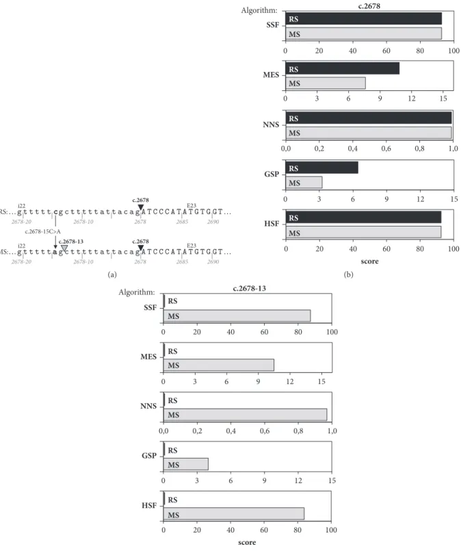
Figure 1: Schematic representation of in silico analysis of the c.2678-15C>A variant identified in intron 22 of theFBN1gene by various
computational tools. (a) Representation of the position of the canonical (c.2678, black triangle) and cryptic (c.2678-13, grey triangle) splice
site identified in the reference (RS) and mutated sequence (MS), respectively. Arrow: the C to A point mutation (c.2678-15C>A). The intron 22
(i22) sequence is shown in lower case and exon 23 (E23) sequence in upper case. (b) The predicted splice score of the canonical site decreases by 29.8% and 49.7% in the mutated sequence (grey parallelogram) versus the reference sequence (black parallelogram) as revealed by the
HiPure plasmid filter purification kit (Invitrogen) according to the manufacturer’s protocol. All theFBN1constructs were verified by full-length insert sequencing (for primers, see Table 1). Plasmid 1283 was selected as theFBN1-Ref minigene, and plasmid 1288 was selected as the FBN1-Mut minigene (Supp. Figure 1B). Plasmids were formulated at a final DNA concentration of 1 mg/ml in sterile isotonic saline.
2.5. Cell Culture and Transfection In Vitro. HeLa (an epithe-lial cell line derived from human cervical epithelioid car-cinoma, passage +4) and COS-7 (a fibroblast-like cell line derived from African green monkey kidney tissue, passage +5) were purchased from the European Collection of Authen-ticated Cell Cultures (Sigma). Cells were trypsinized at 70–80% confluence, cell numbers were determined using an automated cell counter (Countess, Invitrogen), and 100,000 HeLa or 60,000 COS-7 cells/well were plated in 12-well culture plates, allowed to attach overnight, and transiently transfected with 1.0 𝜇g of plasmid DNA. All transfections were carried out with Lipofectamine 3000 (Invitrogen) fol-lowing the manufacturer’s instructions. For each plasmid, four separate transfection assays were employed, and in each assay transfections were performed in duplicate. The transfection efficiency, evaluated by cotransfection with a CMV-EGFP (enhanced green fluorescent protein) vector followed by fluorescent microscopy, was 70-80%. Equivalent transfection efficiency was verified by cotransfection with a plasmid coding for the N-terminal 3xFLAG-tagged bacterial alkaline phosphatase (BAP) fusion protein (p3XFLAG-CMV-7-BAP, Sigma) followed by Western blot detection with an anti-FLAG antibody. Additional controls included mock and empty vector-transfected cells. In some experiments, the transfected cells were incubated with the cell-permeable proteasome inhibitor MG132 (Sigma) or dimethyl sulfoxide (Sigma) vehicle for 7 hours. The cells were harvested at 24–48 hours after transfection and processed for RNA and protein extraction.
2.6. Semiquantitative RT-PCR. Total RNA was extracted by RNeasy-Mini Kit (Qiagen) according to the manufacturer’s protocol, subjected to column digestion of DNA with RNase-free DNase (Qiagen), and reverse transcribed using Super-Script IV (Invitrogen) and oligo-dT primers. Semiquan-titative RT-PCR was performed in a Biometra II system using theRPL19gene (coding for ribosomal L19 protein) for normalization of RT-PCR data [18]. The amount of cDNA and the number of cycles were varied for each primer pair (see Table 1) to ensure amplification within the linear phase. Reactions, including non-RT control and nontemplate con-trol, were performed at least in triplicate. PCR products were visualized on 2% agarose gels by ethidium bromide staining and band intensity was estimated by densitometry (VersaDoc 1000) and Quantity One software (Bio-Rad).
2.7. SDS-PAGE and Western Blotting. Cell samples were homogenized and solubilized in standard 2X Laemmli buffer (Invitrogen) supplemented with complete protease inhibitor cocktail (Roche). Following centrifugation at 20,000 g for 30 minutes, the concentration of supernatant proteins was
analyzed using the Bio-Rad DC Protein Assay Kit accord-ing to the manufacturer’s protocol. Extracted proteins were subjected to SDS-PAGE (Mini-Protean-III, Bio-Rad), stained with Coomassie or blotted onto PVDF membranes (Hybond-P, Amersham Bioscience), and probed with mouse mono-clonal anti-Mycor anti-FLAG antibodies (Sigma). Molecular weight (MW) standards (MARK-12 and SeeBlue Plus2 from Invitrogen) were included on each gel. Equivalence of protein loading was confirmed by Amido Black staining of blots after immunodetection. Blocking, washing, incubation with diluted primary and secondary HRP-conjugated antibodies (Sigma), and visualization of immunodecorated bands by the Super-Signal West Pico PLUS chemiluminescent substrate (Thermo Scientific) were carried out as previously described [19].
3. Results
DNA from patient saliva was used forFBN1 gene targeted NGS analysis with in-house analysis pipeline. Paired end
sequencing covered nearly 34,000 bases of FBN1
encom-passing 66 exons along with their flanking (200 bp) intron regions. A total of 8 variants ofFBN1were identified with high variant-calling stringency including 7 variants localized in intronic noncoding regions and one synonymous exonic vari-ant which does not influence the amino acid structure of the protein (Table 2). All variants were confirmed by Sanger sequencing. It is worth noting that the identified variants, with the exception of the heterozygous c.2678-15C>A, have been previously described in the database of short genetic variation (dbSNP) and classified as benign or uncertain in the ClinVar database. NGS coverage analysis metrics across the region of interest forFBN1allowed for the identification of the heterozygous c.2678-15C>A variant which are shown in Supp. Figure S2. The c.2678-15C>A variant was not identified by Sanger sequencing in any of the three clinically unaffected patient’s relatives. This variant was also not detected in the control population consisting of 4752 individuals with phenotypes other than MFS who were referred to the Health in Code laboratory for NGS analysis. In addition, our NGS screening of a panel of 34 genes related to aortic diseases or involved in the differential diagnosis of MFS did not reveal any pathogenic variant in the case patient.
530 528 521
(PL. 1283)
(PL. 1288)
E22 E23 E24
i22 i23
c.2678-15C>A
1 200 400 600 800 1000 1200 1400 1600 1800 2000 2200 2400 2600 2800
bp
FBN1-Ref
FBN1-Mut F
F
M
M
(a)
500
400
300 (521-530)
bp (Primers)
FBN1, 374 bp
500 600 700
400
(36-65)
RPL19, 480 bp
FBN1, 387 bp
HeLa Cos7
FBN1-Ref
(PL. 1283) FBN1-Mut(PL. 1288) MC FBN1-Ref(PL. 1283) FBN1-Mut(PL. 1288) MC
1 2 3 4 5 6 7 8 9 10
L
(b)
E22
374 bp E23 E24
533
E22
387 bp E23 E24
535
(c)
300
200
300
200 400
(528-533)
(528-535) FBN1, 250 bp
FBN1, 230 bp
HeLa Cos7
FBN1-Ref
(PL. 1283) FBN1-Mut(PL. 1288)
MC FBN1-Ref
(PL. 1283) FBN1-Mut(PL. 1288) MC
1 2 3 4 5 6 7 8 9 10
L
(d)
E22
230 bp E23
E22
250 bp E23
(e)
Figure 2: RT-PCR analysis of minigene-derived transcripts. (a) Schematic representation of FLAG (F) and Myc- (M-) tagged reference (
FBN1-Ref) and mutant (FBN1-Mut) minigene plasmids (PL). Exons (E) are denoted with white boxes and introns (i) with solid black horizontal
lines. The approximate location of the primers for downstream RT-PCR analysis is shown (for primer sequences see Table 1). (b) Expression ofFBN1-Ref andFBN1-Mut minigenes in HeLa and COS-7 cells as revealed by RT-PCR, using primers (521 and 530) targeting E22 and Myc. A
representative of two independent experiments for each transfection is shown.RPL19amplification was carried out as an input RNA control
Table 2: IdentifiedFBN1variants using targeted NGS.
FBN1 variant compared to RS:
Location Zygosity SNP ID Frequency1 Splicing2
NC 000015.10 NM 000138.4
1 g.48610929A>G c.248-103T>C intron 3 HZ rs1018148 0,932343 NA
2 g.48537938C>G c.539-130G>C intron 6 HTZ rs147780575 NR NA
3 g.48494269G>T c.2678-15C>A intron 22 HTZ NR NR A4
4 g.48488453G>A c.3123C>T5 exon 26 HTZ rs576395584 0.00001 NA
5 g.48444487A>T c.6037+54T>A intron 49 HZ rs2303502 0.22331 NA
6 g.48437451T>C c.6314-64A>G intron 51 HZ rs2042746 0.39285 NA
7 g.48428329G>C c.6997+17C>G intron 57 HZ rs363832 0.27742 NA
8 g.48421799G>T c.7571-113C>A intron 61 HZ rs1820488 0,813073 NA
RS: reference sequence; g.: genomic; c.: coding DNA; HZ: homozygous; HTZ: heterozygous; NR: not registered; NA: not affected; A: affected. 1Population frequency from gnomAD (Genome Aggregation Database) except3from dbSNP.
2Alamut Visual v.2.9 predictions.
4Activation of a cryptic acceptor splice site at c.2678-13 (see Figure 3). 5NP 000129.3:p.(His1041=).
activate a cryptic acceptor splice site within intron 22, at position 13 nucleotides (nt) prior to exon 23.
Taken together, these results strongly suggested that the c.2678-15C>A intronic variant induces a cryptic splice site in intron 22 of theFBN1gene that in turn could lead to an inefficient recognition of canonical splice site, giving rise to a frameshift and premature termination codon (PTC) in the
FBN1mRNA.
To investigate the effect of the FBN1splice site variant c.2678-15C>A at the RNA and protein level, we used a minigene approach with reference (Ref) and mutant (Mut) expression plasmids containing the FBN1fragments which were amplified from the patient genomic DNA (see Supp. Figure S1). Each plasmid was transfected into HeLa and COS-7 cells for transient expression. RT-PCR amplifications of the mini-Ref transcript with different primer sets yielded single bands of the expected size (Figure 2(b); 374 bp bands) corresponding to the normal inclusion of exons 22, 23, and 24, with introns 22 and 23 having been removed. The sequencing of these RT-PCR products confirmed that the introns were removed and the exons were correctly ligated together (Figure 2(c) and Supp. Figure S3A). In contrast, slightly longer 387 bp bands were PCR amplified from HeLa and COS-7 cells transfected with the mini-Mut plasmid (see Figure 2(b)). These RT-PCR products were eluted from the gel and sequenced. The 387 bp mini-Mut band included exon 22, a part (13 nt) of intron 22, and exons 23 and 24 (see Fig-ure 2(c) and Supp. FigFig-ure S3B). The results indicated that the FBN1c.2678-15C>A variant affects splicing by promoting the insertion of a 13-nt intron 22-derived sequence in theFBN1 transcript due to activation of a cryptic splice site localized at c. 2678-13. A more detailed PCR analysis using primers located inside the intron 22-derived insertion (Figure 2(c)) revealed that the aberrantly spliced transcript was expressed in HeLa and COS-7 cells transfected with the mini-Mut plasmid being not detected in cell transfectants expressing mini-Ref plasmids (Figures 2(d) and 2(e)). The latter supports the suggestion that theFBN1c.2678-15C>A variant could lead to haploinsufficiency of full-length FBN1 protein.
Overall, the data demonstrated that the c.2678-15C>A variant can repress recognition of the canonical splice site in
intron 22 of the FBN1gene causing a shift from canonical toward cryptic splicing that can lead to insertion of a 13-nt intron 22-derived sequence, frameshift, and the creation of a PTC (see Supp. Figure 3B). Following the standards and guidelines for the interpretation of sequence variants [20], theFBN1c.2678-15C>A mutation was classified as pathogenic (see Supp. Table S1) and submitted to the ClinVar database at NCBI (www.ncbi.nlm.nih.gov/clinvar/) with the accession number SCV000611711.
The relative level of transcripts produced in the mini-Ref-containing cells was comparable with that observed in cells transfected with the mini-Mut construct (see Figure 2(b)), suggesting that the aberrantly spliced transcripts may escape, at least partly, from a nonsense-mediated mRNA decay (NMD) under our experimental conditions.
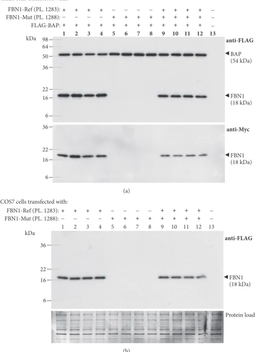
To verify whether these aberrantly spliced transcripts are translated into protein products, we performed SDS-PAGE and Western blotting of protein lysates of HeLa and COS-7 cells transfected with FLAG/Myc-tagged Ref and mini-Mut plasmids. The 18 kDa bands were detected by both anti-FLAG and anti-Myc antibodies in HeLa (Figure 3(a)) and COS-7 (Figure 3(b)) cells expressing the mini-Ref plasmid 1283. Their apparent molecular weight (MW) values (18 kDa) were found to acceptably match the deduced MW sum of the mini-Ref FBN1 sequence, three FLAG and one c-Myc tags (i.e., 16.6 kDa, see Figure 4(b)). Surprisingly, neither the 18-kDa band nor any others were detectable in cells transfected with the mini-Mut plasmid 1288 (see Figure 3) although these cell transfectants produced high levels of the aberrantly spliced transcript (see Figure 2).
50
36
22 16
6 64 98
36
22
16
6
1 2 3 4 5 6 7 8 9 10 11 12 13
FBN1-Ref (PL. 1283): FBN1-Mut (PL. 1288):
+ + + +
_ _ _ _ + + + +
_ _ _ + + + + _
+ + + +
_
_
FLAG-BAP:+ + + + + + + + + + + + _
anti-FLAG
anti-Myc
FBN1 (18 kDa) BAP (54 kDa)
FBN1 (18 kDa) kDa
(a)
36
22
16
6
1 2 3 4 5 6 7 8 9 10 11 12 13
FBN1-Ref (PL. 1283): FBN1-Mut (PL. 1288):
+ + + +
_ _ _ _ + + + +
_ _ _ + + + + _
+ + + +
_
_
anti-FLAG
FBN1 (18 kDa) COS7 cells transfected with:
Protein load kDa
(b)
Figure 3: Western blot analysis of minigene-derived proteins. (a) FLAG/Myc-tagged reference (FBN1-Ref) and mutant (FBN1-Mut) minigene
plasmids (PL) were transfected into HeLa cells as indicated at the top of each lane, and cell lysates were analyzed by Western blotting with a mouse monoclonal anti-FLAG (upper panel) or anti-Myc (lower panel) antibody. The results from experiments performed on four batches of cells for each transfection are shown. Lane 13: cells transfected with empty vector. The Western blot detection of the FLAG-tagged bacterial alkaline phosphatase (BAP) was used as a marker of equivalent transfection efficiency and equal loading. MW values (kDa) of the bands detected are shown in brackets. (b) COS-7 cells were transfected as indicated at the top of each lane and analyzed by Western blotting with a mouse monoclonal anti-FLAG antibody (upper panel). Protein load (lower panel): membrane stained with Amido Black 10B after immunostaining. Other indications as in (a).
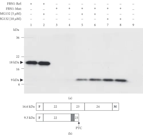
effect was dose-dependent (Figure 4). As might be expected in the presence of a PTC in the aberrant transcript (see Supp. Figure S3B), the corresponding protein band was of a lower MW in comparison with that of the correctly processed mini-Ref construct, i.e., 9 kDa versus 18 kDa, respectively (see
36
22
16
6 kDa
18 kDa
9 kDa
1 2 3 4 5 6 7 8 9
FBN1-Mut: _ _ + + + + + + _
FBN1-Ref: + + _ _ _ _ _ _ _
MG132 [5 휇M]: _ + _ _ + + _ _ _
MG132 [10 휇M]: _ _ _ _ _ _ + + _
(a)
16.6 kDa
9.3 kDa
23 24 M
22
F
23 22
F
PTC
(b)
Figure 4: Treatment of transfected cells with MG132 leads to detection of truncated proteins derived from mutantFBN1constructs. (a)
FLAG/Myc-tagged reference (FBN1-Ref; PL 1283) and mutant (FBN1-Mut; PL 1288) minigene plasmids (PL) were transfected into HeLa
cells as indicated at the top of each lane. Lane 9: mock cells. Either the proteasome inhibitor MG132 or the DMSO vehicle was applied to transfected cells as indicated in Materials and Methods. Whole-cell extracts of transfected cells were analyzed by Western blotting with a mouse monoclonal anti-FLAG antibody. Black arrows: MW values (kDa) of the bands detected. (b) A diagram of the protein products as predicted by sequence analysis of the corresponding cDNAs. Deduced MW values (kDa) are also indicated. Dark grey box: partial inclusion of intron 22; F: FLAG epitope; M: Myc epitope; PTC: premature termination codon.
shown) because theFBN1c.2678-15C>A variant produced, as predicted, a frame shift and PTC within exon 23. Because of the PTC, the Myc-coding region will not be read-through.
4. Discussion
Our study adds to the mutational spectrum of theFBN1gene in MFS. Using a previously unrecognized variant in intron 22 of theFBN1gene (c.2678-15C>A), revealed by targeted NGS in a MFS patient, we demonstrate, as a proof-of-concept, that this variant causes aberrant splicing, frameshift, and PTC. This pathogenic variant induces aberrantFBN1pre-mRNA splicing by the generation of a new cryptic acceptor site that outcompetes the canonical splice site. The single nucleotide C to A change in intron 22 of theFBN1is, therefore, considered clinically relevant to MFS. In contrast, the known C to T nucleotide substitution at the same site (i.e., c.2678-15C>T, rs181681840) is not expected to have any clinical significance because its population frequency (1% in the African pop-ulation; gnomad.broadinstitute.org/variant/15-48786466-G-A) is not consistent with MFS incidence data (approximately 1 case per 5000 individuals [14]), and multiple lines of computational evidence suggest no impact on gene product;
the variant c.2678-15C>T is classified as benign in ClinVar (www.ncbi.nlm.nih.gov/clinvar/variation/137303).
Point mutation variants that affectFBN1pre-mRNA splic-ing represent approximately 10% of reportedFBN1mutations in patients with MFS [12] and are frequently associated with aortic wall alterations [13]. The accumulated data clearly indicate that splice mutations located besides theFBN1 exon-coding area and canonical exon’s splice site motifs also play an important role in MFS [21]. In particular, it was found that patients suffering from MFS can carryFBN1intronic variants resulting in cryptic splicing and exonization of intronic sequences at the transcript level [13–16, 22].
In this work, we identified a new intronic variant (c.2678-15C>A) of the FBN1 gene in a patient with a classic MFS phenotype. The mutation was not identified in his unaffected mother, brother, and paternal aunt and could have been inherited from his deceased father who was retrospectively considered as very likely affected by MFS.
expected to direct the corresponding transcript(s) to NMD, because it is located in a NMD-insensitive region, less than 50 nt upstream of the 3-most exon-exon junction measured after splicing [23]. Our data strongly suggest that aberrantly spliced transcripts carrying this PTC, located 38 nt upstream of the junction between exons 23 and 24 in the spliced mini-gene (Supp. Figure S3B), could escape NMD in transfected cells and express unstable truncated proteins which were only detectable upon proteasome inhibition with MG132.
Although as yet unproved, we believe that the c.2678-15C>A variant would lead to haploinsufficiency of the FBN1 functional protein and structural connective tissue fragility. Our NGS analysis did not reveal any relevant change in a panel of 34 additional genes related to aortic diseases or involved in the differential diagnosis of MFS, supporting the assumption that the c.2678-15C>A variant of the FBN1 can be considered as a main disease-causing variant in the case patient.
5. Conclusion
The intronic c.2678-15C>A substitution has not previously been reported, representing therefore a novel FBN1 gene variant in MFS. Our results demonstrate that the identified intronic variant disrupting normalFBN1mRNA splicing is a MFS-associated mutation that should be taken into account in the diagnosis of patients with MFS. On the basis of our results, we suggest that the clinical phenotype associated with the intronic c.2678-15C>A variant is caused by a reduction of wild-type FBN1 protein. Our findings and others highlight the potential importance of intronicFBN1variants in causing MFS as well as the continued need for identifying noncoding mutations in theFBN1gene.
Data Availability
All data are presented in the manuscript.
Conflicts of Interest
The authors declare no conflicts of interest.
Acknowledgments
The authors thank the test patient and relatives for participat-ing in this study. This work was supported by the Autonomic Government of Galicia, Spain (Grant no. GRC 2013/061).
Supplementary Materials
Supp. Table S1: classifying the FBN1c.2678-15C>A variant as pathogenic. Supp. Figure S1: vector mapping and sequencing analysis of plasmids. Supp. Figure S2: NGS identification of the intronic FBN1c.2678-15C>A variant. Supp. Figure S3: direct sequencing analysis of RT-PCR products amplified with primer sets targeting the minigene transcripts in the transfected HeLa cells.(Supplementary Materials)
[1] S. M. Hammond and M. J. A. Wood, “Genetic therapies for RNA
mis-splicing diseases,”Trends in Genetics, vol. 27, no. 5, pp. 196–
205, 2011.
[2] J. Wang, J. Zhang, K. Li, W. Zhao, and Q. Cui, “SpliceDisease
database: Linking RNA splicing and disease,” Nucleic Acids
Research, vol. 40, no. 1, pp. D1055–D1059, 2012.
[3] F. Ramirez, C. Caescu, E. Wondimu, and J. Galatioto, “Mar-fan syndrome; A connective tissue disease at the crossroads
of mechanotransduction, TGF𝛽signaling and cell stemness,”
Matrix Biology, 2017.
[4] J. R. Cook, L. Carta, J. Galatioto, and F. Ramirez, “Cardiovas-cular manifestations in marfan syndrome and related diseases;
multiple genes causing similar phenotypes,”Clinical Genetics,
vol. 87, no. 1, pp. 11–20, 2015.
[5] V. C˜aadas, I. Vilacosta, I. Bruna, and V. Fuster, “Marfan
syn-drome. Part 1: pathophysiology and diagnosis,”Nature Reviews
Cardiology, vol. 7, no. 5, pp. 256–265, 2010.
[6] P. N. Robinson, E. Arteaga-Solis, C. Baldock et al., “The molecular genetics of Marfan syndrome and related disorders,”
Journal of Medical Genetics, vol. 43, no. 10, pp. 769–787, 2006. [7] M. Arslan-Kirchner, E. Arbustini, C. Boileau et al., “Clinical
utility gene card for: Marfan syndrome type 1 and related
phenotypes [FBN1],”European Journal of Human Genetics, vol.
18, no. 9, p. 1070, 2010.
[8] R. Franken, T. J. Heesterbeek, V. De Waard et al., “Diagnosis
and genetics of Marfan syndrome,”Expert Opinion on Orphan
Drugs, vol. 2, no. 10, pp. 1049–1062, 2014.
[9] C. J. A. Ramachandra, A. Mehta, K. W. Q. Guo, P. Wong, J. L. Tan, and W. Shim, “Molecular pathogenesis of Marfan
syndrome,”International Journal of Cardiology, vol. 187, no. 1,
pp. 585–591, 2015.
[10] L. Y. Sakai, D. R. Keene, M. Renard, and J. De Backer, “FBN1: The disease-causing gene for Marfan syndrome and other genetic
disorders,”Gene, vol. 592, no. 1, pp. 279–291, 2016.
[11] L. Faivre, G. Collod-Beroud, B. L. Loeys et al., “Effect of mutation type and location on clinical outcome in 1,013 probands with Marfan syndrome or related phenotypes and
FBN1 mutations: an international study,”American Journal of
Human Genetics, vol. 81, no. 3, pp. 454–466, 2007.
[12] K. A. Zeyer and D. P. Reinhardt, “Engineered mutations in fibrillin-1 leading to Marfan syndrome act at the protein, cellular
and organismal levels,”Mutation Research - Reviews in Mutation
Research, vol. 765, pp. 7–18, 2015.
[13] L. M. Baudhuin, K. E. Kotzer, and S. A. Lagerstedt, “Increased frequency of FBN1 truncating and splicing variants in Marfan
syndrome patients with aortic events,”Genetics in Medicine, vol.
17, no. 3, pp. 177–187, 2015.
[14] E. Gillis, M. Kempers, S. Salemink et al., “An FBN1 Deep Intronic Mutation in a Familial Case of Marfan Syndrome:
An Explanation for Genetically Unsolved Cases?” Human
Mutation, vol. 35, no. 5, pp. 571–574, 2014.
[15] D.-C. Guo, P. Gupta, V. Tran-Fadulu et al., “An FBN1 pseu-doexon mutation in a patient with Marfan syndrome:
Con-firmation of cryptic mutations leading to disease,”Journal of
Human Genetics, vol. 53, no. 11-12, pp. 1007–1011, 2008. [16] S. Hutchinson, P. B. Wordsworth, and P. A. Handford, “Marfan
syndrome caused by a mutation in FBN1 that gives rise to cryptic splicing and a 33 nucleotide insertion in the coding
[17] L. Tjeldhorn, S. S. Amundsen, T. Barøy et al., “Qualitative and quantitative analysis of FBN1 mRNA from 16 patients with
Marfan Syndrome,”BMC Medical Genetics, vol. 16, no. 1, article
no. 113, 2015.
[18] M. Torrado, A. Centeno, E. L´opez, and A. T. Mikhailov, “In
vivoforced expression of myocardin in ventricular myocardium
transiently impairs systolic performance in early neonatal pig
heart,”The International Journal of Developmental Biology, vol.
53, no. 8–10, pp. 1457–1467, 2009.
[19] M. Torrado, R. Iglesias, A. Centeno, E. L´opez, and A. T. Mikhailov, “Targeted gene-silencing reveals the functional
sig-nificance of myocardin signaling in the failing heart,” PLoS
ONE, vol. 6, no. 10, Article ID e26392, 2011.
[20] S. Richards, N. Aziz, and S. Bale, “Standards and guidelines for the interpretation of sequence variants: a joint consensus recommendation of the American College of Medical Genetics and Genomics and the Association for Molecular Pathology,”
Genetics in Medicine, vol. 17, no. 5, pp. 405–423, 2015.
[21] L. Tan, Z. Li, C. Zhou et al., “FBN1 mutations largely contribute
to sporadic non-syndromic aortic dissection,”Human
Molecu-lar Genetics, vol. 26, no. 24, pp. 4814–4822, 2017.
[22] L. Karttunen, T. Ukkonen, K. Kainulainen, A.-C. Syv¨anen, and L. Peltonen, “Two novel fibrillin-l mutations resulting in pre-mature termination codons but in different mutant transcript
levels and clinical phenotypes,”Human Mutation, vol. 11, no. 1,
pp. S34–S37, 1998.
[23] O. Isken and L. E. Maquat, “Quality control of eukaryotic mRNA: Safeguarding cells from abnormal mRNA function,”
Hindawi www.hindawi.com
International Journal of
Volume 2018
Zoology
Hindawi
www.hindawi.com Volume 2018
Anatomy
Research International
Hindawi
www.hindawi.com Volume 2018
Hindawi
www.hindawi.com Volume 2018
Journal of
Parasitology Research
Genomics
International Journal of
Hindawi
www.hindawi.com Volume 2018
Hindawi Publishing Corporation
http://www.hindawi.com Volume 2013 Hindawi
www.hindawi.com
The Scientific
World Journal
Volume 2018Hindawi
www.hindawi.com Volume 2018
Bioinformatics
Advances inMarine Biology
Journal ofHindawi
www.hindawi.com Volume 2018
Hindawi
www.hindawi.com Volume 2018
Neuroscience
Journal
Hindawi
www.hindawi.com Volume 2018
BioMed
Research International
Cell Biology
International Journal of
Hindawi
www.hindawi.com Volume 2018
Hindawi
www.hindawi.com Volume 2018
Biochemistry Research International
Archaea
Hindawi
www.hindawi.com Volume 2018
Hindawi
www.hindawi.com Volume 2018
Genetics
Research International
Hindawi
www.hindawi.com Volume 2018
Advances in
Virology
Stem Cells
International
Hindawi
www.hindawi.com Volume 2018
Hindawi
www.hindawi.com Volume 2018
Enzyme
Research
Hindawi
www.hindawi.com Volume 2018
International Journal of
Microbiology
Hindawi
www.hindawi.com Volume 2018