University of Valladolid‐INIA
Instituto Universitario de Invest
Universidad de Valladolid‐INIA
igación Forestal Sostenible
Effect
of
putative
mitoviruses
on
growth
of
Gremmeniella
abietina
isolates
in
vitro
and
on
its
pathogenicity
on
Pinus
halepensis
seedlings
Carmen
Romeralo
Tapia
Supervisor:
Julio Javier Diez Casero
Sustainable Use of Forest Systems
MSc in Conservation and
of Valladolid
University
Efecto
de
posibles
mitovirus
sobre
el
crecimiento
de
aislados
de
Gremmeniella
abietina
in
vitro
y
en
su
patogenicidad
en
plántulas
de
Pinus
halepensis
Carmen
Romeralo
Tapia
Supervisor: Professor Julio Javier Diez Casero [email protected] Instituto de Investigación Forestal Sostenible,
Universidad de Valladolid‐INIA
Avda. Madrid 44, Edificio E, 34004 Palencia, Spain
Assistant supervisors: Dr. Leticia Botella Sánchez [email protected] Instituto de Investigación Forestal Sostenible, Universidad de Valladolid‐INIA,
vda. Madrid 44, Edificio E, 34004 Palencia, Spain A
Dr. Óscar Santamaría Becerril [email protected]
Departamento de Ingeniería del Medio Agronómico y Forestal. d de Extremadura). Escuela de Ingenierías Agrarias (Universida
Ctra. de Cáceres, s/n. 06007 Badajoz, Spain
Credits: 30 hec
MSc in Conservation and Sustainable Use of Forest Systems
Place of publication: Palenciaear of publication: 2012 Y
Universidad de Valladolid
INDEX
Abstract
... 6
1.
Introduction
... 8
2.
Objetives
... 11
3.
Materials
and
Methods
... 11
3.1
Fungal
material... 11
3.2
Mycelial
growth... 12
3.3
Culture
conditions
for
monitoring
mycelium
growth ... 12
3.4
DsRNA
extractions... 13
3.5
In
vivo
pathogenicity
tests ... 13
3.6
Statistical
analysis... 15
4.
Results
... 16
4.1
Effect
of
temperature... 16
4.2
Effect
of
pH... 17
4.3
Effect
of
osmotic
potential
(
ψ
π) ... 19
4.4
DsRNA
banding
patterns ... 20
4.5
In
vivo
pathogenicity
tests ... 21
5.
Discussion
... 23
6.
Conclusions
... 25
Acknowledgements
... 25
References
... 26
Abstract
Mitoviruses have been found in several forest pathogens (e.g. Cryphonectria parasitica, Gremmeniella abietina), and because they have been shown to reduce the virulence of host fungi there is a growing interest in studying their use as a biocontrol. This study was carried out to test the effect of temperature (5 ºC, 15 ºC, 25 ºC and 35 ºC), pH (4, 5, 7 and 9) and osmotic potential (-0.6, -1.2, -1.8 and -2.4 MPa) on the mycelial growth of seven G. abietina isolates under controlled laboratory conditions and to observe the effect of the presence of mitoviruses in the pathogenity of G. abietina isolates inoculated to Pinus halepensis seedlings. Four of the isolates hosted mitoviruses and three of them did not. During the in vitro experiment, mycelial growth was recorded every week for a period of 8 weeks. In the greenhouse experiment, once the seedlings started to show symptoms, disease severity was recorded during 5 weeks. At the end of the experiment, plants were carried to laboratory and necrosis length was measured in all of them. Results from in vitro experiment showed that the mitovirus-infected isolates presented larger mycelial growth than the mitovirus-free ones when at the fungi’s optimal growing temperature of 15 ºC. However, no differences in growth behavior were observed between mitovirus infected and non-infected isolates when placed under different pH modifications. When growing at certain osmotic potentials (-0.6 and -1.8 MPa) a reduction in growth of the mitovirus-infected isolates was observed. In the greenhouse experiment, larger necrosis lengths were observed in the plants inoculated with mitovirus infected isolates. The results of this experiment provide further insight into the effects of mitovirus on Gremmeniella abietina isolates.
Key words: mitoviruses, Scleroderris canker, in vitro, in vivo, biological control,
Gremmeniella abietina, dsRNA.
Resumen
invernadero, a partir de que las plántulas mostraron síntomas, se midió la severidad de la enfermedad una vez por semana, durante 5 semanas. Al finalizar el experimento, las plántulas fueron llevadas al laboratorio, donde se midió la longitud de la necrosis producida por el patógeno. Los resultados del experimento in vitro mostraron que los aislados infectados con mitovirus presentaron mayor crecimiento micelial que los que no infectados en la temperatura de crecimiento óptimo del hongo de 15 ºC. No se observaron efectos de la presencia de mitovirus entre los aislados infectados y los no infectados en los tratamientos de modificación del pH. Cuando se modificaron los potenciales osmóticos se observó una reducción del crecimiento micelial de los aislados infectados con mitovirus en comparación con los no infectados en los potenciales osmóticos de -0.6 y -1.8 MPa. En el experimento efectuado en el invernadero, la longitud de las necrosis encontradas en plántulas con aislados infectados por mitovirus fueron mayores que las que presentaron las plántulas inoculadas con aislados sin mitovirus. Este estudio proporciona un conocimiento más profundo de los efectos de las infecciones víricas en aislados españoles de Gremmeniella abietina.
Palabras clave: mitovirus, Scleroderris canker, in vitro, in vivo, control biológico,
1. Introduction
Gremmeniella abietina (Lagerberg) Morelet (anamorph Brunchorstia pinea (P. Karsten) Höhnel) is a pathogenic fungus which has caused destruction in plantations and natural conifer forests in Northern and Central Europe, North America, and Japan (Yokota, 1975; Dorworth, 1979; Kaitera and Jalkanen, 1992) producing symptoms such as stem cankers and shoot dieback (Donaubauer, 1972). This fungus has been divided into three races: European, North American and Asian. Within the European race three biotypes have been determined based on the length of spores, number of septa, disease symptoms, and molecular markers: biotype A (LTT, large tree type), biotype B (STT, small tree type) and alpine biotype (Uotila, 1983; Hamelin et al., 1993; Hellgren and Hogberg, 1995; Kaitera and Jalkanen, 1996; Hantula and Muller, 1997). In Europe, the fungus mostly affects genera Picea spp. and Pinus spp. although it has also been found on genera Abies and Larix. In Spain, it presence on Pinus pinaster was first reported in 1929 (Martínez, 1933) and later on Pinus halepensis in 1999 (Santamaria et al., 2003). Notwithstanding, it has only been isolated from symptomatic Pinus halepensis trees.
A B
E D
C
The symptoms observed generally consist of dry needles, branches with some distortion of terminal twigs and eventual dieback or death of the trees (Figure 1) (Santamaria et al., 2003). Spanish G. abietina is currently recognized as part of the European race (Santamaria et al., 2005) and has recently been related to biotype A, although it has a unique genotype (Botella et al., 2010).
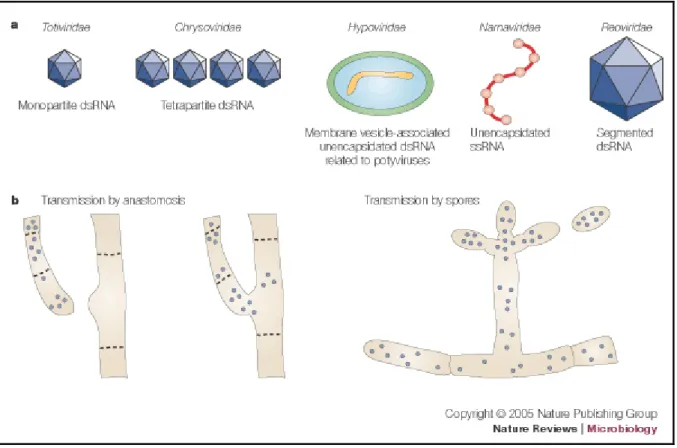
Mycoviruses, which are obligate parasites of fungi, are widespread in all major taxonomic groups of plant pathogenic fungi (Ghabrial and Suzuki, 2009; Pearson et al., 2009). They are transmitted through hyphal anastomosis and/or fungal sporulation (Fig 2b) (Zhang et al., 2010). Fungal viruses differ in their genomes, which can contain DNA, double-stranded (ds) RNA or single-double-stranded (ss) RNA genomes (Fig 2a) (Pearson et al., 2009). Eight families and one genus are currently described in the International Committee on Taxonomy of Viruses (ICTV) (2011): Chrysoviridae, Endornaviridae, Hypoviridae, Narnaviridae, Barnaviridae, Partitiviridae, Reoviridae and Totiviridae and genus Rhizidiovirus (Hausner et al., 2000; Zhang et al., 2010). Mycoviruses usually produce latent infections in nature, affecting sometimes the host’s phenotype and/or its growth (Aoki et al., 2009). Symptoms produced by the presence of mycoviruses may vary from zero to severe effects on host physiology and may lead to attenuation (hypovirulence) or enhancement of fungal virulence (hypervirulence) (Ghabrial and Suzuki, 2009). Because some viruses are capable of reducing virulence of fungal pathogens they can potentially be used for control of fungal diseases (McCabe et al., 1999; Boland 2004; Zhang et al., 2010). However, they must fulfill two requirements in order to be suitable for biological control: firstly, to have the ability to decrease the fitness of the pathogenic fungus and secondly, to transmit the dsRNA efficiently enough to be maintained in a large proportion of the pathogen population (McCabe et al., 1999).
Fig 2a Taxonomic families represented by mycoviruses that are associated with hypovirulence of plant
pathogenic fungi are shown with the virus structure and genome composition. b Mycoviruses are not
infectious by an extracellular route. Transmission is restricted primarily to intracellular routes that include anastomosis (fusion of hyphae) or during the formation of spores (Nuss, 2005)
2. Objetives
Although some strains of G. abietina have been shown to host dsRNA mycoviruses, the effect these agents have on the virulence of this phytopathogenic fungus has not yet been investigated. Accordingly, the main objectives of the present study were:
i. To evaluate the effect of the occurrence of viral dsRNA molecules (the replicative form of Mitovirus) on the in vitro mycelial growth of G. abietina isolates under different temperature, pH and osmotic potential conditions
ii. To observe the effect of the presence of mitoviruses in the pathogenity of G. abietina isolates inoculated on Pinus halepensis seedlings
3. Materials and Methods
3.1 Fungal material
To develop this study seven Spanish isolates of G. abietina (Table 1) were chosen: four isolates were naturally infected by putative mitoviral molecules (P3-12, 00P-07, Hon 3-3 and P1-12) and three were not (Hon 9-2; P1-8 and VAI-13) (Botella et al., 2010). All isolates were selected based on previous studies developed in our laboratory in which RT-PCR and sequencing techniques confirmed the presence or absence of mitoviruses (Botella et al., 2012a, 2012b). The isolates were previously stored in 15% glycerol at -80 º C and were reactivated on modified orange serum agar medium (MOS-agar medium; Müller et al., 1994) before performing the experiment. Thus, four weeks before the experiment fungi isolates were sub-cultured in MOS medium and kept in the dark at 15 ºC in order to obtain sufficient amounts of mycelium.
Table 1: Isolates used in the experiment (1according to Botella et al. 2010).
Isolate Mitoviral
molecules1 Origin Province Year of isolation
00P-07 Yes Valle de Cerrato Palencia 2001
VAI-13 No Villalba de los Alcores Valladolid 2003
Hon 3-3 Yes Hontoria Palencia 2007
Hon 9-2 No Hontoria Palencia 2007
P1-8 No Valle de Cerrato Palencia 2007
P1-12 Yes Valle de Cerrato Palencia 2007
3.2 Mycelial growth
At the bottom of every Petri dish containing 20 ml MOS medium two perpendicular lines were drawn, and a 1mm squared piece of mycelium from each isolate was placed over the intersection of both lines (Fig 3a, 3b). Mycelial growth was measured weekly for a period of 8 weeks. The response variable was the growth area calculated by the following formula: Area = π/4 (d1 x d2) where d1 and d2 were the two diameters measured along the lines.
B A
Fig 3a Petri dishes used in the experiments with the two perpendicular lines. b Boxes where the
Petri dishes were stove in.
3.3 Culture conditions for monitoring mycelium growth
The effect of mitovirus infection on mycelial growth under different laboratory conditions was the main focus of this study. Three experiments were conducted, each taking into account a separate factor: changes in temperature, pH or osmotic potential. Within each experiment four variations were tested: four temperatures (5 ºC, 15 ºC, 25 ºC and 35 ºC), four pH values (4, 5, 7 and 9) and four osmotic potentials (-0.6, -1.2,-1.8 and -2.4MPa). The effect of temperature on mycelial growth was investigated by placing Petri dishes in several stoves at 5 ºC, 15 ºC, 25 ºC and 35 ºC. To examine the effect of pH, HCl or KOH 1N was added to MOS medium until the pH required was reached. All these Petri dishes were placed in the dark at 15 ºC since it is the optimal temperature for fungal development (Santamaria et al., 2004). Finally, in order to evaluate the effect of different osmotic potential on mycelial growth, different concentrations of KCl (250, 500, 750 and 1000mM) were added to MOS medium in order to reach the osmotic potential (ψπ) values of -0.6 MPa, -1.2 MPa, -1.8 MPa
3.4 DsRNA extractions
In order to know if mitoviral molecules remain present after the treatments, extractions of dsRNA were carried out in significative treatments (Fig 4a). Fungal mycelium of mitovirus-infected isolates was incubated in MOS medium covered with cellophane for two weeks. Mycelia were first freeze-dried and then ground for 20 minutes into a fine powder. DsRNA was extracted following a modified version of the protocol described by Morris and Dodds (1979). The dsRNA presence in every isolate was verified by electrophoresis (Fig 4b). Samples were loaded in a 1% agarose gel, which contained 1x TAE buffer and GelRedTM
10.000X. The test was run in a 1x TAE buffer during 60 min at 90V/30 cm, and immediately afterwards observed under UV light and photographed. The marker used to estimate the lengths of the dsRNA molecules was λ-DNA Hind III – ΦX174Hae III(DyNAzymeTM).
B A
Fig 4a dsRNA extraction process b Gel electrophoresis apparatus.
3.5 In vivo pathogenicity tests
C
A B
Fig 5 Followed steps to inoculate the fungus: a Wound with the scapel; b Piece of G. abietina; c Cover with Parafilm.
C
A B
D E F
Fig 6 Severity index a Symtomless; b Chlorosis; c Advanced chlorosis; d Dieback; e Necrotic; f Dead.
3.6 Statistical analysis
All statistical analyses were done with SAS program (SAS Institute Inc., 2004). The response variable in all models was growth area (mm2). A repeated-measures ANOVA for
Results from pathogenicity tests were also analyzed with SAS program. Two models were made to evaluate the presence of mitoviruses (yes/no): first, severity index was used as response variable and second, relative necrosis length. In all the analysis a 95% of significance was considered. Normality, linearity and homocedasticity for the residuals were probed with Shapiro-Wilk test and graphical procedures. Since data did not fulfill these requirements, they were analyzed with a non-parametric test (the two-sample median test).
4. Results
4.1 Effect of temperature
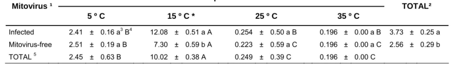
A significant effect of time on the colony growth area (p<0.001) was observed. Although the interaction between time and mitovirus presence was significant (p=0.017) as well as the interaction between time and temperature (p<0.001), only the effect of temperature and mitovirus presence on mycelial growth at the end of the experiment eight weeks after plating is shown in Table 2. The average growth from mitovirus-infected and mitovirus-free isolates is shown in Figure 7 at 5 ºC (A), 15 ºC (B), 25 ºC (C) and 35 ºC (D) throughout the eight weeks. Growth at 25 ºC was minimal and there was no growth at 35 ºC. Mean growth area was significantly different among mitovirus-infected and mitovirus-free isolates (p=0.0030), temperatures (p<0.001) and their interactions (p<0.001). According to the Tukey test, the largest colony areas were found at 15 ºC whereas the smallest were found at 35 ºC. The overall mean colony size of mitoviruses-infected isolates was significantly bigger than that of the mitoviruses-free ones. When temperatures were considered separately, significant differences among infected and mitoviruses-free isolates were found only at 15 ºC (p=0.0043), the temperature that produced the most growth.
Table 2: Mycelial growth (mm²) after 8 weeks at different temperatures. Mean value ± standard error (SE). Treatments tagged with * presented significant differences among isolates. 1If the isolate was naturally-infected with mitovirus. 2Average growth when combining all the temperatures together. 3 Different letters in the same column show values significantly different from p<0.05 (ANOVA Tukey’s HSD Test). 4Different letters in the same row show values significantly different from p<0.05 (ANOVA Tukey’s HSD Test). 5Average growth when combining all the isolates together.
Temperature Mitovirus ¹
5 º C 15 º C * 25 º C 35 º C
TOTAL²
Infected 2.41 ± 0.16 a3 B4 12.08 ± 0.51 a A 0.254 ± 0.50 a B 0.196 ± 0.00 a B 3.73 ± 0.25 a
Mitovirus-free 2.51 ± 0.19 a B 7.30 ± 0.59 b A 0.223 ± 0.59 a C 0.196 ± 0.00 a C 2.56 ± 0.29 b
5º C 0 0.5 1 1.5 2 2.5 3
1 2 3 4 5 6 7 8
Time (weeks) M yc eli al gr ow th ( m m 2) Infected Mitovirus free 15º C 0 2 4 6 8 10 12 14
1 2 3 4 5 6 7 8
Time (weeks) M yc eli al gr ow th ( m m 2) Infected Mitovirus free 25º C 0.2 0.21 0.22 0.23 0.24 0.25 0.26 0.27
1 2 3 4 5 6 7 8
Time (weeks) M yc e lia l gr ow th ( m m 2) Infected Mitovirus free 35º C 0 0.05 0.1 0.15 0.2 0.25
1 2 3 4 5 6 7 8
Time (weeks) M yc e lial grow th (m m 2 ) Infected Mitovirus free (A) (D) (C) (B)
Fig 7 Average growth from mitovirus-infected and mitovirus-free isolates at 5 ºC (a), 15 ºC (b), 25 ºC
(c) and 35 ºC (d) over the eight weeks.
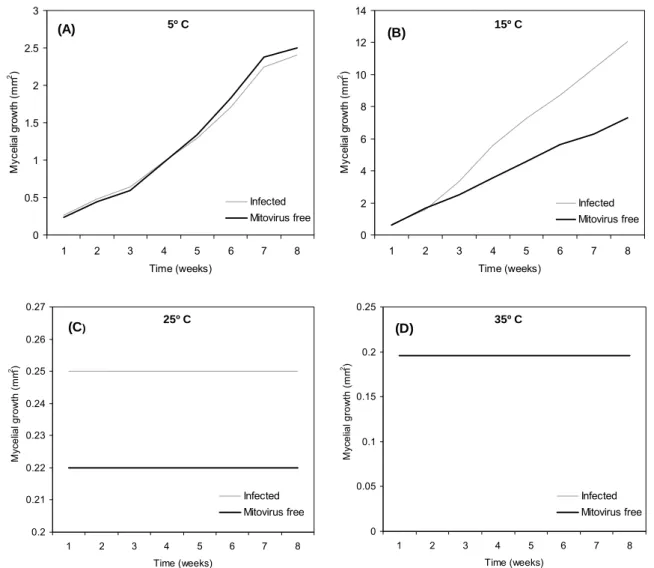
4.2 Effect of pH
Table 3: Mycelial growth (mm²) after 8 weeks at different pHs. Mean value ± standard error (SE). 1If
the isolate was naturally-infected with mitovirus. 2Average growth when combining all the pH values
together. 3Different letters in the same column show values significantly different from p<0.05 (ANOVA Tukey’s HSD Test). 4Different letters in the same row show values significantly different from p<0.05 (ANOVA Tukey’s HSD Test). 5Average growth when combining all the isolates together.
pH value Mitovirus 1
pH 4 pH 5 pH 7 pH 9
TOTAL2
Infected 17.28 ± 1.84 a3 A4 9.96 ± 0.97 a B 9.75 ± 0.88 a B 6.62 ± 0.43 a B 10.90 ± 0.58 a
Mitovirus-free 20.25 ± 2.13 a A 8.92 ± 1.12 a B 8.23 ± 1.02 a B 6.45 ± 0.50 a B 10.96 ± 0.66 a
TOTAL5 18.55 ± 0.88 A 9.51 ± 0.88 B 9.10 ± 0.88 B 6.55 ± 0.88 B
pH 4 0 5 10 15 20 25
1 2 3 4 5 6 7 8
Time (weeks) M yc el ial grow th (m m 2 ) Infected Mitovirus free pH 5 0 2 4 6 8 10 12
1 2 3 4 5 6 7 8
Time (weeks) M yc el ial grow th (m m 2 ) Infected Mitovirus free pH 7 0 2 4 6 8 10 12
1 2 3 4 5 6 7 8
Time (weeks) M yc el ial grow th (m m 2 ) Infected Mitovirus free pH 9 0 1 2 3 4 5 6 7
1 2 3 4 5 6 7 8
Time (weeks) M yc el ial grow th (m m 2) Infected Mitovirus free (A) (D) (C) (B)
Fig 8 Average growth from mitovirus-infected and mitovirus-free isolates at pH 4 (a), pH 5 (b), pH 7(c)
4.3 Effect of osmotic potential (ψπ)
In this experiment, time also affected the growth (p<0.001) and interacted as well with the osmotic potential (p<0.001) and the mitovirus presence (p<0.0447). Data taken in the eight week showed the greatest differences (Table 4). The average growth of mitovirusinfected and mitovirusfree isolates is shown in Figure 9 at 0.6MPa (A), 1.2MPa (B), -1.8MPa (C) and -2.4MPa (D) throughout the eight weeks. The model was significative (p=0.027) although it was not the mitovirus presence (p=0.1378) nor osmotic potential (p=0.0805), but the interaction was significative (p=0.0034), that is, the effect of mitovirus presence was different among the different osmotic potentials. When osmotic potential was considered separately at ψπ of -0.6 MPa (p=0.0167) and at -1.8 MPa (p=0.0387),
mitovirus-free isolates presented a higher mycelial growth than the mitovirus-infected ones which did not happen at the osmotic potentials of -1.2MPa (p=0.7515) and -2.4MPa (p=0.1004).
Table 4: Mycelial growth (mm²) after 8 weeks at different osmotic potentials. Mean value ± standard
error (SE). Treatments tagged with * presented significant differences among isolates. 1If the isolate
was naturally-infected with mitovirus. 2Average growth when combining all the pH values together. 3 Different letters in the same column show values significantly different from p<0.05 (ANOVA Tukey’s HSD Test). 4Different letters in the same row show values significantly different from p<0.05 (ANOVA Tukey’s HSD Test). 5Average growth when combining all the isolates together.
Osmotic potential TOTAL²
Mitovirus ¹
- 0.6 MPa * - 1.2 MPa - 1.8 MPa * - 2.4 MPa
Infected 7.66 ± 0.79 b3 AB4 9.24 ± 0.49 a AB 6.47 ± 0.71 b B 9.96 ± 0.77 a A 8.33 ± 0.35 a
Mitovirus-free 10.74 ± 0.80 a A 9.00 ± 0.56 a AB 8.82 ± 0.82 a AB 7.96 ± 0.89 a B 9.13 ± 0.40 a
-0.6MPa 0 2 4 6 8 10 12
1 2 3 4 5 6 7 8
Time (weeks) M yc eli al gr ow th ( m m 2) Infected Mitovirus free -1.2MPa 0 1 2 3 4 5 6 7 8 9 10
1 2 3 4 5 6 7 8
Time (weeks) M yc el ial grow th (m m 2) Infected Mitovirus free -1.8MPa 0 1 2 3 4 5 6 7 8 9 10
1 2 3 4 5 6 7 8
Time (weeks) M yc e lial grow th (m m 2) Infected Mitovirus free -2.4MPa 0 2 4 6 8 10 12
1 2 3 4 5 6 7 8
Time (weeks) M yc el ial grow th (m m 2) Infected Mitovirus free (A) (D) (C) (B)
Fig 9 Average growth from mitovirus-infected and mitovirus-free isolates at -0.6MPa (a), -1.2MPa (b),
-1.8MPa (c) and -2.4MPa (d) over the eight weeks.
4.4 DsRNA banding patterns
The presence of the different putative mitoviruses was confirmed by dsRNA extraction and gel electrophoresis after significant treatments were carried out (Fig 10). Isolate P3-12 had a 2.5 kb band in treatment 15 ºC (lane 7) and was found to maintain this band despite receiving the treatments of ψπ -0.6MPa (lane 1) and -2.4 MPa (lane 10). It
happened the same with isolates 00P-07 and P1-12: treatments of ψπ -0.6MPa (lane 3, lane
4), -2.4 MPa (lane 11) and temperature of 15 ºC (lane 5). Conversely, the 2.5 kb band was not sustained in isolate Hon3-3 after treatments of temperature 15 ºC (lane 6), ψπ -0.6MPa
(lane 2) and -1.8MPa (lane 8) and in isolate P1-12 after treatment of ψπ -1.8MPa (lane 9).
in P3-12 suggested the occurrence of other putative mycoviruses, which did not seem to be affected by the different treatments either. According to the size of the bands and the previous work developed in the laboratory (Botella et al., 2010) they possibly belonged to genera Totivirus (ca 6kb) and Partitivirus (three bands of ca 1-2 kb).
1
2
3
4
5
6
7
8
9
10
11
2.5
kb
Fig 10 A GelRed-stained 1 % agarose gel showing the dsRNA banding patterns. Lane 1, P3-12
(-0.6MPa); lane 2, Hon 3-3 (-(-0.6MPa); lane 3, 00P-07 (-(-0.6MPa); lane 4, 12 (-(-0.6MPa); lane 5, P1-12 (15 ºC); lane 6, Hon 3-3 (15 ºC); lane 7, P3-P1-12 (15 ºC); lane 8, Hon 3-3 1.8MPa); lane 9, P1-P1-12 (-1.8MPa); lane 10, P3-12 (-2.4MPa); lane 11, 00P-07 (-2.4MPa).
4.5 In vivo pathogenicity tests
The average relative necrosis length (Fig 11) from seedlings inoculated with mitovirus infected and from non-infected isolates is shown in Figure 12. The necrosis length of seedlings inoculated with mitovirus-infected isolates was significantly larger (p=0.0291) than the necrosis from seedlings which were inoculated with non-infected isolates. In the second model, severity index was used as response variable, but it was not significative with a 95% level of significance. Seedlings inoculated with mitovirus infected isolates had a higher disease severity that the non-infected isolates but it was not statistically significant (Fig 13). Gremmeniella abietina was re-isolated from 35% of the symptomatic seedlings that were inoculated.
A B
0 0.2 0.4 0.6 0.8 1 1.2 1.4
Control Mitovirus-free Infected
R
elat
iv
e n
ecr
o
sis
le
n
g
th
Fig 12 Box plot for control seedlings and seedlings inoculated with mitovirus infected and mitovirus-free isolates. Bars and boxes represent: maximum, 3rd quartile, median, 1st quartile and minimum.
0 0.2 0.4 0.6 0.8 1 1.2 1.4 1.6 1.8
0 1 2 3 4 5
Tim e (w eeks)
S
ever
it
y
6 Control
Infected Mitovirus-free
5. Discussion
Mycelial growth depends on the temperature of the environment. In our study, all the isolates showed an optimal growth at 15 ºC, which was in accordance with Santamaría et al. (2004) who demonstrated that Spanish isolates of G. abietina had the best growth at this particular temperature. Furthermore, the presence of mitovirus seemed to have a significant effect on G. abietina isolates at its optimal growing temperature of 15 ºC because the isolates with mitovirus present had higher mycelial growth than isolates without mitoviruses. This increase in the mycelial growth of our isolates could be related to a higher virulence of the pathogen since, in general terms, a suppression of mycelial growth has been reported to be closely associated with hypovirulence of fungi (Ghabrial et al., 2009; Pearson et al., 2009) although it could also be related to other factors (e.g., poor sporulation).
Heat tolerance was previously observed in several fungi among virus-infected and virus-free isolates (Marquez et al., 2007; Herrero et al., 2011) but in our study neither mitovirus-infected nor mitovirus-free isolates were able to endure the heat (few isolates hardly grew at 25 ºC and no growth was observed at 35 ºC). Marquez et al., (2007) observed that plants inoculated with the virus-infected wild type isolate of Curvularia protuberata R.R. Nelson and Hodges, with presence of the virus named CThTV, tolerated soils temperatures as high as 65ºC for two weeks whereas plants inoculated with the virus-free isolate of the fungus dried-up and became chlorotic. Light evidence of heat tolerance was also observed in Tolypocladium cylindrosporum W. Gams due to the different behaviors displayed between virus-infected and virus-free isolates at 30 ºC (Herrero et al., 2011).
In our study, there wasn’t any clear evidence that a decrease in osmotic potential produced a reduction of mycelial growth as previously observed in other fungal species (Imolehin et al., 1980; Lira-Mendez and Mayek-Perez, 2006; Palmero et al., 2008; Armengol et al., 2011). A reduction of the growth of the mitovirus-infected isolates was observed at -0.6 MPa and -1.8 MPa which can be linked to a decrease in the virulence of the isolates. Changes in behavior were also observed in isolates with and without viral infection when growing at certain osmotic potentials for Monosporascus cannonballus (Armengol et al., 2011).
Concerning in vivo pathogenicity test, mitovirus’ infection resulted in any statistically significant difference in visual disease severity among seedlings. Necrosis length of seedlings inoculated with mitovirus-infected isolates was significantly larger than the necrosis from seedlings which were inoculated with non-infected isolates, that is, mitovirus infected isolates did not reduce the pathogen aggressiveness, which is the most desirable feature in control of plant pathogenic fungi (Xu et al. 2005). This behavior could suggest an advantageous to the infected isolates and could lead to a hypervirulence of the pathogen. However additional studies are required to understand the role of this mitovirus in G. abietina features.
and sporulate poorly (McCabe et al. 1999). In other cases, the presence of dsRNAs did not cause any fungal specific symptoms, such as reduced mycelial growth (Aoki et al., 2009). In some Alternaria spp. species there was no correlation between the radial growth of isolates and the presence of the dsRNAs (Zabalgogeazcoa, 1998). In contrast to hypovirulent interactions, there is evidence that some mycoviruses are beneficial to their hosts. Tan et al., (2007) observed statistically significant differences in in vitro growth rates of virus-infected versus uninfected isolates, with the infected cultures growing more rapidly. In our results, an increase of the mycelial growth was observed at treatment 15 ºC, the optimal growing conditions of G. abietina. Although no visual disease severity statistically significant difference was noticed among plants, larger necrosis lengths were recorded in those seedlings infected with mitovirus isolates, suggesting a possible hypervirulence produced by the mitoviruses’ presence.
This study provides additional knowledge on the effects of mitovirus infection on G. abietina isolates. However, further research including other virulence-associated parameters such as sporulation rates are recommended to establish an association between mycovirus infection and fungal virulence in Spanish G. abietina isolates. The development of a biocontrol protocol may create opportunities for biological control of this disease.
6. Conclusions
In our study mycelial growth depended on the treatment and the presence of mitoviruses. The presence of mitoviruses did not reduce mycelial growth of Gremmeniella abietina at its optimal growing temperature of 15 ºC. No effects of the occurrence of mitoviruses were shown among the mitovirus-infected and the mitovirus-free ones at any pH value. When growing at certain osmotic potentials (-0.6 and -1.8 MPa) a reduction in the growth of the mitovirus-infected isolates compared to the mitovirus-free ones was observed. No differences were found in visual disease severity among plants however larger necrosis lengths were observed in the plants inoculated with mitovirus infected isolates, suggesting a possible hypervirulence produced by the mitoviruses’ presence. Further research including other virulence-associated parameters is recommended.
Acknowledgements
References
Aoki N, Moriyama H, Kodama M, Arie T, Teraoka T, Fukuhara T. 2009. A novel mycovirus associated with four double-stranded RNAs affects host fungal growth in Alternaria alternata. Virus Res140, 179-187.
Armengol J, Alaniz S, Vicent A, Beltran R, Abad-Campos P, Perez-Sierra A, Garcia-Jimenez J, Ben Salem I, Souli M, Boughalleb N. 2011. Effect of dsRNA on growth rate and reproductive potential of Monosporascus cannonballus. Fungal Biol115, 236-244.
Boland G, 2004. Fungal viruses, hypovirulence, and biological control of Sclerotinia species. Can J Plant Pathol26, 6-18.
Botella L, Tuomivirta TT, Kaitera J, Navarro VC, Diez JJ, Hantula J. 2010. Spanish population of Gremmeniella abietina is genetically unique but related to type A in Europe. Fungal Biol 114, 778-789.
Botella L, Tuomivirta TT, Hantula J, Diez JJ. 2012a. Presence of viral dsRNA molecules in the Spanish population of Gremmeniella abietina. Journal of Agricultural Extension and Rural Development 4 (9), 211-213.
Botella L, Tuomivirta TT, Vervuur S, Diez JJ., Hantula J. 2012b. Occurence of two different species of mitoviruses in the European race of Gremmeniella abietina var abietina, both hosted by the genetically unique Spanish population. Fungal Biol
http://dx.doi.org/10.1016/j.funbio.2012.05.004
Brasier CM.1983. A cytoplasmically transmitted disease of Ceratocystis ulmi. Nature 305, 220-223.
Carlile, M.J.; Watkinson, S. C.; Gooday, G.W., 2001. The Fungi 2nd ed. Academic press,
London, UK.
Castro M, Kramer K, Valdivia L, Ortiz S, Castillo A. 2003. A double-stranded RNA mycovirus confers hypovirulence-associated traits to Botrytis cinerea. FEMS Microbiol Lett 228, 87-91.
Chu Y, Jeon J, Yea S, Kim Y, Yun S, Lee Y, Kim K. 2002. Double-stranded RNA mycovirus from Fusarium graminearum. Appl Environ Microb68, 2529-2534.
Deng, F., Xu, R. Boland, G. J. 2003. Hypovirulence associated double-stranded RNA from Sclerotinia homoeocarpa is conspecific with Ophiostoma novo-ulmi mitovirus 3a-Ld. Phytopathology 93, 1407-1414.
Deng, F., Boland, G.J. 2004. A satellite RNA of Ophiostoma novo-ulmi mitovirus 3a in hypovirulent isolates of Sclerotinia homoeocarpa. Phytopathology 94, 917-923.
Donaubauer, E. 1972. Distribution and hosts of Scleroderris lagerbergii in Europe and North America. Eur J Forest Pathol 2, 6-11.
Dorworth CE. 1979. Influence of inoculum concentration on infection of red pine seedings by Gremmeniella-abietina. Phytopathology69, 298-300.
Ghabrial S.A, Suzuki N. 2009 Viruses of plant pathogenic fungi. Annu Rev Phytopathol 47, 353-384.
Hamelin R, Ouellette G, Bernier L. 1993. Identification of Gremmeniella-abietina races with Random Amplified Polymorphic DNA markers. Appl Environ Microb59, 1752-1755.
Hantula J, Muller MM, 1997. Variation within Gremmeniella abietina in Finland and other countries as determined by Random Amplified Microsatellites (RAMS). Mycol Res 101, 169-175.
Hausner, G., Belkhiri, A., Klassen, G.R. 2000. Phylogenetic analysis of the small subunit ribosomal RNA gene of the hyphochytrid Rhizidiomyces apophysatus. Can J Botany 78, 124-128.
Hellgren M, Hogberg N, 1995. Ecotypic Variation of Gremmeniella-abietina in Northern Europe - Disease patterns reflected by DNA variation. Can J Botany 73, 1531-1539.
Herrero N, Perez-Sanchez R, Oleaga A, Zabalgogeazcoa I, 2011. Tick pathogenicity, thermal tolerance and virus infection in Tolypocladium cylindrosporum. Ann Appl Biol 159, 192-201.
Imolehin ED, Grogan RG, Duniway JM.1980. Effect of temperature and moisture tension on growth, sclerotial production, germination and infection by Sclerotinia minor. Phytopathology 70, 1153–1157
Kaitera J, Jalkanen R, 1992. Disease history of Gremmeniella-abietina in a Pinus-sylvestris Stand. Eur J Forest Pathol22, 371-378.
Kaitera J, Jalkanen R, 1996. In vitro growth of Gremmeniella abietina isolates (European race) at different temperatures. Scand J Forest Res11, 159-163.
Lee K, Yu J, Son M, Lee Y, Kim K, 2011. Transmission of Fusarium boothii mycovirus via protoplast fusion causes hypovirulence in other phytopathogenic fungi. PloS ONE 6, e21629.
Lira-Méndez, K., Mayek-Pérez, N. 2006. Potencial osmótico variable en el crecimiento in vitro y la patogenicidad en frijol (Phaseolus vulgaris L.) de Fusarium spp. Rev Mex Fitopatol 24 (2), 88-97.
Marquez LM, Redman RS, Rodriguez RJ, Roossinck MJ. 2007. A virus in a fungus in a plant: three-way simbiosis required for themal tolerance. Science 315, 543-515.
Martínez J. 1933. Una grave micosis del pino observada por primera vez en España. Bol Soc Española de Historia Natural 33, 25-29.
McCabe P, Pfeiffer P, Van Alfen N. 1999. The influence of dsRNA viruses on the biology of plant pathogenic fungi. Trends Microbiol7, 377-381.
Morris T.J., Dodds J.A. 1979. Isolation and analysis of double-stranded-RNA from virus-infected plant and fungal tissue. Phytopathology 69, 854-858.
Müller Mm, Kantola R, Kitunen V (1994) Combining sterol and fatty acid profiles for the characterization of fungi. Mycol Res 98, 593–603.
Nuss D.L. 2005. Hypovirulence: Mycoviruses at the fungal-plant interface. Nat Rev Microbiol 3, 632-642.
Palmero D, de Cara M, Iglesias C, Ruiz G, Tello JC. 2008. Effects of water potential on spore germination and viability of Fusarium species. J Ind Microbiol Biotechnol 35,1405–1409.
Park Y, Chen X, Punja ZK. 2006. Molecular and biological characterization of a mitovirus in Chalara elegans (Thielaviopsis basicola) Phytopathology 96, 468-479.
Pearson MN, Beever RE, Boine B, Arthur K. 2009. Mycoviruses of filamentous fungi and their relevance to plant pathology RID D-3988-2011. Mol Plant Pathol10, 115-128.
Perez J, Martinez B, Rivas E, Diaz MA. 2000. Efecto del pH sobre la germinación conidial, crecimiento y esporulación de Didymella bryoniae (Awersw) Rehn. Rev Protección Veg Vol 15 (3), 185-187.
Polashock JJ, Hillman BI. 1994. A small mitochondrial double-stranded (ds) RNA element associated with hypovirulent strain of the chesnut blight fungus and ancestrally related to yeast cytoplasmic T and W dsRNAs. Proc Natl Acad Sci U.S.A. 91, 8680-8684.
Polashock J.J, Bedker PJ, Hillman BI.1997 Movement of a small mitochondrial double-stranded RNA element of Cryphonectria parasitica: ascospore inheritance and implications for mitochondrial recombination. Mol Gen Genet 256,566-571.
Robin C, Lanz S, Soutrenon A, Rigling D.2010. Dominance of natural over released biological control agents of the chestnut blight fungus Cryphonectria parasitica in south-eastern France is associated with fitness related traits. Biol control 53, 55-61.
Rogers HJ, Buck KW, Brasier CM. 1987. A mitochondrial target for doubled-stranded RNA in diseased isolates of the fungus that causes Dutch elm disease. Nature 329: 558-560
Santamaria O, Pajares JA, Diez JJ. 2003. First report of Gremmeniella abietina on Pinus halepensis in Spain. Plant Pathol52, 425-425.
Santamaria O, Pajares JA, Diez JJ. 2004. Physiological and morphological variation of Gremmeniella abietina from Spain. Forest Pathol 34, 395-405.
Santamaria O, Alves-Santos FM, Diez JJ. 2005. Genetic characterization of Gremmeniella abietina var. abietina isolates from Spain. Plant Pathol 54, 331-338.
Tan CMC, Pearson MN, Beever RE, Parkes SL. 2007. Why Fungi Have Sex? Abstract: XIVth International Botrytis Symposium, Cape Town, South Africa. October 21th-27th.
Tuomivirta T, Hantula J. 2003. Gremmeniella abietina mitochondrial RNA virus S1 is phylogenetically related to the members of the genus Mitovirus. Arch Virol148, 2429-2436.
Uotila A. 1983. Physiological and morphological variation among Finish Gremmeniella abietina isolates. Commun Inst For Fenn 119, 1-12.
Van Diepeningen A, Debets A, Hoekstra R. 2006. Dynamics of dsRNA mycoviruses in black Aspergillus populations. Fungal Genet Biol 43, 446-452.
Vainio,E. J.; Korhonen,K; Tuomivirta,T.T.; Hantula,J., 2010 A novel putative partitivirus of the saprotrophic fungus Heterobasidion ecrustosum infects pathogenic species of the Heterobasidion annosum complex. Fungal Biol 114, 11–12, 955-965
Vazquez-Garcia A, Santiago-Martinez G, Estrada-Torres A. 2002. Influencia del pH en el crecimiento de quince cepas de hongos ectomicorrizógenos. Anales del Instituto de Biología, UNAM, Serie Botánica 73(1), 1-15.
Wu M, Zhang L, Li G, Jiang D, Hou MS, Huang HC. 2007. Hypovirulence and double stranded RNA in Botrytis cinerea. Phytopathology 97, 1590-1599.
Wu M, Zhang L, Li G, Jiang D, Ghabrial S. 2010. Genome characterization of a debilitation associated-mitovirus infecting the phytopathogenic fungus Botrytis cinerea. Virology 406, 117-126.
Xu P, Fang C, Mannas J P, Feldman T, Sumner Ll W, Roossinck M J. 2008. Virus infection improves drought tolerance. New Phytol 180, 911-921.
Yokota, S. 1975. Scleroderris canker of todo-fir in Hokkaido, Northern Japan. III. Dormant infection of the causal fungus. Eur J Forest Pathol 5, 7-12.
Zhang L, De Wu M, Li GQ, Jiang DH, Huang HC. 2010. Effect of Mitovirus infection on formation of infection cushions and virulence of Botrytis cinerea. Physiological and Mol Plant Pathol75, 71-80.