HIV-1 immune activation induces Siglec-1 expression and enhances viral transmission in myeloid cells
200
0
0
Texto completo
(2) Department of Cellular Biology, Physiology and Immunology Universitat Autònoma de Barcelona. HIV-1 immune activation induces Siglec-1 expression and enhances viral transmission in myeloid cells MARIA PINO CLAVERIA. Institut de Recerca de la SIDA – IrsiCaixa Hospital Universitari Germans Trias i Pujol 2016. Thesis to obtain the PhD degree in Immunology of the Universitat Autònoma de Barcelona. Directors: Dr. Javier Martinez-Picado Dra. Nuria Izquierdo-Useros Tutor:. Dra. Dolores Jaraquemada.
(3)
(4) The Spanish Secretariat of Research (Grants SAF2010-21224 and SAF2013-49042-R) supported this thesis. Maria Pino Claveria was supported by grant BES-2011-044268 (Formación de Personal Investigador – FPI) from the Spanish Ministry of Science and Innovation and the Spanish Ministry of Economy and Competitiveness. The Spanish AIDS Network “Red Temática Cooperativa de Investigación en SIDA” provided additional support (RIS)..
(5)
(6) El Dr. Javier Martínez-Picado, investigador principal i professor de recerca ICREA a l’Institut de Recerca de la SIDA, IrsiCaixa (Hospital Universitari Germans Trias i Pujol).. Certifica:. Que el treball experimental i la redacció de la memoria de la Tesi Doctoral titulada “HIV-1 immune activation induces Siglec-1 expression and enhances viral transmission in myeloid cells” han estat realitzades per na Maria Pino Claveria sota la seva direcció i considera que és apta per ser presentada per optar al grau de Doctora en Immunologia per la Universitat Autònoma de Barcelona.. I per tal que quedi constància, signa aquest document.. Badalona 12 de Maig de 2016.. Dr. Javier Martínez-Picado.
(7)
(8) La Dra. Nuria Izquierdo-Useros, investigadora a l’Institut de Recerca de la SIDA, IrsiCaixa (Hospital Universitari Germans Trias i Pujol).. Certifica:. Que el treball experimental i la redacció de la memoria de la Tesi Doctoral titulada “HIV-1 immune activation induces Siglec-1 expression and enhances viral transmission in myeloid cells” han estat realitzades per na Maria Pino Claveria sota la seva direcció i considera que és apta per ser presentada per optar al grau de Doctora en Immunologia per la Universitat Autònoma de Barcelona.. I per tal que quedi constància, signa aquest document.. Badalona 12 de Maig de 2016.. Dra. Nuria Izquierdo-Useros.
(9)
(10) La Dra. Dolores Jaraquemada Pérez de Guzmán, catedràtica d’Immunologia de la Universitat Autònoma de Barcelona.. Certifica:. Que el treball experimental i la redacció de la memoria de la Tesi Doctoral titulada “HIV-1 immune activation induces Siglec-1 expression and enhances viral transmission in myeloid cells” han estat realitzades per na Maria Pino Claveria sota la seva direcció i considera que és apta per ser presentada per optar al grau de Doctora en Immunologia per la Universitat Autònoma de Barcelona.. I per tal que quedi constància, signa aquest document.. Badalona 12 de Maig de 2016.. Dra. Dolores Jaraquemada Pérez de Guzmán.
(11)
(12) A la meva familia i amics que m’aguanten.
(13)
(14) SUMMARY. Myeloid cells are key players in the recognition and response of the host against invading viruses. Paradoxically, upon HIV-1 infection, myeloid cells might also promote viral pathogenesis through trans-infection, a mechanism that promotes HIV-1 transmission to target cells via viral capture and storage. The receptor Siglec-1 (CD169) potently enhances HIV-1 trans-infection and is regulated by immune activating signals present throughout the course of HIV-1 infection, such as interferon α (IFNα). Here we show that IFNα-activated dendritic cells, monocytes and macrophages have an enhanced ability to capture and trans-infect HIV-1 via Siglec-1 recognition of viral membrane gangliosides. Monocytes from untreated HIV-1-infected individuals transinfect HIV-1 via Siglec-1, but this capacity diminishes after effective antiretroviral treatment. Furthermore, Siglec-1 is expressed on myeloid cells residing in lymphoid tissues, where it can mediate viral trans-infection. Our results strongly support that Siglec-1 is an important molecule that could accelerate HIV-1 transmission in the crowded cellular environment of lymphatic tissues, where many T-cells can contact myeloid cells. Since no current antiviral therapy blocks Siglec-1-mediated viral dissemination, we hypothesize that designing a novel generation of therapeutic agents against Siglec-1 could pose an interesting approach to reduce viral spread and limit the settlement of viral reservoirs, bringing us closer to a functional cure of HIV-1. We have followed different approaches to obtain Siglec-1 blocking compounds and tested small or multivalent sialyllactose-like compounds and mAbs against Siglec-1. Optimization of these approaches will aid to identify potent Siglec-1 inhibitors to complement current antiretroviral treatments and enhance future therapeutic strategies.. 15.
(15) 1.
(16) RESUM. Les cèl·lules mieloides són actors clau en el reconeixement i resposta de l'hoste contra els virus invasors. Paradoxalment, després de la infecció del VIH-1, les cèl·lules mieloides també poden promoure la patogènesis viral a través de la trans-infecció, un mecanisme que promou la transmissió del VIH-1 a les cèl·lules diana a través de la captura i emmagatzematge del virus. El receptor Siglec-1 (CD169) augmenta potentment la trans-infecció del VIH-1 i es regulat per senyals immune activadores presents al llarg del curs de la infecció del VIH-1, com és l'interferó α (IFNα). Aquí demostrem que cèl·lules dendrítiques, monòcits i macròfags activats amb IFNα tenen l'habilitat de capturar i trans-infectar el VIH-1 via el reconeixement per Siglec-1 de gangliòsids presents en la membrana viral. Monòcits de pacients infectats pel VIH-1 sense tractar trans-infecten el virus via Siglec-1, però aquesta capacitat es veu disminuïda després d'un tractament antirretroviral efectiu. A més, Siglec-1 s'expressa en cèl·lules mieloides residents en teixits limfoides, on poden mediar la trans-infecció viral. Els nostres resultats suporten fermament que Siglec-1 és una mol·lècula important que pot accelerar la transmissió del VIH-1 en l'entorn multicel·lular dels teixits limfàtics, on les cèl·lules T poden contactar amb les cèl·lules mieloides. Degut a que actualment no existeix una teràpia antiviral que bloquegi la disseminació del VIH-1 mediada per Siglec-1, nosaltres hipotetitzem que el disseny d'una nova generació d'agents terapèutics dirigits a Siglec-1 poden plantejar un enfocament interessant per a reduir la propagació viral i limitar l'establiment de reservoris virals apropant-nos a una cura funcional del VIH-1. Hem seguit diferents estratègies per a obtenir un compost que bloquegi Siglec-1, hem testat compostos petits o multivalents similars a la sialillactosa i anticossos monoclonals contra Siglec-1. La optimització de aquestes estratègies ens ajudaran a identificar inhibidors potents de Siglec-1 per a complementar els tractaments antirretrovirals actuals i millorar futures estratègies terapèutiques.. 17.
(17) 1.
(18) ABBREVIATIONS COMMONLY USED α2-3Gal: α2-3 galactose Ab: antibody ABS: antibody binding sites AIDS: acquired immunodeficiency syndrome APCs: antigen presenting cells APC: allophycocyanin (flow cytometry) APC-Cy7: allophycocyanin-Cyanine7 conjugate (flow cytometry) ART: antiretroviral treatment AZT: azidothymidine BDCA: blood dendritic cells antigen CCR5: C-C chemokine receptor type 5 CLR: C-type lectin receptor CTL: Cytolitic CD8+ T lymphocytes CXCR4: C-X-C chemokine receptor type 4 DAPI: 4, 6-diamidino-2-phenylindole DC: dendritic cell DC LPS: dendritic cells LPS activated DC IFN: dendritic cells IFN activated DC-SIGN: DC-specific ICAM-3-grabbing non-integrin DMEM: Dulbecco’s Modified Eagle Medium DMSO: dimethyl sulfoxide DNA: deoxyribonucleic acid DsRNA: double-stranded deoxyribonucleic acid eGFP: enhanced Green Fluorescent Protein ELISA: enzyme-linked immunosorbent assay Env: viral envelope glycoprotein ESCRT: endosomal sorting complexes required for transport FACS: flow activating cytometer sorting FBS: fetal bovine serum 19.
(19) 1. Fc: constant fraction (antibodies) FcR: receptor of constant fraction (antibodies) Fig: Figure FITC: fluorescein isothiocyanate FSC: forward scatter channel GALT: gut-associated lymphoid tissue GFP: green fluorescent protein GM-CSF: granulocyte-macrophage colony-stimulating factor GSL: glycosphingolipid GM3: 5-acetyl-alpha-neuraminic acid (2-3) beta-D-galactopyranose (1-4) beta-Dglucopyranose (1-1) ceramide HAART: highly active antiretroviral therapy HIV: human immunodeficiency virus HIV-1: human immunodeficiency virus type 1 HIV-2: human immunodeficiency virus type 2 HLA: human leukocyte antigen Ig: immunoglobulin ICAM-1: intercellular adhesion molecule 1 ICAM-3: intercellular adhesion molecule 3 iDC: immature dendritic cell IFNα: interferon alfa IL-4: interleukin 4 IL-6: interleukin 6 ISGs: interferon-stimulated genes ITIP: interleukin-1 β, tumor necrosis factor α, interleukin-6 and prostaglandin E2 LC: Langerhan cells LFA-1: leukocyte function-associated antigen LPS: lipopolysaccharide LTR: long terminal repeats LUV: large unilamellar vesicles MØ: macrophages 20.
(20) MA: matrix mAb: monoclonal antibody M-CSF: macrophage colony stimulating factor mDC: mature dendritic cell MDDC: monocyte-derived dendritic cell MFI: mean fluorescence intensity MHC: major histocompatibility complex MLV: murine leukemia virus Mo: monocytes MOI: multiplicity of infection MPS: macrophage phagocyte system mRNA: messenger Ribonucleic acid MxA: myxovirus resistance protein A myDC: myeloid dendritic cell NC: nucleocapsid Neu5Ac: n-acetylneuraminic acid NHP: non-human primates Ni-Nta: nickel-nitrilotriacetic acid (agarose gel) NK: natural killer cell NNRTI: non-nucleoside reverse transcriptase inhibitors NRTI: nucleoside reverse transcriptase inhibitors NVP: nevirapine PAMP: pathogen-associated molecular patterns PBMC: peripheral blood mononuclear cells PBS: phosphate buffered saline PCR: polymerase chain reaction pDC: plasmacytoid dendritic cell PE: phycoerythrin PE-Cy5: phycoerythrin-cyanine 5 conjugate PerCP: peridinin chlorophyll protein PerCP-Cy5.5: peridinin chlorophyll protein-cyanine 5.5 conjugate 21.
(21) 1. PGE2: prostaglandin E2 PHA: phytohemagglutin Poly I:C: polyinoisic:polycytidylic complex PRR: pattern-recognition receptors PRRSV: porcine respiratory and reproductive syndrome virus PS: phosphatidylserine RLUs: relative light units RNA: ribonucleic acid RPMI: Roswell Park Memorial Institute medium RT: room temperature SDS-PAGE: sodium dodecyl sulfate polyacrylamide gel electrophoresis SEM: standard errors of the means Siglec-1: sialic acid-binding Ig-like lectin 1 SIV: simian immunodeficiency virus SFV: semliki forest virus SP1: spacer peptide 1 SP2: spacer peptide 2 SQV: saquinavir SSC: side scatter chanel ssRNA: single stranded ribonucleic acid TCR: T-cell receptor TGFβ: transforming growth factor β Th: T helper cells TLR: Toll like receptor TNFα: tumor necrosis factor α TSL: thymic stromal lymphopoietin VL: viral load VLP: viral like particles VSV: vesicular stomatitis virus. 22.
(22) TABLE OF CONTENTS SUMMARY ..................................................................................................................15 RESUM........................................................................................................................17 Chapter 1 – INTRODUCTION .......................................................................................27 1. The immune system and the immune responses .....................................................29 1.1. Innate immunity .......................................................................................29 1.2. Adaptive immunity....................................................................................30 2. The myeloid cell lineage or mononuclear phagocyte system (MPS) .........................31 2.1. Monocytes ................................................................................................33 2.2. Macrophages ............................................................................................34 2.3. Dendritic Cells (DCs) ..................................................................................35 2.3.1. Plasmacytoid DCs (pDCs) ............................................................35 2.3.2. Myeloid DCs (myDCs)..................................................................36 3. Human Immunodeficiency Virus (HIV) .....................................................................38 3.1. Epidemiology ............................................................................................38 3.2. HIV classification .......................................................................................39 3.3. HIV-1 morphology .....................................................................................39 3.4 HIV-1 replication cycle ...............................................................................40 3.5. Transmission and stages of HIV-1 infection ...............................................43 4. HIV-1 Interactions with Myeloid Cells ......................................................................46 4.1. Cell-free virus infection or cis- infection ....................................................47 4.2. Cell-mediated viral transmission via virological synapse............................49 4.3. Cell-mediated viral transmission via infectious synapse ............................49 5. Identification of Siglec-1 as the HIV receptor implicated in trans-infection ..............52 5.1 Siglec-1 has a unique structure that favors trans-infection .........................54 5.2. Siglec-1 is an inducible receptor ................................................................56 5.3. Siglec-1 in HIV-1 pathogenesis ..................................................................56. Chapter 2 – HYPOTHESIS & OBJECTIVES .....................................................................63.
(23) 1. Chapter 3 – RESULTS I Siglec-1 mediates HIV-1 uptake and trans-infection by IFNα-treated myeloid cells67 Introduction ................................................................................................................69 Materials and methods ...............................................................................................70 1. Primary cells ................................................................................................70 2. Cell lines ......................................................................................................73 3. Immunophenotype ......................................................................................74 4. Plasmids and viral stocks..............................................................................75 5. Siglec-1 induction by supernatants from HIV-1-infected pDCs......................76 6. VLP and HIV-1 binding and uptake assays ....................................................77 7. HIV-1 trans-infection assays .........................................................................79 8. Confocal immunofluorescence microscopy ..................................................79 9. Statistical analysis ........................................................................................80 Results ........................................................................................................................80 1. Siglec-1 is up-regulated on dendritic cells even at low doses of IFNα ...........80 2. Siglec-1 is up-regulated on myeloid cells upon IFNα treatment ....................81 3. Siglec-1 mediates HIV-1 capture by IFNα-treated myeloid cells ....................83 4. Siglec-1 mediates viral uptake into a storage compartment and enhances HIV-1 trans-infection especially in IFNα-treated monocytes and DCs ...............85 5. IFNα antiviral activity does not inhibit Siglec-1 mediated HIV-1 transmission to CD4+ T cells ..................................................................................................89 Chapter 4 – RESULTS II Siglec-1 enhances HIV-1 capture and trans-infection on monocytes from HIV-1 infected patients ........................................................................................................93 Introduction ................................................................................................................95 Materials and methods ...............................................................................................96 1. Ethics statement ..........................................................................................96 2. Monocytes from HIV-1-infected patients .....................................................96 3. Siglec-1 quantitation, viral uptake and trans-infection assays ......................98 4. Analysis of Siglec-1 induction by plasmas .....................................................98 5. Statistical analysis ........................................................................................98 Results ........................................................................................................................99 1. Siglec-1 is up-regulated on monocytes from HIV-1-infected individuals, and its expression is reduced upon successful antiretroviral treatment ..................99 2. The plasma of untreated HIV-1-infected individuals stimulates Siglec-1 expression and signals via the type I IFN receptor .......................................... 101 3. Expression of Siglec-1 on monocytes correlates with clinical parameters ... 103 Chapter 5 – RESULTS III Siglec-1 is detected on myeloid cells from lymphoid tissues and enhances HIV-1 capture and trans-infection ...................................................................................... 107.
(24) Introduction .............................................................................................................. 109 Materials and methods ............................................................................................. 110 1. Paraffinized tissues and immunoenzyme staining ...................................... 110 2. Human tonsillar cells.................................................................................. 111 3. Transcriptome RNA-seq analysis ................................................................ 113 4. Immunophenotype of Siglec-1 tonsillar myeloid cells ................................ 114 5. Viral uptake and trans-infection assays ...................................................... 114 6. Statistical analysis ...................................................................................... 115 Results ...................................................................................................................... 115 1. Siglec-1 positive cells accumulate in inflamed lymphoid tissues in areas enriched in CD4+ T cells ................................................................................. 115 2. Transcriptomic analyses indicate that Siglec-1 from lymphoid tissue present a unique myeloid antigen presenting cell profile .............................................. 117 3. Siglec-1 mediates HIV-1 capture and is stored in Siglec-1+ compartments by myeloid cells isolated from lymphoid tissue .................................................. 120 4. Siglec-1 enhances trans-infection by myeloid cells isolated from lymphoid tissue ............................................................................................................. 123 Chapter 6 – RESULTS IV Small inhibitory compounds or antibodies against Siglec-1 peptides do not block viral capture ..................................................................................................................... 125 Introduction .............................................................................................................. 127 Materials and methods ............................................................................................. 128 1. Siglec-1 expressing cell line ........................................................................ 128 2. Screening for new Siglec-1 blocking syalillactose-like compounds .............. 128 3. Blockade of VLP capture with syalillactose-multivalent compounds ........... 130 4. Mice immunization with Siglec-1 immunogenic peptides ........................... 132 5. Design and production of a recombinant V-set domain protein of Siglec-1 134 Results ...................................................................................................................... 137 1. Small sialyllactose-like molecules do not block Siglec-1-mediated viral capture .......................................................................................................... 137 2. Sialyllactose glycodendrons do not block Siglec-1-mediated viral capture.. 138 3. Recognition of small immunogenic selected Siglec-1 peptides by antibodies from immunized mice is not sufficient to block Siglec-1 ................................. 139 Chapter 7 – DISCUSSION........................................................................................... 147 1. Immune activation is a driver of HIV-1 pathogenesis and is fueled by Siglec-1....... 149 2. HIV-1 exploits myeloid cells for viral spread via Siglec-1 receptor .......................... 151.
(25) 1. 3. HIV-induced immune activation regulates Siglec-1 expression on monocytes from HIV-1-infected patients ............................................................................................. 153 4. Siglec-1+ myeloid cells contribute to HIV-1 spread on activated lymphoid tissues . 158 5. Siglec-1 blocking compounds as a new strategy to combat HIV-1 cell-to-cell transmission mediated by myeloid cells .................................................................... 162. Chapter 8 – CONCLUSIONS ....................................................................................... 167 Chapter 9 – REFERENCES .......................................................................................... 171 Chapter 10 – PUBLICATIONS ..................................................................................... 191 Chapter 11 – ACKNOWLEDGEMENTS ....................................................................... 195.
(26) Chapter 1 INTRODUCTION.
(27)
(28) INTRODUCTION. The immune system coordinates innate and adaptive immune responses to eliminate infectious agents in an efficient and specific way. Dendritic Cells (DCs), monocytes and macrophages are components of the myeloid cell lineage that initiate and modulate innate and adaptive immune responses, and thus have a critical role in limiting invading pathogens. In this thesis we will focus on how myeloid cells confront invading pathogens and how those pathogens, such as the Human Immunodeficiency Virus (HIV), have developed strategies to evade these defenses. Our work deciphers the interaction between HIV and myeloid cells, and sheds light into the molecular mechanism that HIV uses to exploit myeloid cells through a myeloid cell receptor called Siglec-1. Siglec-1 allows HIV to dodge the immune response, favoring viral dissemination throughout the body and influencing HIV pathogenesis.. 1. The immune system and the immune responses. The immune system is composed of a group of tissues, cells and molecules, which have as their main function to protect the organism against infectious agents such as fungi, bacteria and viruses. To fight back against the wide variety of invading pathogens, the immune system has developed multiple strategies to eliminate infectious agents without altering the individual homeostasis by distinguishing self- from non-self. The global responses coordinated by the immune system against infectious agents are both innate and adaptive immune responses [1, 2].. 1.1. Innate immunity The innate immune response is the first line of host defense against infection and tumor cells and has a profound effect on the establishment of adaptive immunity. The innate immune system evolved several strategies of self/non-self discrimination that are based on the recognition of molecular patterns from infectious non-self, as well as normal and abnormal self. These patterns are deciphered by receptors that either induce or inhibit an immune response, depending on the meaning of these signals. These receptors are called pattern recognition receptors (PRRs) (reviewed by [3]). PRRs 29. 1.
(29) Chapter 1. such as Toll-like receptors (TLRs), RIG-I-like receptors (RLRs), Nod-like receptors (NLRs), AIM2-like receptors (ALRs), C-type lectin receptors (CLRs) and other DNA sensors, recognize invariant molecular structures called pathogen-associated molecular patterns (PAMPs) (e.g., Lipopolysaccharide -LPS-, peptidoglycan, unmethylated CpGDNA, bacterial lipoprotein, and yeast mannans) that are shared by numerous pathogens, but are not normally expressed in host tissues [1, 4]. TLRs are type I transmembrane glycoproteins that play a key role in the immune response against microbes. TLRs are expressed in various immune cells including macrophages, monocytes, neutrophils, mast cells, eosinophils, DCs, and T cells. Ten human TLRs have been identified to date and are localized on the cell surface such as the case of TLRs 1, 2, 4, 5, 6, 10 or have an endosomal location as TLRs 3, 7, 8, 9. Important TLRs expressed on myeloid cells are TLR4 that recognizes LPS through association with soluble protein MD-2, and endosomal TLR7/9 that recognize viral single stranded RNA (ssRNA) [3, 5]. On pathogen invasion, PRRs trigger the activation of NF-κB, type I interferon (IFN), or other inflammasome signaling pathways on myeloid cells, such as DCs or macrophages. In turn, this leads to the production of a variety of proinflammatory and antiviral cytokines and chemokines, as well as the maturation and migration of antigen-presenting cells (APCs), such as DCs, macrophages and monocytes. The activation of APCs is a prerequisite for the induction of adaptive immunity [6].. 1.2. Adaptive immunity DCs, monocytes, and macrophages are specialized in antigen processing and presentation, which initiate and regulate immune responses. Thus, they are often referred to as APCs [7]. Myeloid cells in an immature state act as sensors that capture encountered pathogens into the peripheral tissues. Captured pathogens are shuttled to lysosomes where proteins are chopped to peptides and loaded onto Major Histocompatibility Complex (MHC) class-II molecules that are retained intracellularly for later presentation [8]. Innate responses create an inflammatory microenvironment that prompts antigen presenting cell activation and migration to secondary lymphoid tissues. This process 30.
(30) INTRODUCTION. culminates with cell-maturation; when MHC class II-peptide complexes are transported to the membrane and co-stimulatory molecules (e.g. CD80 and CD86) are expressed on the surface, preparing APCs for competent T cell priming [8][9]. Then, fully mature APCs present processed antigens that are recognized specifically by CD4 +T lymphocytes via T Cell Receptor (TCR) [7]. DCs are also able to present antigens derived from endogenous proteins degraded mainly in the cytosol by the proteasome, presenting them to CD8+ T cells in MHC class I molecules, and triggering cytotoxic responses. However, DCs also have the unique capability of presenting exogenous antigens through MHC class I. This process is called cross-presentation, and is necessary for immunity against tumors and viruses (as reviewed in [10]). By these means, APCs from the myeloid cell lineage are coordinators of the innate and adaptive immune responses, and thus have a critical role in limiting invading pathogens.. 2. The myeloid cell lineage or mononuclear phagocyte system (MPS) Myeloid and lymphoid progenitor cells are the two major cell lineages of the immune system originating from common hematopoietic stem cells in the bone marrow and giving rise to the myeloid cells (monocytes, macrophages, DCs, neutrophils and others) and lymphoid cells (T cells, B cells, natural killer (NK) cells and others), respectively [11]. In the early 1970s, Ralph van Furth, James Hirsch, and Zanvil Cohn described that the mononuclear phagocyte system (MPS) is constituted by monocytes and macrophages and assumed the premise that all macrophages are derived from monocytes [12]. Later, Ralph Steinman and Zanvil Cohn identified and characterized DCs, which were incorporated into the MPS [13]. Since then, monocytes, macrophages and DCs have been grouped together, and named on the basis of their morphology, function and origin. Advances in flow cytometry technology have enabled the study of different surface markers and the discovery of a wide variety of distinct subsets of DCs, monocytes and macrophages. However, it has also revealed that many of the 31. 1.
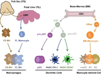
(31) Chapter 1. proposed unique subset markers are in fact shared between distinct cell types [14]. Furthermore, markers of a particular cell subset are not always consistent between mice and humans. This has led to much confusion and debates regarding which subsets represent distinct cell types and which are simply modified versions of the same cell type [15]. Ontogeny studies have recently demonstrated that the MPS are not derived exclusively from monocytes as originally proposed. Indeed, macrophages also arise during embryonic development and DCs also derive from an adult hematopoietic common DC precursor (CDP) too [16–18]. Based on their cellular origin, MPS is composed of three broad families of cells: (i) embryonic progenitor-derived Macrophages, (ii) CDP-derived DCs, which would be subdivided into plasmacytoid DCs (pDCs) and myeloid DCs (myDCs), and (iii) Monocyte-derived Cells, which would include monocyte-derived DC and monocyte-derived macrophages (Figure 1). In addition to this precursor-based classification, functional specialization, cellular activation state, micro-anatomical localization, and surface marker expression should also be taken into account to identify cells in a particular study [14, 19, 20]. In this thesis, we have worked with monocyte-derived cells, including monocytes, monocytederived DCs and monocyte-derived macrophages, and tissue isolated APCs. Based on functional specialization, we refer to these cells as monocytes, DCs, macrophages, or tonsillar myeloid cells.. 32.
(32) INTRODUCTION. MyDC CD1c. +. MyDC CD141. +. Figure 1. Mononuclear phagocytes and their precursors. EMP: erythro-myeloid progenitors, MØ: macrophage, HSC: haematopoietic stem cells, CDP: common Dendritic cell progenitor, pre-pDC: pre-plasmacytoid dendritic cell, pre-cDC: pre-conventional dendritic cell, pDC: plasmacytoid dendritic cell, myDC: myeloid dendritic cell, cMop: common monocyte progenitor, moDC: monocyte-derived dendritic cell, M1: macrophage type 1, M2: macrophage type 2. Figure from [19].. 2.1. Monocytes Monocytes constitute 2% to 10% of all the leukocytes and are originated from bone marrow precursors (Figure 1). They are normally circulating in peripheral blood and their fate depends on the local microenvironment in tissues [20]. Monocytes attach to the endothelium, roll and diapedese into extravascular tissues, where they differentiate into macrophages and DCs, or transiently traverse tissues, maintaining a monocytic profile [21]. In an in vitro model of endothelial trafficking without addition of exogenous cytokines, it was observed that monocytes differentiate into DCs, moving from endothelium to the lumen. Those monocytes that remained in the endothelium became macrophages [22, 23]. Monocytes are multifunctional, and have roles in homeostasis, immune defense, and tissue repair (Table 1). This cell population is very heterogeneous, has an enormous plasticity, and is able to migrate to tissues. 33. 1.
(33) Chapter 1. Three major subsets of blood monocytes have been described in humans, characterized by differential expression of CD14 and CD16 markers. Classical monocytes are described as CD14++ CD16- or CD14+ CD16-, intermediate are defined as CD14++ CD16+, and non-classical monocytes as CD14+ CD16++. This latter population is expanded in various infectious diseases, including HIV infection [24]. Monocytes differentiate into DCs upon exposure to granulocyte-macrophage colonystimulating factor (GM-CSF) plus interleukin-4 (IL-4), or differentiate to macrophages upon exposure to macrophage colony-stimulating factor (M-CSF, also called colony stimulating factor 1 CSF-1; M2 polarization), or upon exposure to GM-CSF (M1 polarization). Therefore, depending on the different cytokines and endothelium milieu, monocytes have three potential fates [20, 25].. 2.2. Macrophages Discovered in 1880 by Elie Metchnikoff, macrophages were the first cellular component of the innate immune system identified. They are originated from the yolk sac or fetal liver precursors, but can also be derived from monocytes (Figure 1). Macrophages are a terminally differentiated cell type distributed in almost every tissue of the body: Kupffer cells in the liver, microglia in the central nervous system, metallophilic, and marginal zone macrophages in the spleen, as well as osteoclasts in the bone and alveolar macrophages in the lungs [26]. Macrophages are distinguished as large vacuolar cells that excel in the clearance of apoptotic cells, cellular debris and pathogens. They present pseudopodia, non-specific esterases, and phagocytic granules, which give them a foamy appearance. Their function and phenotype depend on which tissue they reside and the surrounding cytokine milieu. They contribute to several functions during homeostasis, but also during innate and adaptive immune responses. As implied by their name, derived from the Greek makros (large) and phagein (eat), they are highly efficient phagocytes, and their tasks involve engulfing death cells and clearance of pathogens (Table 1) [27]. Owing to the difficulties in isolating primary macrophages in large quantities, in this thesis we have generated macrophages in vitro by monocyte differentiation. M-CSF. 34.
(34) INTRODUCTION. cultured peripheral blood monocytes remain the predominant in vitro system used to generate macrophages [25].. 2.3. Dendritic Cells (DCs) DCs were first identified and characterized in a series of studies examining mouse splenic adherent cells that were published in the 1970s by Ralph Steinman and Zanvil Cohn [13]. They are originated from common DC precursors (CDP) or from bone marrow monocytes (Figure 1). DCs, whose name derive from the Greek Dendron (treelike), are defined as cells with a stellate morphology that can efficiently present antigens on MHC molecules and activate naive T cells [28]. DCs orchestrate innate and adaptive immune responses to infection. DCs are a heterogeneous population that can be divided into subsets based on expression of cell-surface markers, anatomical distribution, and immunological function. There are two major subtypes of human DCs: myeloid DCs (myDCs, including myeloid CD1c+ DCs and myeloid CD141+ DCs) and plasmacytoid CD303+ DCs (pDCs) [24, 29, 30].. 2.3.1. Plasmacytoid DCs (pDCs) pDCs are a distinct DC lineage based on morphology, gene expression and ability to secrete high levels of type I IFN following viral encounter (Table 1) [31–33]. pDCs are mainly located in blood, lymphoid tissues, and cerebrospinal fluid, but can be recruited to sites of inflammation. In contrast to the “dendritic” appearance of DCs, pDCs exhibit a spherical shape characteristic of antibody-secreting plasma cells. Functionally, pDCs are not phagocytic and have low MHC class II expression, rendering them low ability to present exogenous antigens to CD4+ T cells [34]. They express very high levels of TLR7 and TLR9 that transduce signals from sensing viral nucleic acids that triggers the release of large amounts of type I IFN [29, 35, 36]. pDCs are distinguished from DCs by the lack of CD11c, but the expression of Blood Dendritic Cell Antigen-2 (BDCA2/CD303/CLEC4C), CD123 (IL3-R) and BDCA-4 (CD304/neuropilin-1) [37]. No in vitro model for pDCs is available, and thus they are directly isolated from blood [38].. 35. 1.
(35) Chapter 1. 2.3.2. Myeloid DCs (myDCs) myDCs are highly phagocytic, specialized antigen-processing and -presenting cells. A key feature of myDCs is their continuous replacement from bone marrow precursors and their ability to secrete high levels of IL-12 (Table 1) [7, 39]. DCs express the myeloid cell surface marker CD11c, but lack CD14 or CD16. Blood myeloid DCs can be subdivided depending on differential expression of BDCA cell surface markers as BDCA1+ (CD1c+) or BDCA3+ (CD141+) cells. Unlike pDCs, myeloid DCs express all TLRs except for TLR7 and TLR9, responding well to LPS (TLR4 agonist), flagellin (TLR5 agonist), poly I:C (TLR3 agonist), and R848 (TLR7 agonist) [30]. As regards the anatomical distribution of myDCs in tissues, we distinguish Langerhans cells (LC) and dermal interstitial DCs, which are distributed throughout the mucosal surfaces. LC are located in epidermis and express CD1a and Langerin (CD207), which is absent in dermal interstitial DCs. Dermal interstitial DCs express the mannose receptor (CD206) and DC-SIGN (CD209) [40]. Due to the low frequencies of myDCs in vivo (0.5% – 2% of Peripheral Blood Mononuclear Cells, PBMCs), they have been mostly studied using monocyte-derived DCs by culturing monocytes in the presence of GM-CSF and IL-4. Monocyte-derived DCs are a good myeloid DC model, sharing both similar cell surface markers and response to most stimuli. In this thesis, we have used monocyte-derived DCs but will refer to these cells as DCs.. 36.
(36) INTRODUCTION Table 1. Myeloid cell subsets. Adapted from [41, 42].. Cell type. Monocytes. Macrophages. Cellular markers. Primary locations. CD14++ CD16CD14+CD16+ CD14+/CD16++. Peripheral blood. CD68+ EMR1+ CD14+. Mucosal surface Tissues, Brain (Microglia), Lungs (Alveolar), Liver (Kupffer cells), Bone (Osteoclasts). Average Life Span. Functions/Features. Few days. Patrol blood for surveillance Precursor to macrophages Precursor to DCs Interact with LPS via CD14 Phagocytosis. Week to months. Phagocytose cellular debris and pathogens Antigen presentation M1/M2 polarization Tissue remodeling. CD11c CD123+ BDCA2+ BDCA4+. Blood, cerebrospinal fluid, and lymph nodes. Myeloid Dendritic Cells. CD11c+ CD123– BDCA1+. Blood, mucosal surfaces, lymph nodes, and cerebrospinal Fluid. Days to weeks. Langerhans Cells. CD1a+ Langerin+. Epidermis, and mucosal Epithelia. Week to months. ¯. Plasmacytoid Dendritic Cells. Days to weeks. Antigen presentation IFNα production upon HIV exposure IDO production Antigen presentation Phagocytosis DC migration Induce T cells in cell-mediated immunity Phagocytosis Pathogen degradation CD8+ T-cell priming and B-cell activation. 37. 1.
(37) Chapter 1. Despite the presence of an immune surveillance system elegantly coordinated by APCs, only effective antiretroviral treatment can suppress HIV replication, clearly demonstrating that our defenses have failed to control and eliminate HIV infection.. 3. Human Immunodeficiency Virus (HIV). HIV, the etiological agent of the Acquired Immune Deficiency Syndrome (AIDS), is an infectious agent that can cause persistent disease by attacking the immune system, avoiding normal host defense mechanisms, and even subverting these barriers to promote its own replication. 3.1. Epidemiology HIV has spread worldwide and is now a pandemic infection. According to the World Health Organization, at the end of 2014, 36.9 million of people were living with HIV, of whom 25.8 million were living in Sub-Saharan Africa. That same year, there were 2 million new HIV infections and 1.2 million died of AIDS-related diseases [43]. In 1981, the first clinical observations of AIDS were reported in the United States and then quickly around the world [44]. In 1983, a new human retrovirus, at the time named lymphadenopathy-associated virus (LAV) was isolated from a lymph node biopsy of a patient with generalized lymphadenopathy [45]. Within a year, two distinct groups isolated the novel retrovirus and a serological test was developed to carry out large sero-epidemiological studies, which confirmed that HIV causes AIDS [46–48]. They called it human T-lymphotropic virus type III (HTLV-III) and AIDS-associated retrovirus. To eliminate the multiplicity of names, in 1986 the International Committee on Taxonomy of Viruses recommended that the retrovirus identified should be renamed as HIV. The first HIV therapy appeared in 1987, when a clinical trial showed that azidothymidine (AZT) decreased mortality and opportunistic infections in patients with AIDS [49]. However, viral resistance was quickly developed, and new drugs had to be developed [50]. It was not until 1996 that a combination of a protease inhibitor plus two nucleoside reverse transcriptase inhibitors markedly reduced the AIDS morbidity 38.
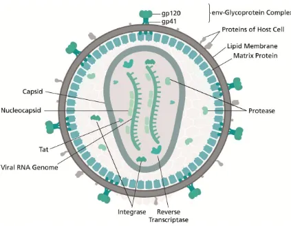
(38) INTRODUCTION. and mortality [51]. This effective treatment was called highly active antiretroviral treatment (HAART). Unfortunately, although HAART provides viral control, HIV has a built-in survival mechanism, creating reservoirs of latent virus that are invisible to both HAART and the immune system. Furthermore, in spite of antiretroviral treatment, HIV infection is associated with abnormal chronic inflammation, and immune activation, which might result in accelerated immunosenescence [52].. 3.2. HIV classification HIV belongs to the group VI of reverse transcribing viruses, Retroviridae family, Orthoretrovirinae subfamily, and Lentivirus genus. It includes the HIV-1 and HIV-2 species, which share many similarities, including basic gene arrangement, mode of transmission, and clinical consequences [53]. However, HIV-2 is characterized by lower transmissibility and reduced progression to AIDS. From an epidemiological point of view, HIV-2 remains largely confined to West Africa, whereas HIV-1 extends worldwide [54]. HIV-1 is classified into three major groups: M (major), O (outlier) and N (new). M group accounts for the majority of infections and can be divided into clades: A, B, C, D, F, G, H, J, K, and Circulating Recombinant Forms (CRF).. 3.3. HIV-1 morphology HIV-1 matured virions are spherical particles of approximately 145 nm of diameter. The outer part of the virus is composed of a lipid bilayer derived from the host cell during the budding of newly formed virions (Figure 2). This lipid bilayer exposes the envelope glycoprotein (Env; gp160) consisting of 3 molecules of gp120 anchored to the membrane by the gp41 transmembrane protein [55]. It also contains several cellular membrane proteins derived from the host cell membrane, including MHC molecules, adhesion molecules, such as leukocyte function-associated antigen (LFA-1) and intercellular adhesion molecules (ICAMs), and co-stimulatory molecules such as CD80, CD86, etc. [56]. Several matrix proteins (MA; p17) lie in the inner surface of the viral membrane, and a conical capsid core (CA; p24) is located in the center of the virus. The capsid encapsulates two copies of the unspliced viral genome, which are stabilized as a ribonucleoprotein complex along with the nucleocapsid protein (NC; p7). Virus 39. 1.
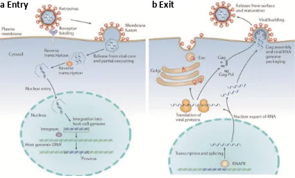
(39) Chapter 1. particles also contain: protease (PR), reverse transcriptase (RT) and integrase (IN) enzymes; accessory proteins: Nef, Vif and Vpr; and three additional accessory proteins: Rev, Tat and Vpu (as reviewed by [55, 57, 58] (Figure 2).. Figure 2. Schematic representation of the structure of HIV. Adapted from "HIV-structure en" by Thomas Splettstoesser (www.scistyle.com) - Own work. Licensed under CC BY-SA 4.0 via https://commons.wikimedia.org/wiki/File:HI-virionCommons structure_en.svg#/media/File:HI-virion-structure_en.svg. Each viral protein acts sequentially and has an important role in allowing viral replication, mediating all the steps involved between viral entry into the host cell until the budding and maturation of the new virion.. 3.4 HIV-1 replication cycle HIV-1 uses the machinery of the target cells to multiply and spread throughout the body. This process, which includes several steps, is called the HIV-1 replication cycle or HIV-1 life cycle. a) Viral entry: It is a complex and intricate process that facilitates delivery of the viral genome to the host cell (Figure 3A). The main viral surface protein implicated is Env, 40.
(40) INTRODUCTION. which is composed of a trimer of gp120 and gp41 heterodimers. First, the virion binds to the host cell, which can be facilitated by distinct cell attachment factors, allowing gp120 Env subunit to bind the host protein receptor CD4. CD4 binding induces conformational changes in Env that enables engagement of a chemokine coreceptor, such as CCR5 or CXCR4, and triggers membrane fusion. Finally, the gp41 subunit of Env enables fusion of the viral and host membranes allowing delivery of the viral cargo [57] (Figure 3A). b) Reverse transcription: Membrane fusion leads to uncoating of viral capside delivering replication enzymes Reverse Transcriptase, Integrase, as well as the viral genomic RNA to the cytoplasm of the cell (Figure 3A). RT retro-transcribes the RNA into a double-stranded linear DNA that complexes with other proteins, forming the pre-integration complex. This pre-integration complex is translocated to the nucleus where the viral DNA is inserted into the host genome by the integrase activity [55, 59, 60]. Depending on the activation status of the target cells, the integrated viral DNA can remain latent in the host genome, or otherwise, it can proceed to the transcription and translation step. c) Transcription and translation: After integration into the host genome, HIV-1 produces sequentially different mRNAs which encode for the viral proteins and the virion genomic RNA: early transcripts encoding Tat and Rev, which control the transcription process; then Env and the HIV-1 accessory genes Vif, Vpr and Vpu; and late-transcripts that encode the virion genomic RNA and the mRNA for the Gag-Pol polyprotein are synthesized. Rev transports the mRNAs encoding the structural proteins from the nucleus to the cytoplasm, where they are translated [61] (Figure 3B). d) Viral assembly, budding and maturation: These events all appear to occur simultaneously at the plasma membrane, where Gag couples membrane binding, virion assembly, and RNA packaging (Figure 3B). HIV-1 virion assembly occurs at the plasma membrane, within specialized cholesterol-enriched micro-domains [62] and share glycosphingolipids and various membrane proteins that reside in lipid rafts [63]. The Gag polyprotein initially assembles into spherical immature particles, in which the membrane-bound Gag molecules project radially towards the interior of the virion. As the immature virion buds, protease is activated and cleaves Gag into its constituent 41. 1.
(41) Chapter 1. matrix, capsid, nucleocapsid, and p6 proteins. Proteolysis is required for conversion of the immature virion into its mature infectious form [64]. Although Gag itself can bind membrane, and assemble into spherical particles, the budding event that releases the virion from the plasma membrane is mediated by the host endosomal sorting complexes required for transport (ESCRT) machinery. Importantly, Viral Like Particles (VLPs) containing only the Gag protein fused to a reporter protein, such as Green Fluorescence Protein (GFP), can be produced and easily released to use them in viral capture assays.. a Entry. b Exit. Figure 3. Schematic representation of the general HIV replication life cycle. A. Viral entry into cells involves the following steps: binding to a specific receptor on the cell surface; membrane fusion either at the plasma membrane or from endosomes (not shown); release of the viral core and partial uncoating; reverse transcription; transit through the cytoplasm and nuclear entry; and integration into cellular DNA to generate a provirus. B. Viral exit involves the following steps: transcription by RNA polymerase II (RNAPII); splicing and nuclear export of viral RNA; translation of viral proteins, Gag assembly and RNA packaging; budding through the cell membrane; and release from the cell surface, and virus maturation. Figure from [65].. HIV-1 mainly infects cells of the immune system, preferentially CD4+ T lymphocytes, but also monocytes, macrophages, and DCs. HIV-1 infection course is characterized by a gradual decline of CD4+ T cells, chronic immune activation, and the impairment of innate and adaptive immune responses that will eventually end in AIDS and lastly, 42.
(42) INTRODUCTION. death. The natural course of untreated HIV-1 infection is characterized by a period that lasts an average of 11 years before progression to AIDS [66]. Once HIV-1 is transmitted, HIV-1 infection course can be divided within 4 stages: eclipse phase, acute or primary infection, chronic infection, and AIDS.. 3.5. Transmission and stages of HIV-1 infection HIV-1 can be transmitted via the exchange of a variety of body fluids from infected individuals, such as blood, breast milk, semen and vaginal secretions. These body fluids must come into contact with a mucosal membrane or damaged tissue or be directly injected into bloodstream (by a needle or syringe) for transmission to occur. The vast majority of HIV-1 infections result from mucosal transmission, with heterosexual transmission being the main route of viral dissemination accounting for 80% of HIV-1 infections worldwide [67]. Clinically, we could distinguish the following phases in HIV-1 untreated infection: a) Eclipse phase (1–2 wk): Cell-free and cell-associated virions cross genital mucosa barrier and infect susceptible target cells, such as CD4+ T cells, macrophages, Langerhans cells and myeloid DCs, which reside within the epithelium or lamina propia [68]. HIV-1 can then spread from the site of infection to other parts of the body by entering the blood and lymphatic vessels in the mucous membrane tissue. The virus must not only establish a small founder population of infected cells at the portal of entry, but also expand that local infection to continue disseminating the virus and the newly infected cells via lymphatic drainage. This establishes a self-propagating infection in the genital draining lymph nodes [69][70]. Throughout this phase, viremia is undetectable, and neither immune response nor symptoms of infection are yet visible (Figure 4). b) Acute (or primary) infection (2–4 wk): infection spreads to establish systemic infection throughout the secondary lymphatic tissues: spleen, gut-associated lymphatic tissue (GALT), and peripheral lymph nodes [69][70]. Acute phase is characterized by high levels of viremia (up to 107 or more copies of viral RNA per milliliter of blood), and the presence of large proportions of infected CD4+ T cells in GALT, blood and lymph nodes. This phase is often, but not always, accompanied by 43. 1.
(43) Chapter 1. “flu-like” symptoms—fever, enlarged lymph nodes, throat inflammation, etc. Around the time of peak viremia, the immune response starts with antibodies against viral proteins and with CD8+ T -cell responses against HIV-1 antigens expressed on infected cells. At the end of the acute phase, the level of viremia declines sharply, 100-fold or more, as a result of partial control by the immune system and the exhaustion of activated target cells. This phase is also characterized by a transient decline in the numbers of CD4+ T cells in blood (Figure 4). c) Chronic infection (1–10 yr): Chronic infection, or “clinical latency,” is characterized by a constant increase in viremia, usually in the order of 1–100,000 copies/mL, referred as the viral load “set point,” and a steady, close to normal, or gradually falling level of CD4+ T cells. Patients in this phase are asymptomatic and usually unaware that they have been infected (Figure 4). d) AIDS: The number of CD4+ T cells declines to the point where immune control is no longer maintained (<200 cells/μL), and opportunistic infections appear. Immune control over HIV-1 is also lost, and the level of viremia rises, culminating in the death of infected individuals. Indeed, untreated HIV-1 infection is one of the most lethal infectious diseases known, with a mortality rate over 95% [60] (Figure 4).. 44.
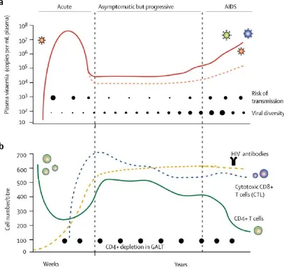
(44) INTRODUCTION. a. b. Figure 4. Course of HIV-1 infection. A. Plasma viremia and B. dynamic changes of lymphocytes and produced specific antibodies. Acute infection is characterized by high plasma viremia (red line), low CD4+ T cell count (green line) and absence of HIV-1 specific antibodies (yellow line). Viremia drops as cytotoxic CD8+ T-lymphocytes develop (blue line) and an individual viral-load set point is reached during chronic infection. It follows an asymptomatic but progressive phase or also called chronic infection characterized by stable high viral load, a gradual depletion of CD4+ T cells and inefficient HIV antibody production and cytotoxic CD8+ T cell responses. AIDS is characterized by an uncontrolled viremia and a decline in CD4+ T cell count to below 200 cells/μl. Viral diversity increases throughout the disease (closed circles, A). The risk of transmission is highest in the first weeks when viremia peaks and during AIDS phase (closed circles, A). CD4+ T cell depletion in GALT occurs during acute phase and is maintained throughout the HIV infection course. GALT=gut-associated lymphoid tissues. Figure from (Simon, Ho, and Abdool Karim 2006).. Thus, infection with HIV-1 results in prolonged, continuous viral replication within the infected host. Remarkably, viral persistence is not prevented by the presence of apparently vigorous virus-specific immune responses. Introduction of successful antiretroviral treatment results in viremia suppression, CD4+ T cell recovery, and 45. 1.
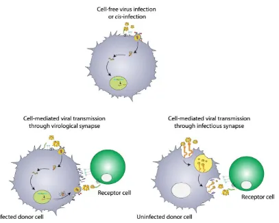
(45) Chapter 1. reduction in systemic immune activation. Despite lowering morbidity and mortality, therapy must continue throughout life, and virus rapidly rebounds if HAART is stopped. Several factors are thought to contribute to persistent viral replication, most notably the destruction of virus-specific T helper cells, the emergence of antigenic escape variants, the antibody inaccessibility to the envelope protein, the impairment of antigen presentation, and the integration and latency of HIV-1 genome into anatomical reservoirs [71]. In addition, HIV-1 evades the immune system by exploiting the myeloid cell function. Myeloid cells contribute to viral pathogenesis through trans-infection, a mechanism that promotes viral capture and transmission to target cells, which become effectively infected [72]. Defining the interactions between HIV-1 and myeloid cells and understanding the role of antigen presenting cells during the course of HIV-1 infection are thus essential to fight against HIV-1.. 4. HIV-1 Interactions with Myeloid Cells Myeloid cells are one of the first cells to encounter the virus, and the specific interaction that occurs between these cells and HIV-1 is critical for HIV-1 to establish infection. Most importantly, HIV-1 is able to efficiently transfer the virus to its primary target cell, the CD4+ T lymphocyte, in which it replicates explosively. Based on viral source, we can distinguish two types of HIV-1 interactions with myeloid cells: cell-free virus infection or cell-mediated transmission. Cell-free virus infection is produced by free-floating viruses that are able to directly infect myeloid cells. Cellmediated transmission takes place when myeloid cells (donor cells) transfer virus to uninfected CD4+ T cells (receptor cells). This cell-to-cell viral transmission to CD4+ T cells can be mediated through virological synapses, where donor myeloid cells are productively infected, or through infectious synapses, where donor myeloid cells are not productively infected but harbor the virus within the cell (Figure 5).. 46.
(46) INTRODUCTION. Figure 5. Different HIV-1-myeloid cell interactions: cell-free virus infection and cell-mediated viral transmission. Cell-mediated viral transmission can be mediated through virological synapses (donor cells are infected), or through infectious synapses (donor cells are not infected).. 4.1. Cell-free virus infection or cis- infection Although CD4+ T cells are the main HIV-1 targets, myeloid cells are also susceptible to HIV-1 infection. Studying HIV-1 infection of myeloid cells in vivo is complex, owing to the low frequency of these cells in HIV-1 infection, their constant migration from mucosal to lymphoid tissues, and the difficulty to obtain cells from these tissues [41]. Cell-free HIV-1 binds myeloid cells via additional specific receptors other than CD4. The C-type lectin receptors (CLRs), such as the dendritic cell-specific intercellular adhesion molecule-3 (ICAM)-grabbing non-integrin (DC-SIGN or CD209) (Figure 5) [73, 74] and dendritic cell immunoreceptor (DCIR) [75], interact with virus particles by binding highmannose oligosaccharides on the heavily glycosylated HIV-1 Env [76–78]. While DCIR is expressed on all myeloid cells, DC-SIGN is expressed only on DCs and macrophages. HIV-1 Env can also bind to immature DCs by interacting with the charged residues of 47. 1.
(47) Chapter 1. heparin sulfate proteoglycans (HSPG) [79] or Syndecan-3 [80]. These mechanisms of HIV-1 binding in myeloid cells facilitate viral interactions with CD4 and viral fusion events leading to cis- infection. All myeloid cells express CD4 and the chemokine co-receptors CCR5 or CXCR4 rendering them susceptible to infection with HIV-1 [81–83]. However, cell-free HIV-1 infection in myeloid cells is generally less productive compared to CD4 + T cells [41]. Limited productive viral infection in myeloid cells could be attributed to lower expression of CD4, CCR5 and CXCR4 [81], and to the internalization of HIV-1 trough phagocytic processes, leading to rapid and extensive viral degradation [73, 84]. R5 HIV1 strains can replicate more efficiently than X4 HIV-1 strains in myeloid cells, being a possible strainer of viral tropism during HIV-1 transmission. HIV-1 infection permissiveness of myeloid cells can vary greatly, depending on their tissue localization, cytokines surrounding, and other environmental factors. For example, maturation of DCs is associated with a marked decline in HIV-1 fusion [85], leading to a decreased in HIV-1 replication in mature DCs, compared to immature DCs [83, 86]. As regards monocytes, it appears that a non-classical monocyte population CD14+ CD16++, which are expanded in HIV-1 infection, are more susceptible to HIV-1 infection and preferentially harbor the virus for long periods, compared to CD14++ CD16- classical monocytes [87, 88]. While sub-epithelial macrophages from vaginal mucosa are susceptible to HIV-1 infection due to high expression of CCR5 co-receptor, jejunum intestinal macrophages are resistant to HIV-1 infection because they do not express CCR5. Infected macrophages have a low but constant viral replication capacity, emphasizing the importance of this viral source. Viral infectiveness in myeloid cells is also modulated by the expression of host restriction factors, such as tetherin [89], apolipoprotein B mRNA-editing enzymecatalytic polypeptide-like 3G (APOBEC3G) [90] and SAM domain and HD domain containing protein 1 (SAMHD1) [91], which represent important innate immune mechanisms against retroviral infection [92]. In contrast to cell-free virus infection, cell-to-cell viral transmission has been shown to be a more rapid and efficient mechanism, where virus and viral receptors are found concentrated at the cell-to-cell synapse [93]. Thus, mathematical models predict that 48.
(48) INTRODUCTION. only ∼ 10% of the infections in the lymphoid tissue are spread by cell-free virions, while the remaining ∼ 90% are transmitted by cell-to-cell transmission [94].. 4.2. Cell-mediated viral transmission via virological synapse Despite being less effective than HIV-1 infection of CD4+ T cells, HIV-1 can infect myeloid cells that could produce progeny virions that are released to infect new target cells via the close contact and formation of a virological synapse (Figure 6A). The primary signal for assembly of virological synapse between an infected donor and an uninfected target cell, is Env-CD4 binding. Subsequent stabilization is achieved by interactions between ICAM-1, ICAM-3 and LFA-1 [95]. Finally, the virus buds at the interface and infects the target cell in a CD4-dependent manner [96–98]. Cell-mediated viral transmission that requires de novo synthesis of new virus has been explained in immature DCs as a delayed phase that occurs for 1 to 2 days upon viral exposure [73]. Although there is limited published data about viral transmission by HIV-1-infected monocytes, circulating monocytes harboring HIV-1 provirus that migrate and differentiate to macrophages could promote HIV-1 replication and cellmediated viral transmission to uninfected cells [99]. HIV-1 replication in macrophages is characterized by the assembly and storage of viral particles in internal cytoplasmic vacuoles [100]. These viral compartments are non-classical endosomal compartments that allow the budding virus to traffic to uninfected CD4 + T cells, mediating viral transmission through infected cells to uninfected cells via virological synapses [101].. 4.3. Cell-mediated viral transmission via Infectious synapse Another cell-mediated viral transmission consists in that virus retained at or near the cell surface is transmitted to a target cell through infectious synapses, a structure analogous to the immunological synapse [102, 103]. Unlike the virological synapse, the infectious synapse does not rely on the productive infection of the donor cell, but also allows for viral transmission to target CD4 + T cells [104] (Figure 6B). The process of viral transmission via formation of infectious synapse is called trans-infection. HIV-1 trans-infection has been mostly studied in DCs [105],. 49. 1.
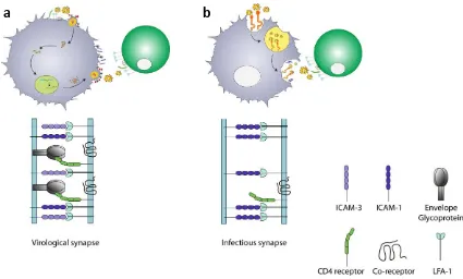
(49) Chapter 1. but other myeloid cells such as monocytes [106] have the potential to establish infectious synapses with target cells [106].. a. b. Figure 6. Comparison between cell-mediated viral transmission through A. virological synapse and B. infectious synapse (also called trans-infection) by DCs. A. In the virological synapse, the primary signal for assembly between an infected donor and an uninfected target cell, is Env-CD4 binding. Subsequent stabilization is achieved by interactions between ICAM-1, ICAM-3 and LFA-1. Finally, the virus buds at the interface and infects the target cell in a CD4dependent manner. B. In the infectious synapse, first, adhesion molecules ICAM-1 and LFA-1 participate in the formation of uninfected DC-CD4+ T-cell conjugates. Then, there is a recruitment of HIV-1 CD4 receptor and CXCR4 and CCR5 co-receptors on the T cell, forming the infectious synapse. Finally, the virus storage compartment in DCs is shifted towards this contact zone, facilitating rapid and efficient infection of the neighboring T cell.. The trans-infection mechanism involves uptake of HIV-1, storage of viruses in a nondegrading compartment, and final release of the virions at the cell to cell contact zone, which enables infection of the target cell [77, 103, 107, 108]. Direct visualization using GFP-tagged HIV-1 revealed that DCs trans-infect HIV-1 by first binding and then concentrating the intact virus at the cellular surface of the donor cell [108, 109]. This viral accumulation leads to the formation of a non-classical endosomal, non-acidic compartment enriched in tetraspanins and MHC class II, which can be under constant remodeling over time [110]. Adhesion molecules, such as ICAM-1 and LFA-1, participate first in the formation of DC-CD4+ T-cell conjugates [102, 111]. Then, there is 50.
(50) INTRODUCTION. a recruitment of HIV-1 CD4 receptor and CXCR4 and CCR5 co-receptors on the T cell, forming the infectious synapse [103]. Finally, the virus storage compartment in DCs is shifted towards this contact zone, facilitating rapid and efficient infection of the neighboring T cell [103]. 3D electron microscopy images of virological synapses showed that membrane extensions are originated from uninfected CD4+ T cells, either as membrane sheets or as filopodial bridges, accessing viral compartments and being involved in HIV-1 transmission from DCs harboring virus to uninfected CD4 + T cells [112]. Although this non-conventional compartment has been mostly studied in DCs, an interesting similar structure exists in HIV-1-infected macrophages [113, 114]. These virus-containing compartments in infected macrophages were initially thought to be late endosomes or multivesicular bodies, but it has been shown that they are separated from the endocytic pathway, co-localize with several tetraspanins (CD63, CD53, CD9, CD81 and CD82), adhesion molecules, MHC classe II and present a nonacidic pH, similar to the DC storage compartment [114, 115]. This compartment also has the ability to allow virus trafficking to a T-cell contact zone, thus facilitating cell-tocell viral transfer via virological synapse, suggesting that this compartment targeted by HIV-1 is similar in macrophages and DCs. The main difference between the systems is that in macrophages virus undergoes assembly on this compartment, while in DCs it gains access through endocytosis [116]. In both cell types, this virus-containing compartment is connected to the extracellular milieu, and is accessible to small membrane-impermeable dyes such as ruthenium red, horseradish peroxidase and lucifer yellow [100, 109, 113, 117]. However, high molecular weight substances, including broadly neutralizing antibodies are excluded [118, 119]. Thus, infectious HIV-1 retained within these compartments in macrophages and DCs seem to be protected from neutralizing antibodies [120]. In sum, this viruscontaining compartment represents a hideout for HIV-1 in myeloid cells, that serves both as an HIV-1 reservoir and as a potent disseminator of the virus within tissues [118, 121]. The precise nature and origin of the capture and storage compartment is currently unknown. It has been demonstrated that capture and transfer of HIV-1 by mature DCs 51. 1.
(51) Chapter 1. converges with a pre-existing traffic pathway of exosomes, which are small secreted vesicles bearing antigens that traffic between APCs [122]. Exosome trafficking augments antigenic presentation and amplifies the immune response [122]. Thus, HIV1 and exosomes compete for DC capture, indicating that they utilize the same pathway [123]. Therefore, HIV-1 is exploiting a pre-existing immune pathway to spread its infection to new target cells.. 5. Identification of Siglec-1 as the HIV-1 receptor implicated in transinfection The mechanism by which DC capture HIV-1 and promote trans-infection of CD4+ T cells has been topic of debate over the last decade. The initial Trojan horse hypothesis suggested that HIV-1 captured by immature DCs in the mucosa may protect the virus from degradation and allow its transport to secondary lymphoid organs, facilitating trans-infection of CD4+ T cells and viral spread [104, 124, 125]. DC-SIGN expressed on immature DCs was described as the main receptor responsible for viral capture and trans-infection of uninfected CD4+ T cells (Figure 7A) [104]. However, subsequent reports demonstrated that most of the captured virions by DC-SIGN are rapidly degraded and presented via MHC class I and MHC class II to T cells, eliciting adaptive immune responses [126, 127]. The limited capacity of immature DCs to sustain transinfection [73] and the limited contribution of DC-SIGN to viral transmission was argued in several independent studies [126, 128–131]. In 2009, viral lipid composition was shown to be involved in viral capture by mDCs. HIV-1 particles produced from cells where glycosphingolipid (GSL) synthesis was inhibited, produced virions with impaired capture and transfer [123]. Furthermore, by modifying the lipid composition of liposomes that mimic the size of HIV-1, but lack any proteins, it was discovered that gangliosides, which are glycosphingolipids with one or more sialic acids (Figure 7B), are involved in viral capture [132]. Specifically, the sialyllactose molecule present in some gangliosides was identified as the determinant moiety for mDC HIV-1 uptake (Figure 7C) [132].. 52.
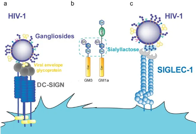
(52) INTRODUCTION. Figure 7. HIV-1 binding to DC receptors. A. HIV-1 can bind to DC-SIGN via recognition of the viral envelope glycoprotein. B. Several gangliosides in the HIV-1 lipid membrane expose a sialyllactose moiety. C. Siglec-1 can capture HIV-1 through recognition of sialyllactose moieties of viral membrane gangliosides. Abbreviations: Cer (ceramide), Gal (galactose), GalNAc (Nacetylgalactosamine), Glu (glucose), SiA (sialic acid). Figure from [133].. Once it was discovered that the sialyllactose motif from viral membrane gangliosides is the attachment factor that mediates viral capture by mature DCs, it was much easier to identify the attachment receptor present on DCs. The main candidates under study were the Siglec family, because these type I transmembrane proteins have an aminoterminal V-set domain that interacts with sialylated ligands [134]. Among all candidates, Siglec-1 was identified as the key factor for HIV-1 spread via infectious DC/T-cell synapses, highlighting a novel mechanism responsible for HIV-1 dissemination (Figure 7C) [135], which was later confirmed in an independent publication [136]. Siglec-1 structure, function and its role as a pathogen receptor, is summarized below.. 53. 1.
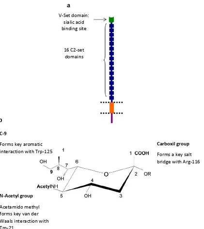
(53) Chapter 1. 5.1 Siglec-1 has a unique structure that favors trans-infection Siglec-1, also known as Sialoadhesin or CD169, is a type I lectin transmembrane glycoprotein that belongs to the Ig superfamily. It consists of an unusually large extracellular region of 17 domains, comprising an N-terminal V-set domain that contains the sialic acid binding site followed by 16 Ig-like C2-set domains [137, 138] (Figure 8A). Siglec-1 has an unusual pattern of conserved cysteine residues characteristic of the Siglec family that gives rise to an intrasheet disulfide bond within domain 1 and a disulfide bond between domains 1 and 2 [134]. Human Siglec-1 is a highly conserved protein within mammals, which is 72% identical to murine Siglec-1. Alignment of the N-terminal region of human and mouse Siglec-1 showed that amino acids important for sialic acid binding are identical, as well as the pattern of cysteine residues [134, 137]. Siglec-1 specifically recognizes N-acetylneuraminic acid (Neu5Ac, one of the most common sialic acids in mammals) linked at α2–3Gal in N- and O-glycans (Figure 8B). Studies that investigated the interaction between Siglec-1 and Neu5Ac, demonstrated by site-directed mutagenesis [139], X-ray crystallography [134], and nuclear magnetic resonance (NMR) [138] that amino acids important for sialic acid binding include an arginine residue at position 116 (which is conserved in all species) and two conserved tryptophans at positions 21 and 125. Use of synthetic sialosides revealed significant but low affinity, for monovalent ligands (kD in the range of 1 mM). Thus, most likely, simultaneous multivalent low affinity associations create sufficient high avidity and lead to biologically meaningful interactions of Siglec-1 for sialic acids on cells or pathogens [140].. 54.
(54) INTRODUCTION. V-Set domain: sialic acid binding site. 16 C2-set domains. b C-9 Forms key aromatic interaction with Trp-125 salt bridge with Arg-116. Carboxil group Forms a key salt bridge with Arg-116. N-Acetyl group Acetamido methyl forms key van der Waals interaction with Trp-21 Figure 8. The features of Siglec-1. A. Schematic diagram showing the domain organization of Siglec-1 protein. B. N-acetylneuraminic acid showing the functional groups that make key contacts with the binding site of Siglec-1. Figure adapted from [141].. Because Siglec-1 possesses a long extracellular domain that can extend the ligandbinding site far from the cell-surface preventing Siglec-1 binding to sialic acids from the same cell (cis- binding), it is suggested that Siglec-1 mediates cell–cell interactions [137]. Furthermore, in contrast to the majority of other Siglecs, Siglec-1 does not contain any inhibitory tyrosine-based motifs in its relatively short cytoplasmic tail, suggesting a primary role as binding partner in cell-to-cell interactions rather than in 55. 1.
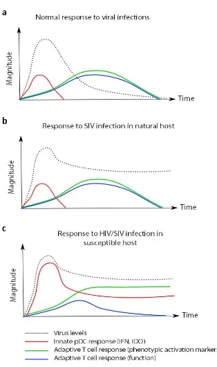
(55) Chapter 1. cell signaling [120, 142]. The long extended Siglec-1 protein structure also can mediate recognition and uptake of sialic acid covered pathogens. Thus, Siglec-1 protrudes outside the cell glycocalyx and can recognize sialyllactose motifs exposed on the HIV-1 membrane.. 5.2. Siglec-1 is an inducible receptor Human Siglec-1 is constitutively absent from monocytes and other peripheral blood leukocytes, but expressed by tissue macrophages in the spleen, lymph node, bone marrow, liver, colon, and lungs [143]. However, Siglec-1 was also found on inflammatory monocytes and macrophages during chronic inflammation, as occurs during autoimmune diseases or viral infections [143–146], where type I IFN levels are up-regulated. That is the case of HIV-1 infection [145], systemic lupus erythematosus [147] and systemic sclerosis [144]. Although there is no in vivo evidence of Siglec-1 expression on DC, it can be induced in vitro after treatment of blood myDC and monocyte-derived DC with inactivated human rhinovirus [148] or after LPS or type I IFN stimulation [135, 136]. The regulation of Siglec-1 is broadly inflammatory and particularly subjected to type I IFN induction [136, 144]. Siglec-1 induction on human PBMCs and various macrophages subsets can be achieved by incubation with either type I IFN or TNFα [106, 146, 149]. Molecules that induce IFNα secretion, such as LPS and poly I:C throughout stimulation of TLRs involved in viral and bacterial sensing (via MyD88independent pathway) also increase Siglec-1 expression [144]. Overall, this strongly suggests a function for Siglec-1 in anti-viral and anti-bacterial activities. 5.3. Siglec-1 in HIV-1 pathogenesis HIV-1 infection results in a storm of different pro-inflammatory stimuli that might control and limit HIV-1 infection [150]. However, Siglec-1 induction by released IFNα could favor HIV-1 transmission in an otherwise antiviral environment. IFNα is a potent antiviral cytokine mainly produced by pDCs in response to HIV-1 challenge. IFNαproducing pDC represents the first line of immune defenses against viral infections [151–153]. Despite its antiviral effect, continuous pDC activation and IFN secretion 56.
Figure
+7
Documento similar
The first objective of this thesis has been to determine if DME expression in DLD-1 human colorectal cancer cells initiates a process of active DNA
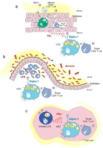
The discovery of Siglec-1 expression on myeloid cervical DCs (Figure 19) and the capacity of Siglec-1 + cells to capture viruses in vivo (Figure 25) help to understand how
Because immune cells in the nasal mucosa can control and regulate viral infections via killing infected res- piratory epithelial cells, low numbers of NK and or T cells in
We found that, at steady state, PSGL-1 knockout mice showed altered proportions of innate and adaptive immune population, as well as an activated phenotype of macrophages
Here we report that infection of human cells with HIV-1 conveys the proteolytic cleavage of GCN2 and that purified HIV-1 and HIV-2 proteases produce direct proteolysis of GCN2 in
The regulation of more than thousand vitamin D target genes in colon carcinoma cells, normal stem cells, cancer stem cells, stromal NFs and CAFs as well as immune cells of the
Tim-3 and CD25 activation/exhaustion markers were upregulated in final NKG2D CAR memory T cell products compared to starting CD45RA − cells; however, PD-1 expression was
Integrated HIV DNA was quantified by q-PCR in cross- sectional samples from different groups of patients: (1) HIV+ group: HIV-monoinfected patients that had never been in contact
