Genetic and molecular analysis or Sanfilippo C syndrome. Generation of a neuronal model using human induced pluripotent stem (iPS) cells and therapeutic strategies
255
0
0
Texto completo
(2) Genetic and molecular analysis of Sanfilippo C syndrome. Generation of a neuronal model using human induced pluripotent stem (iPS) cells and therapeutic strategies. Anàlisi genètic i molecular de la Síndrome de Sanfilippo C. Generació d’un model neuronal utilitzant cèl·lules pluripotent induïdes (iPS) humanes i estratègies terapèutiques Memòria presentada per Isaac Canals Montferrer per optar al títol de Doctor per la Universitat de Barcelona Programa de Genètica Departament de Genètica Tesi realitzada sota la direcció de la Dra. Lluïsa Vilageliu Arqués i el Dr. Daniel Grinberg Vaisman al Departament de Genètica de la Facultat de Biologia de la Universitat de Barcelona. Dra. Lluïsa Vilageliu Arqués. Dr. Daniel Grinberg Vaisman. Isaac Canals Montferrer Barcelona, 2014.
(3)
(4) “Ever tried. Ever failed. No matter. Try again. Fail again. Fail better.”.
(5)
(6) PRESENTATION Sanfilippo C syndrome is a rare inherited monogenic disease that presents a pronounced neurodegenerative course since early stages of patient’s life. It is caused by mutations in the HGSNAT gene, localized few years ago in the chromosome 8, which codes for the enzyme acetyl-CoA α-glucosaminide N-acetyltransferase, a lysosomal membrane protein. Its function is to transfer an acetyl group to glucosamine residues in the heparan sulfate chain that is being degraded. The abnormal action of this protein in patients promotes the accumulation of partially degraded heparan sulfate chains inside the lysosomes, causing their dysfunction. Because of this reason, Sanfilippo C syndrome is a lysosomal storage disease, particularly a mucopolysaccharidoses due to the nature of the stored substrate. Heparan sulfate is a glycosaminoglycan (previously known as mucopolysaccharid) found in the extracellular matrix as part of proteoglycans. These molecules participate in many different and important cellular functions such as migration or adhesion. The impairment of their homeostasis leads to the dysfunction of many cellular processes. This thesis makes an important contribution to the field of Sanfilippo C molecular study. Mutational analysis and the consequent mutation characterization have been performed in order to extend the knowledge of the disease. Moreover, several different therapeutic approaches have been tested as a first step in the achievement of a successful therapy for this devastating neurodegenerative disorder, for which no effective treatment exists. Finally, a neuronal model for Sanfilippo C syndrome have been generated using induced pluripotent stem cells, which may help understand the molecular processes that contribute to the development of the neural disease and will be a valuable tool for the pursuit of successful treatments..
(7)
(8) INDEX.
(9)
(10) INTRODUCTION Lysosomal Storage Disorders. 3. The lysosome. 3. Lysosomal storage disorders. 4. Mucopolysaccharidoses. 8. Sanfilippo Syndrome. 10. General aspects. 10. Clinical aspects. 12. Diagnosis. 14. Incidence. 15. Subtype A. 15. Subtype B. 17. Subtype C. 18. Subtype D. 20. Heparan Sulfate. 22. Heparan sulfate and heparan sulfate proteoglycans. 22. Heparan sulfate biosynthesis. 23. Heparan sulfate degradation. 25. Therapeutic Approaches. 28. Enzyme replacement therapy. 28. Substrate reduction therapy. 29. Coenzyme Q10. 32. Pharmacological chaperones for enzyme-enhancement-therapy. 33. Suppression of premature termination codons. 34. Overexpression of transcription factor EB. 35. Gene therapy. 36. Stem cell therapy. 40.
(11) RNA Splicing. 43. The splicing process. 43. The nonsense mediated decay quality control. 46. Therapies to correct splicing mutations. 48. RNA interference. 50. History and mechanisms. 50. Therapy based on this mechanism. 52. RNA interference therapies for Sanfilippo syndrome. 53. Induced Pluripotent Stem Cells Reprogramming from fibroblasts to induced pluripotent stem cells. 55 55. Use of human induced pluripotent stem cells in lysosomal storage disorders. 57. Direct conversion of fibroblasts to neurons or neural stem cells. 58. Cellular models of Sanfilippo syndrome. 59. OBJECTIVES Objectives. 65. RESULTS Report of the thesis supervisors about the contribution of the PhD student to the articles in this doctoral thesis. 69. Article 1. 75. Article 2. 87.
(12) Article 3. 113. Article 4. 127. Annex. 173. DISCUSSION Discussion. 177. CONCLUSIONS Conclusions. 199. REFERENCES References. 203. RESUM Resum. 231. AGRAÏMENTS Agraïments. 235.
(13) ABBREVIATIONS AAV – Adeno-associated virus. IdoA – Iduronic acid. AON – Anti-sense oligonucleotide. IGFII – Insulin-like growth factor II. BBB – Blood-brain barrier. iNSC – Induced neural stem cells. BMT – Bone marrow transplantation. iPSC – Induced pluripotent stem cell. cDNA – Complementary. ISE – Intronic splicing enhancer. deoxyribonucleic acid. ISS – Intronic splicing silencer. CHX – Cycloheximide. LSD – Lysosomal storage disorder. CoQ10– Coenzyme Q10. M6P – Mannose-6-phosphate. CNS – Central nervous system. miRNA – Micro ribonucleic acid. CS – Chondroitin sulfate. MPS – Mucopolysaccharidoses. DNA – Deoxyribonucleic acid. mRNA – Messenger ribonucleic acid. DMMB – 1,9-dimethylmethylene blue. MSC – Mesenchymal stem cells. DS – Dermatan sulfate. NMD – nonsense-mediated decay. dsRNA – Double strand ribonucleic acid. NSC – Neural stem cells. EGFR – Epidermal growth factor receptor. PTC – premature termination codon. EJC – Exon junction complex. RISC – Ribonucleic acid-induced silencing. ER – Endoplasmic reticulum. complex. ERT – Enzyme replacement therapy. RNAi – Ribonucleic acid. ESC – Embryonic stem cells. RNAi – Ribonucleic acid interference. ESE – Exonic splicing enhancer. shRNA – Small hairpin ribonucleic acid. ESS – Exonic splicing silencer. siRNA – Small interfering ribonucleic. GA – Golgi apparatus. acid. GAG – Glycosaminoglycan. SNM – Spherical neural mass. GlcNAc – N-acetylglucosamine. SNP – Single nucleotide polymorphism. GlcA – glucuronic acid. snRNA – Small nuclear ribonucleic acid. hnRNP – Heterogeneous nuclear. SRT – Substrate reduction therapy. ribonucleoprotein. ss – Splice site. HS – Heparan sulfate. TFEB – Transcription factor EB. HSCT – Hematopoietic stem cell. UTR – Untranslated region. HSPG – Heparan sulfate proteoglycan. WT – Wild-type.
(14) FIGURES. INTRODUCTION Figure 1.. Lysosomes. 3. Figure 2.. Pathways with lysosomal involvement. 4. Figure 3.. Organelles affected in LSDs. 5. Figure 4.. Typical MPS cell vacuolization. 9. Figure 5.. Sanfilippo syndrome lysosomes. 10. Figure 6.. LSDs inside the neuron. 12. Figure 7.. Sanfilippo patients. 13. Figure 8.. SGSH gene. 16. Figure 9.. NAGLU gene. 17. Figure 10.. HGSNAT gene. 19. Figure 11.. HGSNAT mutations grouped by type. 19. Figure 12.. GNS gene. 21. Figure 13.. HSPGs functions. 22. Figure 14.. HS biosynthesis. 23. Figure 15.. Endocytosis of HS prior to its degradation. 25. Figure 16.. HS degradation. 26. Figure 17.. SRT basis. 29. Figure 18.. Mechanism of action of genistein. 30. Figure 19.. Chaperone treatment possibilities. 33. Figure 20.. Read-through mechanism. 35. Figure 21.. M6P-mediated enzyme excretion. 39. Figure 22.. Conserved splice sites. 44. Figure 23.. Splicing reaction. 45. Figure 24.. NMD mechanism models. 47. Figure 25.. U1 snRNA structure. 49. Figure 26.. siRNA structure. 51. Figure 27.. RNAi mechanism. 51. Figure 28.. iPSC basis and applications. 55.
(15) Figure 29.. Disease-modelling using iPSC. 57. Figure 30.. Neuronal-disease-modelling. 58. Figure 31.. Neuronal storage. 59. Figure 32.. Percentage of alleles for each mutation in Spanish patients. 179. Figure 33.. Origin and migration of the c.234+1G>A allele. 180. Figure 34.. Intronic deletion. 185. Figure 35.. Modified U1 snRNAs used. 187. Figure 36.. Alternative ss near the natural one. 188. Figure 37.. SNMs in suspension. 191. Figure 38.. Neuronal cultures. 192. Table 1.. Lysosomal Storage Disorders. 7. Table 2.. ERT strategies for Sanfilippo syndrome in animal models. 28. Table 3.. Gene therapy strategies for Sanfilippo syndrome. 37. Table 4.. RNAi delivery systems. 53. DISCUSION. TABLES INTRODUCTION.
(16) INTRODUCTION.
(17)
(18) INTRODUCTION. Lysosomal Storage Disorders The lysosome Lysosomes are subcellular organelles (figure 1) first described more than 60 years ago (De Duve et al., 1955). They are responsible for the turnover of cellular and extracellular constituents and organelles (De Duve and Wattiaux, 1966) and participate in some important cellular processes such as membrane repair, autophagy, cellular death via signal transduction, apoptosis, receptor recycling, neurotransmission regulation, skin pigmentation, bone biology and play an important role in cellular homeostasis and the endolysosomal system (Boustany, 2013; Saftig and Klumperman, 2009). Molecules degraded into lysosomes come either from outside the cell by phagocytosis, pinocytosis or receptor-mediated endocytosis, or from inside by microautophagy or macroautophagy (Ciechanover, 2005). In addition, the organelle is important for antigen presentation and phagocytosis, which are essential for the control of inflammation and autoimmunity. A scheme of some pathways in which lysosomes are involved can be seen in figure 2.. Figure 1. Lysosomes. Lysosomes display in a transmission electron microscopy image (dark vesicles).. Lysosomes present a single membrane and contain about 50-60 hydrolytic enzymes, integral membrane proteins and transporters. These hydrolases are active at the acidic pH (around 5) inside the organelle and most of them are soluble and localized in the lysosomal lumen. There are proton transporters located in the lysosomal membrane to maintain the acid pH at which the enzymes are functional. Lysosomal enzymes are synthesized in their inactive form at the endoplasmic reticulum (ER) and then glycosylated and transported to the Golgi apparatus (GA). Once in there, most of them acquire a 3.
(19) INTRODUCTION. mannose-6-phosphate (M6P), which direct the protein to the lysosome via the M6P receptor. The low pH inside the lysosome allows the dissociation of the hydrolytic enzyme from the M6P receptor. Finally, the enzymes are maturated and become functional. Some enzymes are not bound to the M6P receptor and are excreted outside the cell, where surrounding cells can take them. Alternative trafficking signals have been proposed for some lysosomal proteins (Lefrancois et al., 2003; Reczek et al., 2007). For instance, lysosomal membrane proteins are not directed via M6P. They present alternative signal peptides at the C-terminal end of the protein, which direct them to the lysosome.. Figure. 2.. Pathways. with. lysosomal involvement. Different pathways in which an appropriate lysosomal. function. plays. an. important role such as autophagy, extracellular turnover or exocytosis (Schultz et al., 2011).. Lysosomal storage disorders Lysosomal storage disorders (LSDs) are a group of inherited metabolic diseases caused by mutations in genes encoding lysosomal proteins (from lumen and membrane), activator proteins, ER or GA modifying proteins that lead to an accumulation of partially degraded macromolecules inside the lysosomes. This accumulation promotes a cascade of events that affects not only the endosomal–autophagic–lysosomal system, but also other organelles (figure 3). 4.
(20) INTRODUCTION. Figure. 3.. Organelles. affected in LSDs. Examples of LSDs affecting different organelles. (Platt. et. al.,. 2012).. More than 50 different LSDs have been reported until today (Table 1). All of them are monogenic disorders, and the majority present an autosomal recessive inheritance. Some present allele heterogeneity and some others present gene heterogeneity. It is thought that some of them remain to be described; since maybe one LSD exist for each lysosomal protein, but some of them could be lethal at early developmental stages (Futerman and van Meer, 2004). The first description of a LSD was that of Tay-Sachs disease in 1881 although the first link between an enzymatic deficiency and a LSD was not established until later, in 1963, for Pompe’s disease (Hers, 1963). Even though individually each LSD can be considered as a rare disease due to their low incidence in population ranging from 1:57000 (Gaucher disease) and 1:4200000 (sialidosis) live births, all together, LSDs present an incidence about 1:4000-8000 (Ortolano et al., 2014). LSDs can be classified either by the defective protein or by the storage material (reviewed in Futerman and van Meer, 2004), being that last criteria the most. used.. LSDs. include. sphingolipidoses,. mucolipidoses,. glycoproteinoses,. oligosaccharidoses or mucopolysaccharidoses (table 1). Clinical symptoms are variable among different LSDs from mild symptoms to severe disorders affecting a wild range of tissues. Despite the broad variability in the symptoms, some of them are common in most of the LSDs such as bone and central nervous system (CNS) affectation, dysmorphology and visceromegaly. 5.
(21) INTRODUCTION. Several therapies have been tested for different LSDs including selective treatments as enzyme replacement therapy (ERT), substrate reduction therapy (SRT), use of chemical chaperones and cell therapy using bone marrow transplantation (BMT), mesenchymal stem cells (MSC) or neural stem cells (NSC) (reviewed in Boustany, 2013; Hollak and Wijburg, 2014; Kim, 2014; Ortolano et al., 2014; Shayman, 2014). Despite all these attempts, until today no effective therapy has been developed for most of LSDs, especially for those affecting CNS, so only therapies aimed at improving patient’s quality of life are available. To study and test new therapeutic approaches for these LSDs the development of neuronal models would be very useful. Some animal models have been described or obtained for many LSDs (reviewed in Ellinwood et al., 2004). In the recent years, induced pluripotent stem cells allowed scientist to obtain cellular models for several LSDs (Higuchi et al., 2014; Huang et al., 2011; Lemonnier et al., 2011; Maetzel et al., 2014; Mazzulli et al., 2011; Panicker et al., 2012; Park et al., 2008; Tiscornia et al., 2013; Tolar et al., 2011; Trilck et al., 2013). This approach allows generating different cell types relevant for the disease. Moreover, these cells present exactly the same genome of the patients. Thus, cells that cannot be obtained directly from the patient can be generated by this technology. It also offers the advantage to test different therapeutic strategies in those cell types most affected by the disease.. 6.
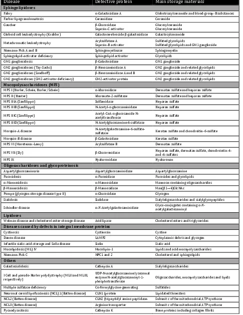
(22) INTRODUCTION. Table 1. Lysosomal Storage Disorders. Defective enzyme and main storage material are indicated for each LSD (adapted from Futerman and van Meer, 2004). Disease. Defective protein. Main storage materials. Fabry. α-Galactosidase A. Globotriasylceramide and blood-group-B substances. Farber lipogranulomatosis. Ceramidase. Ceramide. Gaucher. β-Glucosidase Saposin-C activator. Glucosylceramide Glucosylceramide. Globoid cell leukodystrophy (Krabbe ). Galactocerebroside β-galactosidase. Galactosylceramide. Niemann-Pick A and B. Arylsulfatase A Saposin-B activator Sphingomyelinase. Sulfated glycolipids Sulfated glycolipids and GM1 ganglioside Sphingomyelin. Sphingolipid-activator deficiency. Sphingolipid activator. Glycolipids. GM1 gangliosidosis. β-Galactosidase. GM1 ganglioside. GM2 gangliosidoses (Tay-Sachs). β-Hexosaminidase A. GM2 ganglioside and related glycolipids. GM2 gangliosidoses (Sandhoff). β-Hexosaminidase A and B. GM2 ganglioside and related glycolipids. GM2 gangliosidoses (GM2-activator deficiency). GM2-activator protein. GM2 ganglioside and related glycolipids. MPS I (Hurler, Scheie, Hurler/Scheie). α-Iduronidase. Dermatan sulfate and heparan sulfate. MPS II (Hunter). Iduronate-2-sulfatase. Dermatan sulfate and heparan sulfate. MPS IIIA (Sanfilippo). Sulfamidase. Heparan sulfate. MPS IIIB (Sanfilippo). N-Acetyl-α-glucosaminidase. Heparan sulfate. Sphingolipidoses. Metachromatic leukodystrophy. Mucopolysaccharidoses (MPS). MPS IIIC (Sanfilippo) MPS IIID (Sanfilippo). Acetyl-CoA:α-glucosamide Nacetyltransferase N-Acetylglucosamine-6-sulfatase. Heparan sulfate Heparan sulfate. Morquio-B disease. N-Acetylgalactosamine-6-sulfatesulfatase β-Galactosidase. MPS VI (Maroteaux-Lamy). Arylsulfatase B. MPS VII (Sly). β-Glucoronidase. MPS IX. Hyaluronidase. Heparan sulfate, dermatan sulfate, chondrointin-4and -6-sulfates Hyaluronan. Aspartylglucosaminuria. Aspartylglucosaminidase. Aspartylglucosamine. Fucosidosis. α-Fucosidase. Fucosides and glycolipids. α-Mannosidosis. α-Mannosidase. Mannose-containing oligosaccharides. β-Mannosidosis. β-Mannosidase. Man(β1→4)GlcNAc. Pompe (glycogen-storage-disease type II). α-Glucosidase. Glycogen. Sialidosis. Sialidase. Sialyloligosaccharides and sialylglycopeptides. Schindler disease. α-N-Acetylgalactosaminidase. Glyco-conjugates containing α-Nacetylgalactosaminyl. Acid lipase. Cholesterol esters and triglycerides. Morquio-A disease. Keratan sulfate and chondroitin-6-sulfate Keratan sulfate Dermatan sulfate. Oligosaccharidoses and glycoproteinosis. Lipidoses Wolman disease and cholesterol-ester-storage disease. Diseases caused by defects in integral membrane proteins Cystinosis. Cystinosin. Cystine. Danon disease. LAMP2. Cytoplasmic debris and glycogen. Infantile sialic-acid-storage and Salla disease. Sialin. Sialic acid. Mucolipidosis (ML) IV. Mucolipin-1. Lipids and acid mucopolysaccharides. Niemann-Pick C. NPC1 and 2. Cholesterol and sphingolipids. Galactosialidosis. Cathepsin A. Sialyloligosaccharides. I Cell and pseudo-Hurler polydystrophy (MLII and MLIII, respectively). UDP-N-acetylglucosamine:lysosomal enzyme N-acetylglucosaminyl-1phosphotransferase. Oligosaccharides, mucopolysaccharides and lipids. Multiple sulfatase deficiency. Cα-formylglycine-generating. Sulfatides. Neuronal ceroid lipofuscinosis (NCL)1 (Batten disease). Lipidated thioesters. NCL2 (Batten disease). CLN1 (protein enzyme CLN2 (tripeptidyl amino peptidasepalmitoylthioesterase-1). NCL3 (Batten disease). Arginine transporter 1). Subunit c of the mitochondrial ATP synthase. Pycnodysostosis. Cathepsin K. Bone proteins including collagen fibrils. Others. 7. Subunit c of the mitochondrial ATP synthase.
(23) INTRODUCTION. Mucopolysaccharidoses Mucopolysaccharidoses (MPS) are a LSD group characterized by the impairment in glycosaminoglycans (GAGs, previously known as mucopolysaccharides) degradation. Each MPS is due to mutations in genes coding for a lysosome enzyme involved in GAG catabolism. The enzyme deficiency causes the storage of partially degraded GAGs into lysosomes (and their increased excretion in urine) leading to progressive cellular damage that affects different tissues and organs (Neufeld and Muenzer, 2001). MPS consist of a group of seven different diseases with an autosomal recessive inheritance pattern, except for MPS II, which presents X-linked recessive inheritance. Some reports differentiate among 11 different MPS since MPS III presents four different subtypes and MPS IV presents two subtypes. In each MPS, one or more GAGs are accumulated depending on the enzyme deficiency. Defective enzymes and the main storage products can be found in table 1. The incidence of MPS vary among populations from 1.9 to 4.5 for each 100.000 live births being MPS III, as a group, the most common (Applegarth et al., 2000; Baehner et al., 2005; Héron et al., 2011; Meikle et al., 1999; Nelson et al., 2003; Pinto et al., 2004; Poorthuis et al., 1999; Poupetová et al., 2010). MPS present a large variety of symptoms, being organomegaly and coarse facial features the most frequent. Other organs and functions such as hearing, vision, cardiovascular system and joint mobility are usually affected. In severe MPS, the CNS function is also impaired. These clinical symptoms are chronic and course progressively during the patient’s life. They are due to the accumulation of undegraded GAGs inside lysosomes that increase their size and number (figure 4) affecting other organelles and cellular functions.. 8.
(24) INTRODUCTION. Figure 4. Typical MPS cell vacuolization. Characteristic appearance of an MPS patient’s cell (Neufeld and Muenzer, 2001).. Early diagnosis is important for the treatment of MPS without neurological affectation and available ERT (MPS I, II and VII). Usually diagnosis is delayed because of the wide clinical presentations. Typically they are diagnosed by the analysis of urinary GAGs that allows the differentiation between each MPS but not among the subtypes. For an accurate diagnosis, enzyme activity assays in fibroblasts, leukocytes, blood or plasma should be carried out (Neufeld and Muenzer, 2001). Many therapies have been tried for MPS with different results such as ERT, SRT, pharmacological chaperones, stop-codon read-through, gene therapy and cell therapy (reviewed in Giugliani et al., 2012).. 9.
(25) INTRODUCTION. Sanfilippo Syndrome General aspects Sanfilippo syndrome, also known as mucopolysaccharidoses III (MPS III), is a group of LSDs with an autosomal recessive inheritance pattern that was first described more than 50 years ago (Sanfilippo et al., 1963). Later MPS III was classified in four different subtypes (A: OMIM 252900, B: OMIM 252920, C: OMIM 252930, D: OMIM 252940), caused by mutations in four different genes, which code for the following enzymes: A, heparan Nsulfatase;. B,. α-N-acetylglucosaminidase;. C,. acetyl-CoA. α-glucosaminide. N-. acetyltransferase; and D, N-acetylglucosamine 6-sulfatase. These enzymes are involved in the degradation pathway of the glycosaminoglycan heparan sulfate (HS) (Neufeld and Muenzer, 2001). The deficient enzymes were identified during the 70’s decade and in the last 20 years, so were the genes coding for these enzymes (Fan et al., 2006; Hřebíček et al., 2006; Robertson et al., 1988; Scott et al., 1995; Zhao et al., 1996). The lack of activity for one of these enzymes leads to the accumulation of partially degraded HS chains into the lysosomes. In patient’s neurons and animal models, lysosomes increase in number and size due to this storage (Kurihara et al., 1996; Wilkinson et al., 2012) as showed in the cells of the mouse model of Sanfilippo subtype B (figure 5).. Figure 5. Sanfilippo syndrome lysosomes. A. WT mice lysosomes. B. MPS IIIB mice lysosomes. C. Typical lysosome from affected mice (Vitry et al., 2010).. HS is one of the most common and important GAGs located in the extracellular matrix as a part of proteoglycans, which participate in many different cellular functions (Sarrazin et al., 2011). HS accumulation causes an alteration in the lysosomal environment 10.
(26) INTRODUCTION. since it can bind to various hydrolases reducing their activity (Walkley, 2004). It causes the secondary accumulation of glangliosides and dermatan sulfate (DS) that may contribute to the central nervous system pathology (Lamanna et al., 2011). GAGs are important components of brain and peripheral nerves. The fact that these tissues present a limited capacity of regeneration, a high sensitivity to damage and a need of long cellular survival could explain the severe neural affection in Sanfilippo patients. It has been demonstrated in MPS IIIB mice model that not only the CNS is severely affected but also the peripheral nervous system (Fu et al., 2012). It is possible that the HS fragments released to the extracellular matrix interfere with many HS functions, favouring the disease development. The lysosomal storage of GAGs in neurons can lead to brain atrophy (Palmucci et al., 2013). Since GAGs act as ligands for different factors, the abnormal storage can affect different signalling pathways. The injury in neurons activates microglia and the constant release of inflammatory mediators. The accumulation in storage vesicles has been detected also in microglial cells in a mouse model of Sanfilippo B disease (Li et al., 1999). These cells play an important role in the brain defence and may release different toxic products. Thus, affection of the glial cells together with the inflammation may contribute to neuronal degeneration in MPS III (Ohmi et al., 2003). It has been established that the storage of gangliosides (secondarily accumulated in Sanfilippo disease) leads to a decreased uptake of calcium by the ER with the consequent increase of the cytosolic levels that promotes neuronal apoptosis, favouring neurodegeneration. The decreased levels of calcium in the ER and other alterations activate the unfolded protein response, which also leads to an increased apoptosis (reviewed in Boustany 2013) that is an important contributor to the severe neurodegeneration. Astrocyte dysfunction also plays a role in neuronal degeneration as proved in a mouse model of multiple sulfatase deficiency, another LSD (Di Malta et al., 2012), and for other neurological disorders (Scuderi et al., 2013). It has been reported that in MPS IIIA mice there is an increase in the number of autophagosomes due to an impairment in the autophagosome-lysosome function, which probably leads cells to death. Thus, LSDs could be considered autophagy disorders (Settembre et al., 2008). Some of the processes involved in Sanfilippo-depending neuronal toxicity are summarized in figure 6. 11.
(27) INTRODUCTION. Figure 6. LSDs inside the neuron. Lysosomal storage and secondary mechanisms leading to cell death in neurons (Boustany, 2013). Clinical aspects The four Sanfilippo subtypes present similar clinical symptoms, with phenotypic variation and probably with some very mild forms that are difficult to recognize (Neufeld and Muenzer, 2001). The most significant trait is the severe central nervous system degeneration, unique among MPS disorders, that leads to motor deterioration, learning difficulties, hyperactivity, aggressive behaviour, sleeping problems and pronounced mental retardation (Valstar et al., 2008). It is accompanied by mild somatic manifestations such as hirsutism, mild hepatosplenomegaly, joint stiffness, dysphagia, hypertrichosis, severe hypoacusia, speech loss, mild skeletal involvement, in some cases coarse facial features although not very pronounced and sometimes diarrhoea (Neufeld and Muenzer, 2001). Epileptic seizures (Muenzer, 1986) and retinitis pigmentosa (Berger-Plantinga et al., 2004; Ruijter et al., 2008) can also occur in old patients and an early onset of puberty has been described in some MPS III patients of different subtypes. 12.
(28) INTRODUCTION. These mild somatic manifestations usually lead to a delayed diagnostic after the onset of symptoms. First symptoms appear in early life, among two and six years in apparently normal children, but can occur earlier or later and usually consist in developmental delay and behavioural problems, sometimes accompanied by speech loss (Valstar et al., 2008). Severe neurodegeneration occurs between six and ten years in most patients, together with a rapid deterioration in social skills and a profound mental retardation. The progressive degeneration leads to a severe cortical atrophy, progressive dementia, motor deterioration, sleep disturbances and severe behaviour problems with possible physical aggression. Death usually occurs at the second or third decade in severe patients while in mild patients, life may extend until the fifth or the sixth. It is difficult to differentiate patients from different subtypes because the symptoms are similar for all of them and patients present a significant heterogeneity in the symptomatology (Neufeld and Muenzer, 2001). Even so, it has been found that subtype A is the most severe form of Sanfilippo syndrome, while subtype B and specially subtype C present a slower progress of the symptoms (van de Kamp et al., 1981). Many attenuated cases have been described for subtypes A, B and C with late onset (Coppa et al., 2013; Meyer et al., 2008; Moog et al., 2007; Ruijter et al., 2008; Valstar et al., 2011). At present, Sanfilippo patients are usually classified into severe, intermediate and attenuated cases depending on the severity of the symptoms (Valstar et al., 2010). Appearance of typical MPS patients can be appreciated in figure 7.. Figure 7. Sanfilippo patients. MPS IIIA patient (left image) and MPS IIIB patient (right image) both at seven years old (Neufeld and Muenzer, 2001).. 13.
(29) INTRODUCTION. Diagnosis Diagnosis usually begins with the measure of the increased GAGs in urine by different methods. The most frequent consists of a 1,9-dimethylmethylene blue (DMMB) test. DMMB binds GAGs and the complex can be quantified by spectrophotometry. This method was described more than 20 years ago but was improved later (Barbosa et al., 2003). Other methods include cetyltrimetylammonium bromide screening, toluidine blue spot test or alcian blue method. All these techniques allow the detection of the increase in urinary GAGs. Next step specifically detects the type of GAGs that are increased, either by a thin-layer chromatography (Humbel and Chamoles, 1972) or by electrophoretic separation (Stone, 1998). These methods are useful for the differential diagnosis of MPS III from other types of MPS, but still do not provide information about the MPS III subtype. In order to confirm diagnosis and establish the MPS III subtype, loss of specific enzyme activity in fibroblasts, leukocytes or plasma should be assessed with fluorogenic (4methylumbelliferyl), spectrophotometric or radiolabeled substrates, and recently, new tandem mass spectrometric assays (Wolfe et al., 2012). Fluorogenic substrates are preferable over radiolabeled substrates because their sensitiveness and stability are higher (Kleijer et al., 1996). Once the MPS III subtype is established, next step consists in the identification of mutations causing the disease in each patient. It is useful to analyze related carriers and to perform prenatal diagnosis. Since there are prevalent mutations in some populations for subtypes A, B and C (Bunge et al., 1997; Di Natale et al., 1998; Mangas et al., 2008; Montfort et al., 1998; Ruijter et al., 2008; Tanaka et al., 2002; Weber et al., 1998), the presence of these mutations can be tested first for patients belonging to these populations. Prenatal diagnosis can be performed in families with an affected child or an affected relative. Amniotic fluid or chorionic villi are usually used and the previously described assays, such as enzyme activity assay or mutation analysis, allow the diagnosis (Hopwood, 2005). Recently, a new technique has been proposed for newborn screening of MPS types I, II and III (Ruijter et al., 2012a). This new approach based on the detection of 14.
(30) INTRODUCTION. disaccharides derived from HS and DS could make possible the earlier diagnosis of MPS III patients, which could be important for future therapeutic approaches to improve prognosis and delay the appearance of degeneration symptoms. Even so, further studies are needed to establish the potential of this new test.. Incidence Some studies in different populations indicated that Sanfilippo syndrome as a group, is the most prevalent MPS, with incidences between 0.29 and 1.89 for each 100000 live births (Applegarth et al., 2000; Baehner et al., 2005; Meikle et al., 1999; Nelson et al., 2003; Pinto et al., 2004; Poorthuis et al., 1999; Poupetová et al., 2010)(Meikle et al., 1999; Nelson et al., 2003; Pinto et al., 2004; Poorthuis et al., 1999). These incidences may underestimate the actual prevalence of different MPS III types because of the high heterogeneity in symptomatology and the difficulties in the correct diagnosis of mild forms. According to published data, subtype A is more prevalent in Northern and Central European countries such as Germany, France, the Netherlands, United Kingdom, Sweden or the Czech Republic, and also other countries like Australia, while subtype B is more prevalent in Southern European countries, such as Portugal and Greece and other countries like Taiwan. In contrast, a recent study in Spain showed Sanfilippo syndrome type A as the most frequent subtype in Spanish Sanfilippo patients (62% of the cases), while type B (20%) and type C (18%) were less frequent (Delgadillo et al., 2013). Generally, subtype C and mainly subtype D are less common in all populations (Baehner et al., 2005; Emre et al., 2002; Héron et al., 2011; Lin et al., 2009; Malm and Månsson, 2010; Michelakakis et al., 1995; Pinto et al., 2004; Poorthuis et al., 1999; Poupetová et al., 2010).. Subtype A MPS IIIA or Sanfilippo syndrome type A is caused by mutations in the SGSH gene, coding for sulfamidase (also known as heparan sulfate sulfatase or N-sulfoglucosamine 15.
(31) INTRODUCTION. sulfohydrolase, EC 3.10.1.1). This enzyme releases sulfate groups linked to the amino group of glucosamine. The gene is localized at 17q25.3 (Scott et al., 1995) with an approximated length of 11 Kb and contains eight exons (figure 8). It codes for a protein of 502 aminoacids with five possible glycosylation sites. A total of 139 mutations have been identified until now, including 106 missense/nonsense mutations, 17 small deletions, nine small insertions, three gross deletions, two splicing mutations, one small indel and one gross insertion/duplication (HGMD® Professional 2014.3). Prevalent mutations have been described for different populations such as p.R74C in Polish patients, accounting for 56% of mutant alleles (Bunge et al., 1997), p.R245H representing the 35% of German mutated alleles (Bunge et al., 1997), p.S66W as the 33% of Italian mutant alleles (Di Natale et al., 1998) or c.1091delC accounting for 45% of mutation alleles in Spanish patients (Montfort et al., 1998).. Reverse strand. 11.14 kb. Figure 8. SGSH gene. SGSH gene structure, with 11.14 Kb of lenght and containing eight exons (www.ensembl.org).. Patients are divided in three phenotypic groups depending on the course of the disease: severe, intermediate and attenuated. Severe patients present the typical course of the disease, where intellectual and motor abilities are lost during teenage years. In contrast, intermediate patients show a slower progression of the disease and their lifespan is extended until adulthood. Finally, attenuated patients present speech and motor abilities until adulthood and could live until fifties (Valstar et al., 2010). Genotypephenotype correlations have been established for Sanfilippo type A despite the different polymorphisms that can modify the enzyme activity making difficult these predictions. A strong correlation has been established between homozygous or compound heterozygous for p.R245H, p.Q380R, p.S66W or c.1080delC mutations and a severe phenotype. In contrast, heterozygous patients for p.S298P and one of the mutations linked to the severe phenotype present intermediated course of the disease. Finally, patients homozygous for p.S298P mutation and presumably carriers of p.L12Q, p.P180L, and 16.
(32) INTRODUCTION. p.T421R mutations showed an attenuated phenotype (Valstar et al., 2010). Other studies have also demonstrated that p.G122R, p.R206P, p.I322S and p.E369K mutations produce an attenuated phenotype (Gabrielli et al 2005; Yogalingam and Hopwood 2001). Two different natural animal models were described for Sanfilippo A syndrome, a Dachshund dog presenting a 3 base-pair deletion resulting in the loss of a threonine in position 246 (Fischer et al., 1998) and a mouse with a point mutation resulting in the change p.D31N (Bhattacharyya et al., 2001; Bhaumik et al., 1999). Both animal models mimic the human phenotype, with a progressive neurodegeneration, loss of motor abilities and mild somatic symptoms such as hepatosplenomegaly, and the urinary excretion of GAGs (Bhattacharyya et al., 2001; Fischer et al., 1998; Hemsley and Hopwood, 2005). Impairment in the autophagy function has also been detected in the mouse model (Settembre et al., 2008).. Subtype B MPS IIIB or Sanfilippo syndrome type B is caused by mutations in the NAGLU gene, which encodes N-acetyl-α-glucosaminidase (EC 3.2.1.50), a lysosomal enzyme of 720 aminoacids with six possible glycosylation sites. The function of the enzyme is the hydrolysis of the linkage between N-acetylglucosamine (GlcNAc) and the uronic acid, the two saccharides that conform HS. The gene maps to 17q21.1 (Zhao et al., 1996), spans 8.3 Kb and contains six exons (Figure 9). Until now, 153 mutations have been identified including 104 missense/nonsense mutations, 23 small deletions, 13 small insertions, five splicing mutations, four gross deletions, three gross insertions/duplications and one small indel (HGMD® Professional 2014.3). Only one mutation, p.R565P, has been found as a common mutation in one population (Tanaka et al., 2002). Sanfilippo B presents a high heterogeneity of mutations in the rest of the affected populations.. 8.28 kb. Figure 9. NAGLU gene. NAGLU gene estructure, with 8.28 Kb of length and containing six exons (www.ensembl.org).. 17. Forward strand.
(33) INTRODUCTION. Genotype-phenotype correlations are difficult to establish because of the wide spectrum of phenotypes, the high number of described mutations with very low frequencies and the different polymorphisms that can modify the enzyme activity. Nevertheless p.F48L, p.G69S, p.S612G, and p.R643C mutations have been associated with a less severe phenotype in Sanfilippo B patients (Yogalingam and Hoopwood, 2001). A recent work correlates many missense mutations with an attenuated phenotype of Sanfilippo B syndrome, while nonsense mutations are correlated with a severe phenotype of the disease (Moog et al., 2007). Two natural animal models were described for Sanfilippo B syndrome, a Schipperke dogs (Ellinwood et al., 2003) and an avian model, Emu (Aronovich et al., 2001). Furthermore, one mouse model has been generated (Li et al., 1999). Both natural animal models presented similarities with the human disease, such as motor deterioration, low enzyme activity and storage of GAGs in different tissues. The mutation in the avian model was found to be a 2 base-pair deletion, c.1098-1099delGG, in exon 6 (Aronovich et al., 2001). The mouse model was generated by the disruption of exon 6 of the mouse orthologous Naglu gene. These mice presented some symptomatology that mimics the human disorder, such as HS accumulation in different tissues, vacuolization in different cell types, secondary storage of gangliosides, hearing loss and a shorter lifespan (Li et al., 1999).. Subtype C MPS IIIC or Sanfilippo syndrome type C is caused by mutations in the HGSNAT gene, which encodes the acetyl-CoA α-glucosaminide N-acetyltransferase (EC 2. 3.1.78), a lysosomal membrane protein. The HGSNAT gene was identified by two independent groups in 2006 (Fan et al., 2006; Hřebíček et al., 2006). The gene is located at chromosome 8p11.1, spans about 62.5 Kb, contains 18 exons and presents two possible initiation codons giving rise to proteins of 663 and 635 amino acids respectively (Figure 10). There was controversy about the real initiation codon. It was proposed that both were used at the same time (Durand et al., 2010), but a later publication suggested that only the second ATG codon worked as an initiation codon (Fan et al., 2011). 18.
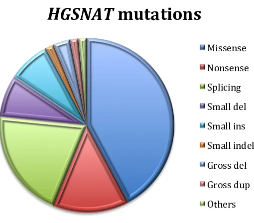
(34) INTRODUCTION. 62.40 kb. Forward strand. Figure 10. HGSNAT gene. HGSNAT gene estructure, with 62.4 Kb of length and containing 18 exons (www.ensembl.org).. Until now, 64 mutations have been identified (figure 11), including 27 missense mutations, 13 splicing mutations, nine nonsense mutations, five small deletions, five small insertions, one small indel, two gross deletions, one gross duplication and one complex rearrangement (HGMD®Profesional 2014.3). Most of the missense mutations affect amino acid residues adjacent to or within a transmembrane domain, presumably interfering with the correct folding of the protein.. HGSNAT mutations Missense Nonsense Splicing Small del Small ins Small indel Gross del Gross dup Others. Figure 11. HGSNAT mutations grouped by type. Missense, nonsense and splicing mutations represent more than 75% of total mutated alleles, while other type of mutations are found in a small proportion.. Sanfilippo C syndrome presents a very low frequency and many different mutations have been identified. Even so, different prevalent mutations have been reported such as p.R344C (accounting for 22%) and p.S518F (accounting for 29.3%) in the Dutch 19.
(35) INTRODUCTION. population (Ruijter et al., 2008), c.852-1G>A that represent 30% of Italian alleles (Fedele et al., 2007) and c.525dupT that accounts for 62.5% of Portuguese mutated alleles (Coutinho et al., 2008). Genotype-phenotype correlations have not been established, although p.G262R and p.S539C mutations are probably associated with an attenuated phenotype with late onset of symptomatology (Ruijter et al., 2008). The enzyme encoded by the HGSNAT gene catalyses the acetylation of the terminal glucosamine residues of HS prior to its hydrolysis by α-N-acetyl glucosaminidase (Klein et al., 1978). The protein contains 11 transmembrane domains and has a molecular weight of about 62 KDa. After the cleavage of the signal peptide in the ER, this enzyme is processed in two subunits into the lysosome by a cleavage in the first lysosomal loop, an α chain of about 12-27 KDa containing two glyscosylation sites and no transmembrane domains and a β chain of about 38-44 KDa containing three glycosylation sites and the 11 transmembrane domains (Fan et al., 2011). A ping-pong mechanism involving a big heterocomplex was proposed to explain the functional action of the enzyme (Durand et al., 2010) but it was demonstrated that the enzyme can work as a monomer even in the precursor form (Fan et al., 2011). No animal model has been described for Sanfilippo C syndrome so far, which means that it is the only Sanfilippo syndrome subtype for which neither a natural nor an artificially generated animal model is available.. Subtype D MPS IIID or Sanfilippo syndrome type D is caused by mutations in the GNS gene, which codes for N-acetylglucosamine-6-sulfatase (EC 3.1.6.14), a lysosomal enzyme of 552 amino acids and 13 potential glycosylation sites (Robertson et al., 1988). This enzyme catalyses the removal of the sulfate in the N-acetylglucosamine residues. The gene is located at 12q14.3, is 46-Kb long and contains 14 exons (Figure 12). Until now, 23 mutations have been described, including seven missense/nonsense mutations, four small 20.
(36) INTRODUCTION. deletions, four small insertions, three splicing mutations, two gross deletions, two complex rearrangements and one small indel (HGMD®Profesional 2014.3).. Reverse strand. 46.00 kb. Figure 12. GNS gene. GNS gene estructure, with 46 Kb of length and containing 14 exons (www.ensembl.org).. Due to the extremely low frequency of this subtype and the low number of patients described, no genotype-phenotype correlations have been established until today. Furthermore, and considering again the low number of patients for Sanfilippo type D, no prevalent mutations have been found. One natural animal model was described for Sanfilippo type D, a Nubian goat (Thompson et al., 1992), carrying a nonsense mutation. This model presented neurological and histological manifestations similar to those of human patients.. 21.
(37) INTRODUCTION. Heparan Sulfate Heparan sulfate and heparan sulfate proteoglycans HS is one of the most common and important GAGs located at the cell surface and in the extracellular matrix as a part of proteoglycans (Sarrazin et al., 2011). HS is composed of many disaccharide units, comprised of one type of uronic acid and one derivate of GlcNAc that can be sulfated (Esko and Selleck, 2002). The composition among different HS chains can differ, giving rise to different types of HS. The HS chains formed by these disaccharides repetitions are attached to a core protein being part of HS proteoglycans (HSPGs). A great variety of HSPGs can be found in the cell surface and extracellular matrix based on the core protein and the type and number of HS chains linked to it (reviewed in Esko and Selleck, 2002). HSPGs participate in many different cellular functions and systems (Figure 13) such as cell migration, vesicle secretion system, endocytic system, cell adhesion and motility, membrane basement structure and recognition of different factors and molecules as receptors or co-receptors (reviewed in Sarrazin et al., 2011).. Figure 13. HSPGs functions. Different. activities. of. HSPGs. located in the extracellular matrix and the cell surface (Sarrazin et al., 2011). 22.
(38) INTRODUCTION. Heparan sulfate biosynthesis The first step in the HSPGs synthesis is the formation of the different core proteins in the ER. The amount of these proteins, which could compete for the HS synthesis, could be the limiting factor in the synthetic pathway. HS synthesis per se, is widely accepted to take place in the GA, despite the first enzyme (xylosyltransferase) involved in this pathway is found in ER. All enzymes involved in HS synthesis are transmembrane proteins except for one sulfotransferase.. Figure 14. HS biosynthesis. HS structure and three-steps biosynthesis scheme, including linkage region formation, HS chain elongation and HS chain modification. (Kreuger. and. Kjellén, 2012).. HS synthetic pathway (figure 14) starts with the formation of the linkage region, a tetrasaccharide that binds the HS chain to the core protein. A xylose is transferred to a specific serine residue next to a glycine residue and flanked by acidic and hydrophobic residues in the core protein (Esko and Zhang, 1996; Zhang et al., 1995) by the action of one of the two xylosyltransferases (coded by XYLT1 and XYLT2). Afterwards, two galactose residues are attached to the xylose by the sequential action of galactosyltransferases I and II (coded by GALT1 and GALT2, respectively). Finally, the attachment of one glucuronic acid by a glucoronosyltransferase (GlcAT-1) completes the formation of the linkage 23.
(39) INTRODUCTION. tetrasaccharide. This linkage region is common to HS, chondroitin sulfate (CS), DS and heparin. It has been proven that the xylose may be phosphorylated while the galactoses may be sulfated. These modifications could have an effect in the GAG to be synthesized. For instance the sulfation of the second galactose residue was reported to be associated with the CS synthesis (Ueno et al., 2001). After the linkage region synthesis, the next step consists on the chain elongation. In the case of HS, elongation starts with the addition of one GlcNAc to the linkage region, step under control of the EXTL genes (EXTL1, EXTL2 and EXTL3). While EXTL1 is the only EXTL gene that is not ubiquitously expressed and whose function remains unclear, the EXTL2 and EXTL3 proteins have been proved to present GlcNAc-TI activity. In the case of EXTL2, the enzyme has been reported to have also a low level of GlcNAc-TII activity, but only in vitro (Kitagawa et al., 1999). Inhibition of the EXTL2 gene in fibroblasts from patients affected of MPSI and III using shRNAs resulted in a decrease in the HS synthesis and storage (Kaidonis et al., 2010). On the other hand, EXTL3, which also presents GlcNAc-TI activity, shows more ability to transfer the GlcNAc residue to the linkage region (Kim et al., 2001). EXTL3 also presents in vitro GlcNAc-TII activity but it remains unclear if this activity also exists in vivo. When EXTL3 is inhibited using specific siRNAs in 293 cells, longer HS chains are found, strongly suggesting that EXTL3 participates in the initiation process. When EXTL3 is inhibited, less linkage regions would have the GlcNAc residue necessary for the HS elongation, so the few HS chains available would grow longer (Busse et al., 2007). In contrast, its overexpression did not show any effect on HS chains, remaining unclear its GlcNAc-TII activity in vivo. After this initiation step with participation of the EXTL genes, the elongation of the HS chain takes place by the action of the EXT1-EXT2 complex, which adds alternative glucuronic acid (GlcA) and GlcNAc residues to the chain, forming polymers of different length. Inhibition of both genes using siRNAs resulted in shorter HS chains, while the overexpression of EXT1 or both EXT genes resulted in longer HS chains and the overexpression of EXT2 did not affect the HS chain length. It has been demonstrated that EXT1 presents both activities in vitro, GlcA-TII and GlcNAc-TII, while the presence of EXT2 24.
(40) INTRODUCTION. is essential in vivo, suggesting that EXT2 assists EXT1 in its translocation from ER to GA and plays a fundamental role for HS elongation even when it does not present any transferase activity (reviewed in Busse-Wicher et al., 2013). Mutations in EXT1 and EXT2 cause hereditary multiple exostoses (HME), a common (1:100000) autosomal dominant disorder affecting skeleton with a risk of malignant transformation. Mutations in the EXT genes result in reduced or absence of HS in the cartilage, producing a disorganization of chondrocytes (reviewed in Zak et al., 2002). Finally, the last step in HS biosynthesis is the modification of the HS chain. These modifications take place while the chain is being synthesized and involve six different steps: deacetylation of the GlcNAc residues in some chain regions, sulfation of these residues, epimerization of many GlcA residues next to a modified GlcNAc residue to form iduronic acid (IdoA), sulfation of some IdoA and GlcA residues, 6-O-sulfatation and 3-Osulfatation of some glucosamine residues in specific group contexts. Four different types of sulfotransferases and one epimerase are responsible for these modifications (reviewed in Esko and Selleck, 2002). All these modifications are important for the HS interactions and recognition of different factors and molecules.. Heparan sulfate degradation. Figure 15. Endocytosis of HS prior to its degradation.. Internalizing. of. HSPGs. from. extracellular matrix to lysosomes to be degraded (www.glycoforum.gr.jp).. 25.
(41) INTRODUCTION. In the extracellular matrix, some endosulfatases and secreted heparanase could partially degrade HS chains giving rise to smaller and potentially active fragments (Gong et al., 2003). These fragments can be recycled with the same core protein, being internalized and brought under partial degradation and new rounds of biosynthesis and finally, exocyted again to the cell membrane and the extracellular matrix (Fransson et al., 2004). Final HS degradation takes place inside the lysosomes after internalization of HSPGs (figure 15) through the stepwise action of nine different enzymes (figure 16) (Neufeld and Muenzer, 2001). HS consists of GlcA and IdoA residues that can be sulfated or not, and glucosamine residues that can be acetylated or sulfated.. Figure 16. HS degradation. Stepwise degradation. of. HS. with. an. scheme. including all steps and all enzymes participating in the pathway (Neufeld and Muenzer, 2001).. 26.
(42) INTRODUCTION. The first enzyme of the pathway, heparanase, is an endoglucuronidase that cleaves HS chains into smaller fragments to facilitate the polymer degradation. Iduronate sulfatase is the enzyme that desulfates iduronic acid residues previous to their cleavage by the α-Liduronidase. Deficiency in the iduronate sulfatase causes MPS II while deficiency in α-Liduronidase causes MPS I, with consequent accumulation of HS and also DS, indicating their role also in the DS degradation pathway. The next step in the HS degradation consists in the desulfamation of the amino group of the glucosamine by the heparan N-sulfatase (sulfamidase). Deficiency in this enzyme causes MPS IIIA, with HS accumulation. This glucosamine has to be acetylated in its recently exposed amino group previous to its cleavage. This acetylation function corresponds to the acetyl-CoA α-glucosaminide N-acetyltransferase, the only lysosomal enzyme that is not a hydrolase, which takes the acetyl group from acetyl-CoA from cytoplasm and transfers it to the HS chain under degradation in the lysosomal lumen. Deficiency of this enzyme causes MPS IIIC due to the HS storage. Once the glucosamine has been acetylated to obtain GlcNAc, it can be cleaved from the chain by the action of α-N-acetylglucosaminidase. This is the enzyme deficient in MPS IIIB patients, who present HS accumulation. After that, GlcA residue has to be sulfated previous to its cleavage. The enzyme responsible for this sulfation is glucuronate 2sulfatase, while the enzyme responsible for the cleavage of the sulfated residue is βglucuronidase. Deficiency of the first enzyme is not yet associated with any known disease, while deficiency of the second one causes MPS VII, with consequent storage of HS and DS. Finally, the last enzyme taking part in the HS degradation pathway is Nacetylglucosamine 6-sulfatase, which desulfates the non-deacetylated residues of glucosamine previous to their cleavage. This enzyme is deficient in MPS IIID patients, and causes HS storage. It has been suggested that all these enzymes function as a complex in the lysosomes (Freeman and Hopwood, 1992). 27.
(43) INTRODUCTION. Therapeutic Approaches Non-effective therapy has already been developed for Sanfilippo syndrome patients, though different kinds of interesting approaches have been tested in cells and animal models of the disease, focused mainly in the treatment of the CNS involvement.. Enzyme replacement therapy Different approaches are available concerning this type of therapy, differing in the administration strategy (table 2). For a disease affecting the CNS such as MPS III, is important to consider the existence of the blood-brain barrier (BBB) that does not allow the pass of enzymes to the brain. Thus, intravenously administration is not as useful as for other LSDs without CNS affection, in which ERT is currently approved and in use (revised in Desnick and Schuchman, 2012). For Sanfilippo syndrome, intravenously administration was tested in subtype B murine model (Yu et al., 2000) and subtype D caprine model (Downs-Kelly et al., 2000) without positive results in the brain. In contrast, intravenous administration to a mouse model of Sanfilippo type A from birth, when the BBB is not formed, showed a delay in the development of the disease, confirming the usefulness of the ERT when the enzyme can reach the brain (Gliddon and Hopwood, 2004). More recently, intravenously chemically modified enzymes at high doses have been showed as a good option for the treatment of MPS IIIA mice (Rozaklis et al., 2011).. Table 2. ERT strategies for Sanfilippo syndrome in animal models.. Enzyme. Administration. Subtype. Model. N-acetylglucosamine N-acetylglucosamine 6-sulfatase Sulfamidase Chemically modified sulfamidase Sulfamidase Sulfamidase Sulfamidase Sulfamidase + IGFII. Intravenous Intravenous Intravenous Intravenous Intracerebral Intravenous + intracerebral Intracerebral Intraventricular. B D A A A A A A. Murine Caprine Mouse Mouse Mouse Mouse Dog Mouse. 28.
(44) INTRODUCTION. Intrathecal, intracerebral, intracisternal and intracerebrospinal fluid injections of recombinant enzymes have been proved to be a considerable option for the treatment of MPS IIIA dogs (Crawley et al., 2011; Hemsley et al., 2009b; Jolly et al., 2011; Marshall et al., 2014) and MPS IIIA mice (Hemsley et al., 2008; Savas et al., 2004). Combined intravenous administration before adulthood and intracerebrospinal fluid administration during adulthood has been tested in MPS IIIA mice but this combination did not show better results than the use of only intracerebrospinal fluid administration (Hemsley et al., 2009a). It has been proved that this type of therapy is more efficient in early stages of the disease when compare to middle or late stages in mice (Hassiotis et al., 2014). Also the use of fusion protein containing NAGLU and insulin-like growth factor II (IGFII) has been tested in MPS IIIB mice to overcome the BBB. The fusion enzyme persists in the brain for around ten days after intraventricular administration, promoting a decrease in the brain levels of HS as well as in the liver levels to that of the control cases (Kan et al., 2014). Direct brain administration for the treatment of neurological disorders presents some problems. It is an aggressive treatment that needs continued injections and the administration of an enzyme not naturally present in patient’s brain may trigger an immune response.. Substrate reduction therapy Taking into account the handicaps of ERT, SRT has been presented as a valid alternative approach. The objective of this therapy is to find molecular targets to decrease the production of the accumulated substrate and restore the balance between synthesis and degradation (figure 17). For this reason, it is important to remark the fact that the mutant enzyme has to maintain residual activity to achieve this restoration.. Figure 17. SRT basis. Restoration of the balance between synthesis and degradation is the main objective of SRT.. 29.
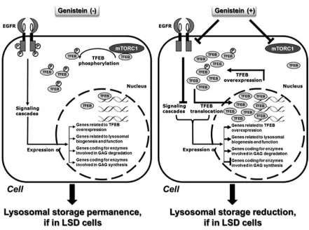
(45) INTRODUCTION. In the case of Sanfilippo disease, the goal of SRT is to decrease HS synthesis. In order to achieve this aim, several approaches have been developed using different types of molecules. All of them should be able to cross the BBB, since the main objective of any therapy for this disease is to reach the CNS. In the case of Sanfilippo syndrome, genistein and rhodhamine B are the two small molecules more frequently studied and tested to check their SRT properties.. Figure 18. Mechanisms of action of genistein. Schematic representation of the putative mechanism of genistein to regulate the lysosomal metabolism-related genes and TFEB. (Moskot et al., 2014).. Genistein is a natural isoflavone that inhibits the kinase activity of epidermal growth factor receptor (EGFR), which is important for complete expression of genes encoding enzymes responsible for GAG production (Jakóbkiewicz-Banecka et al., 2009; Piotrowska et al., 2006). Genistein showed significant results in patients’ fibroblasts (Piotrowska et al., 2006), peripheral tissues of Sanfilippo B mice (Malinowska et al., 2009) 30.
(46) INTRODUCTION. and CNS of Sanfilippo B mice (Malinowska et al., 2010). Surprisingly, the reduction in the synthesis and storage of GAGs was also found in patients’ cells carrying null mutations, indicating a combination of genistein mechanisms of action inside the cell (BaneckaMajkutewicz et al., 2012). A recent study established that genistein not only acts decreasing GAG synthesis, but also promotes transcription factor EB (TFEB) overexpression (figure 18), the lysosomal master regulator that promotes cellular clearance (Moskot et al., 2014). Despite these promising results, low doses of genistein in human patients did not show significant amelioration in neuropathology although some improvement of the somatic symptoms have been described (Delgadillo et al., 2011; Ruijter et al., 2012b). The use of elevated doses of genistein has been proved to be safe in patients (Kim et al., 2013) but HS reduction results were inconclusive (Malinová et al., 2012). Other flavonoids have also shown the ability to reduce the GAG synthesis in fibroblasts, independently of EGFR, and a combination of several of them resulted in a higher inhibition effect on the GAG synthesis (Kloska et al., 2011). Further studies with higher doses of genistein and other flavonoids should be carried out to establish the ability of this group of molecules to ameliorate CNS pathology in Sanfilippo patients. On the other hand, rhodamine B is a colorant used in cosmetics that is thought to inhibit the GAG elongation step in the synthetic pathway (Kaji et al., 1991). Rhodamine B showed the ability to delay the somatic pathology in MPS IIIA mice (Roberts et al., 2006) and improved their behaviour and learning difficulties (Roberts et al., 2007). It is also thought that the effect of rhodamine B is stronger if there is some residual enzyme activity. Even though exposure to high doses of rhodamine B results in liver toxicity and reproductive detriment, none of these problems is detected after continued exposure at low doses, which have been reported to improve neurological defects in MPS IIIA mice (Roberts et al., 2010). Further studies should assess the ability of rhodamine B to ameliorate the neurological symptoms in animal models and patients without toxic effects. Finally, some other molecules have been proposed as useful as SRT for Sanfilippo syndrome. One of them is miglustat, which was developed for the treatment of Gaucher disease type I patients (Cox et al., 2000). Some studies in Niemann-Pick disease type C 31.
(47) INTRODUCTION. patients have shown a reduction in disease progression using miglustat, due to the decrease of the neurotoxic storage of GM2 ganglioside (Patterson et al., 2007). Since this ganglioside is secondarily accumulated in Sanfilippo syndrome patients, miglustat could be an interesting therapeutic drug to reduce the neurodegeneration. Another option is the use of specific RNAi directed to genes involved in the GAG synthesis. RNAi is a mechanism to selectively silence the expression of a particular gene by the specific degradation of the mRNA. Synthetic siRNAs and shRNAs have been widely used to downregulate the expression of a large number of genes. The use of shRNAs to downregulate EXTL2 and EXTL3 genes was found to reduce the GAG synthesis and storage in MPS IIIA fibroblasts (Kaidonis et al., 2010). The same effect was reported for the use of siRNAs to downregulate XYLT1, XYLT2, GALTI and GALTII, genes coding for enzymes responsible of the linkage region formation (Dziedzic et al., 2010).. Coenzyme Q10 Coenzyme Q10 (CoQ10) is a lipophilic molecule synthesized in all the cells and found in all the membranes of eukaryotic organisms. It works as an electron carrier in the mitochondrial respiratory chain, although it participates in many other processes (Turunen et al., 2004). It is known that lysosomal membrane contains similar amounts of CoQ10 than mitochondrial membrane, and plays a role in the acidification of the lysosomal lumen (Gille and Nohl, 2000). A recent study showed that CoQ10 was low in Sanfilippo B patients when compared to healthy individuals (Delgadillo et al., 2011). In order to assess its potential therapeutic effect, CoQ10 and also an antioxidant cocktail were administrated to MPS IIIA and B fibroblasts. The results about the decrease in GAG storage and enzyme activity enhancement were not conclusive, but they showed a slight increase in exocytosis probably due to the treatment (Matalonga et al., 2014). Further studies in cell and animal models are needed to establish whether or not CoQ10 presents a therapeutic effect.. 32.
(48) INTRODUCTION. Pharmacological chaperones for enzyme-enhancement-therapy Many specific mutations, mainly missense, can give rise to misfolding proteins that can retain some residual activity but may be subjected to rapid intracellular degradation (reviewed in Suzuki, 2014). Molecular chaperones are cellular proteins that act on the correct folding of other polypeptides (reviewed in Muchowski and Wacker, 2005). Pharmacological and chemical chaperones are small compounds that can be used, in a similar way, to avoid the misfolding of mutant proteins (figure 19). They are principally enzyme inhibitors, which at low concentrations interact specifically with the active site of proteins to restore the correct folding and to stabilize them (Bernier et al., 2004). In the case of LSDs, once in the lysosome, the enzyme substrate replaces the chaperone, completing enzyme activity restoration (Sawkar et al., 2002). Amino and iminosugars are the most common pharmacological chaperones used in enzyme-enhancement-therapy.. Figure. 19.. treatment Action. Chaperone possibilities.. mechanisms. of. chaperones to restore correct folding enzyme. of. the. and. lysosomal its. correct. trafficking to the lysosome (www.minoryx.com).. Chaperones may represent an interesting approach for many LSDs. It is known that for these diseases, an enzyme activity above 10-20% is sufficient to preclude the development of clinical symptoms. Many chaperone approaches have been assayed at different levels for LSDs such as Fabry disease, GM1-gangliosidosis, Morquio B disease, Pompe disease, Gaucher disease, Krabbe disease, Niemann-Pick A/B and C diseases, as 33.
(49) INTRODUCTION. well as for other types of disorders such as retinitis pigmentosa, cystic fibrosis, Parkinson disease, Alzheimer disease or cancer (reviewed in Suzuki, 2014). Several compounds were assayed as chaperones at the cellular level for Sanfilippo syndrome (Feldhammer et al., 2009b). One of them, glucosamine, which is a competitive inhibitor of the HGSNAT enzyme that is not expected to be highly toxic, showed to significantly increase enzyme activity in eight out of nine patients’ fibroblasts, indicating its therapeutic potential. Further studies should be done in order to establish its efficacy and lack of toxicity in other cellular and animal models.. Suppression of premature termination codons This therapy is based on the stop-codon read-through technology. It can be used in the cases where mutations introduce a premature termination codon (PTC) that gives rise to a truncated protein without proper enzymatic activity Usually this PTC in the mRNA would activate the nonsense-mediated decay (NMD) response with a rapid degradation of the transcript. The use of small molecules that can read through the PTC would allow the cells to produce protein with a change in one amino acid but at least non-truncated. The read-through process occurs naturally in the cells at low frequencies (reviewed in Brooks et al., 2006). Aminoglycosides are one type of molecules that have been tested for their readthrough properties, especially gentamicin. Unfortunately, it promotes read-through also over well-positioned stop codons, resulting in unspecific effects. Moreover, long-term treatments using gentamicin can result in nephrotoxicity (Prayle et al., 2010). Aminoglycosides derivatives have been developed in order to reduce their toxicity and improve their efficiency. Many different works have been reported using aminoglycosides or derivatives as a treatment for nonsense mutations in many cellular and animal models of different disorders (reviewed in Keeling et al., 2014). For example, NB54 aminoglycoside derivative was tested in MPS I fibroblasts (Nudelman et al., 2009) and mouse model (Wang et al., 2012) and showed improved properties when compared to 34.
(50) INTRODUCTION. gentamicin. Gentamicin did not show a correction for MPS VI nonsense mutations (Bartolomeo et al., 2013).. Figure. 20.. Read-through. mechanism. Example of readthrough-mechanism of a PTC after PTC124. (Ataluren). treatment. (watcut.uwaterloo.ca).. Other non-aminoglycoside antibiotics have been tested for this type of therapy such as PTC-124, also known as Ataluren (figure 20). Different works have been reported with different success (reviewed in Keeling et al., 2014). Ataluren has been tested in MPS VI (Bartolomeo et al., 2013) and MPS I (Peltz et al., 2013) with some degree of success. Nowadays, there is some controversy about the ability of Ataluren to restore enzyme activity. Finally, the combination of PTC suppression together with NMD inhibition can result in an increase of the functional protein. One study in an MPS I mouse showed higher levels of enzyme activity in mice treated with an inhibitor of the NMD process together with a PTC suppression drug (Keeling et al., 2013). Until now, no reports have been published for Sanfilippo syndrome regarding this type of therapy.. Overexpression of transcription factor EB TFEB has been proved to be a master regulator in the lysosome biogenesis and function. It is considered a target for any disorder that affects the lysosomal-autophagic 35.
(51) INTRODUCTION. pathway since its overexpression promotes cellular clearance in cultured cells (Sardiello et al., 2009). It also reduces GAG levels and vacuolization in NSC from multiple sulfatase deficiency and MPS IIIA mouse models (Medina et al., 2011). Overexpression of TFEB in muscle cell cultures and Pompe mice enhance the lysosomal-autophagosomes fusion and result in an increase in the exocytosis of autolysosomes (Spampanato et al., 2013). TFEB overexpression has been proved to be useful for other neurodegenerative disorders such as Parkinson disease (Dehay et al., 2010), Huntington disease (Tsunemi et al., 2012) and α-1-anti-trypsin deficiency (Pastore et al., 2013). How TFEB modulates cellular clearance should be further studied in order to determine the mechanism that activates the autolysosomal exocytosis. Different molecules that can promote TFEB dephosphorylation or inhibit its phosphorylation, enhancing its nuclear translocation, represent a novel putative therapeutic strategy for LSDs.. Gene therapy Gene therapy consists in the delivery of the correct copy of the gene (mainly the cDNA) to affected cells in order to recover the enzyme function. For Sanfilippo syndrome, many different viral vectors such as retroviruses, lentiviruses, adenoviruses and adenoassociated viruses (AAV) have been used to transduce cells (table 3). Also a gene therapy approach using a non-viral vector (pFAR4) administered via tail vein has been recently tested in MPS IIIA mice with significant results in the increase of the enzyme activity and the decrease of GAGs in different tissues and the reduction of the lysosomes in the brain (Quiviger et al., 2014). In these animals the liver became a sulfamidase secretor that promotes a reduction in GAG accumulation in others tissues (an scheme of the M6P transport of the lysosomal enzyme from a corrected cell to an affected cell is shown in figure 21)... 36.
(52) INTRODUCTION. Table 3. Gene therapy strategies for Sanfilippo syndrome. AAV (adeno-associated virus), LV (lentivirus), IV (intravenous).. Model. Gene. Viral vector. Administration. MPS IIIA Fibroblasts MPS IIIB Fibroblasts MPS IIIA mice MPS IIIA mice MPS IIIA mice MPS IIIB mice MPS IIIB dog MPS IIIB mice MPS IIIB mice MPS IIIA mice MPS IIIA mice MPS IIIA mice MPS IIIA dog. SGSH NAGLU SGSH SGSH SGSH NAGLU NAGLU NAGLU NAGLU + SUMF1 SGSH SGSH + signal peptide SGSH SGSH. Retrovirus LV LV LV Adenovirus 2 AAV 2 AAV 5 AAV 5 + LV AAV 5 AAV 8 AAV 8 AAV 9 AAV 9. IV Intraventricular Intraventricular IV + Intracinsternal Intracerebral Intracranial + IV Intracerebral Intramuscular + IV IV Intracerebrospinal fluid Intracerebrospinal fluid. Retroviral constructs were reported to express the corresponding cDNA for MPS I, II, IIIA, VI and VII (reviewed in Neufeld and Muenzer, 2001). For MPS IIIA fibroblasts, continuous infections were necessary to obtain a high enzyme activity (Bielicki et al., 1996). Lentiviral vectors are another option to deliver the correct copy of the gene into the cells. For Sanfilippo B fibroblasts, high amounts of the enzyme were produced using this strategy, but with a poor uptake of the secreted protein by untreated cells (Villani et al., 2002). Lentiviral gene therapy was also assessed in the mouse model of MPS IIIA by intravenous delivery (McIntyre et al., 2008). Somatic amelioration was detected in treated mice, with high enzyme production and reduction of the GAG storage, while in the CNS the storage was decreased but not significantly, suggesting a poor neural transduction. With intraventricular infusion of this lentiviral vector, a reduction in the GAG storage in the CNS was achieved in the mouse model of Sanfilippo A (McIntyre et al., 2010). Since retroviruses and lentiviruses are integrative viruses and promote an immunogenic response, a canine adenovirus serotype 2, which induces a low level of innate response, was assayed in the mouse model of MPS IIIA (Lau et al., 2010b). 37.
(53) INTRODUCTION. Intraventricular injection of this virus resulted in a long-term enzymatic expression and an improvement in the neuropathology. Lately, the use of AAV, which are non-integrative, non-pathogenic in humans and with capacity to infect non-dividing cells providing long-term expression, have emerged as a good delivery tool for the treatment of neurological disorders. In the last few years several reports have been published concerning this AAV-mediated therapy using different AAV serotypes. A combined administration via intravenous and intracisternal of an AAV2 carrying the NAGLU cDNA was shown to improve the lifespan and behaviour of the mouse model of MPS IIIB with a variable correction of the lysosomal storage pathology in the CNS and somatic tissues, and a restoration of the enzyme activity (Fu et al., 2007). In MPS IIIB dogs, intracerebral administration of AAV serotype 5 plus immunosuppression showed to be a safe and an efficient method for gene therapy with an increase in the enzymatic activity and a decrease in the storage of GAGs (Ellinwood et al., 2011). A combination of intracranial AAV5 and intravenous lentiviral administration was assayed in MPS IIIB mice obtaining an improvement in the lifespan and motor activities (Heldermon et al., 2013). Intracerebral administration of AAV5 carrying the SGSH gene together with the SUMF1 gene (coding for an essential and limiting factor for sulfatases) in the mouse model of MPS IIIA showed an increase in the SGSH activity in the brain, a decrease in the storage and inflammation and an improvement in the motor and cognitive function (Fraldi et al., 2007). After these results, a clinical trial phase I/II for MPS IIIA using AAV serotype 10 expressing the deficient SGSH enzyme and the SUMF1 enzyme was started. It recently finished showing no toxicity or lack of tolerance and a possible slight improvement in the patients’ behaviour (NCT01474343, results at Tardieu et al., 2014). AAV serotype 8 carrying the SGSH gene has been tested in a mouse model of Sanfilippo A, by intramuscular and intravenous administration (Ruzo et al., 2012a). Results showed no amelioration using intramuscular administration, while intravenous administration resulted in liver transduction and an improvement in the somatic tissues and some amelioration in the CNS symptoms, only in male mice. Another work, using AAV8 carrying SGSH together with a signal peptide to promote the secretion of the enzyme 38.
(54) INTRODUCTION. and a BBB-binding domain to promote the cross of the BBB, resulted in a high enzyme activity in the brain together with an improvement in brain pathology and behaviour with minimal invasive treatment (Sorrentino et al., 2013). Other works showed the ability of serotype 9 to transduce neural and some somatic cells and achieve gene correction, both in the mouse and in the canine models of MPS IIIA (Haurigot et al., 2013; Ruzo et al., 2012b). The mouse model, treated intravenously, showed an increase in the enzymatic activity together with a decrease in the GAG storage and neuroinflammation, expanding mice lifespan (Ruzo et al., 2012b). Intracerebrospinal fluid administration in the mouse and in the canine models of MPS IIIA resulted in an increase in the enzymatic activity in brain and serum, leading to whole body correction of GAG storage and lysosomal pathology, as well as a prolonged lifespan with low immune response (Haurigot et al., 2013). These results encourage the application of this approach in human patients.. Figure 21. M6P-mediated enzyme excretion. M6P-mediated transport of lysosomal enzymes from a corrected cell to an affected cell. This is an important process in the case of diseases affecting enzymes located at the lysosomal lumen that are directed to the lysosome via M6P signalling. For gene therapy and cell therapy, where not all affected cells are corrected due to the treatment, this process could play an important role (Kohan et al., 2011).. 39.
Figure


+7


Documento similar
Models and therapeutic approaches for Niemann-Pick (A/B and C) and other lysosomal storage disorders
Most of them aim to increase the functionality of the defective enzyme or protein by gene therapy, enzyme replacement, pharmacological chaperones, cell based therapy and
SECTION 3 - The role of RINGO proteins in the brain 123 RingoA mRNA is expressed in neural stem cells and proliferating progenitor cells 123 Analysis of neural stem cells
human mesenchymal stem cells from bone marrow and adipose tissue under xeno-free conditions for cell therapy. Stem Cell
Serum ferritin as risk factor for sinusoidal obstruction syndrome of the liver in patients undergoing hematopoietic stem cell transplantation. Plasminogen activator inhibitor-1
Human cancer cells show predominant expression of NANOGP8, whereas pluripotent cells (embryonic stem cells and embryonal carcinoma cells) express the NANOG variant
A novel lentiviral vector targets gene transfer into human hematopoietic stem cells in marrow from patients with bone marrow failure syndrome and in vivo in humanized mice.. The
Two human induced pluripotent stem cell (iPSC) lines were generated from fibroblasts of two siblings with methylmalonic acidemia cblB type carrying mutations in the MMAB gene:
Consequently, in this thesis we have generated two cellular models, a mouse model based on NURR1 overexpression in neural stem cells, and a human model based
