J Lizardi-Cervera et al. Hepatitis C virus infection and non-Hodgkin’s lymphoma 257
edigraphic.com
Annals of Hepatology 2006; 5(4): October-December: 257-262Annals of Hepatology
Original article
Hepatitis C virus infection and non-Hodgkin’s
lymphoma: a review and case report of nine patients
Javier Lizardi-Cervera;1 Jorge Luis Poo;1 Karla Romero-Mora;1 Beatriz Castañeda;2 Raúl Pichardo-Bahena;3 Segundo Morán;2 Misael Uribe1
Abstract
The role of hepatitis C virus (HCV) is well established in the development of chronic hepatitis, cirrhosis and hepatic carcinoma, as well as in mixed type II cryoglo-bulinemia, membranoproliferative glomerulonephritis (MPGN) and porphyria cutanea tarda (PCT). Increas-ing evidence has been reported of a close association of HCV infection with autoimmune and hematological processes, mainly cytopenias and lymphoproliferative disorders such as B cell non-Hodgkin’s lymphoma. We describe the demographic, clinical and histopathologi-cal findings of nine patients from the Mexican popula-tion with non-Hodgkin’s lymphoma and HCV infec-tion.
Key words: non-Hodgkin’s lymphoma, hepatitis C vi-rus.
Introduction
The role of hepatitis C virus (HCV) has been well de-fined in the development of chronic C hepatitis, cirrhosis and hepatic carcinoma, as well as in mixed type II cryo-globulinemia. HCV is lymphotrophic and can stimulate clonal proliferation of type B lymphocytes. Moreover, some studies have shown an association between anti-hepatitis C virus antibodies and B cell non-Hodgkin’s lymphoma (NHL). Specific genomic sequences of the vi-rus have also been identified in the lymph nodes of pa-tients with NHL as well as in papa-tients with hyperplasic lymphadenopathies.1,2
1Gastroenterology Clinic and Liver Unit.
2Gastroenterology Center, Centro Médico Nacional Siglo XXI,
IMSS.
3Anatomy Pathology Department, Medica Sur Clinic & Foundation.
Address for correspondence:
Javier Lizardi Cervera MD. Subdirección Académica. Tercer Piso de Hospital, Fundación Clínica Médica Sur. Puente de Piedra 150, Tlalpan 14050, Distrito Federal, México. e-mail:
jlizardi@medicasur.org.mx
Manuscript received and accepted: 16 October, 2006
The aim of this study was to document the clinical and pathologic characteristics of nine patients with HCV in-fection and NHL, and to review the literature about this subject.
Methods
Nine patients with diagnoses of hepatitis C and NHL were included. They were seen at three different institu-tions—Medica Sur Clinic & Foundation, Instituto Nacio-nal de Ciencias Médicas y de la Nutrición Salvador Zu-birán, and Centro Medico Nacional Siglo XXI, all located in Mexico City-between March 1998 and December 2005. Lymphoma localization and demographic and clin-ical findings are described. Laboratory findings as well as treatment and survival are also reported.
Results
Demographic and clinical characteristics
The clinical and demographic characteristics of the patients are shown in Table I. Nine patients (two men and seven women) with an average age of 59.1 years (range, 38-77 years) were included. All patients had a history of blood transfusion before the diagnosis of NHL and HCV infection. The average time between the transfusion and the diagnoses was 22 years. Four patients had cirrhosis, three patients had chronic hepatitis, and two were asymp-tomatic.
Laboratory tests (Table II).
Six patients had abnormal liver function tests at the time of diagnosis of NHL. One patient had normal liver enzyme activities but a low albumin concentration. Two patients had increased liver enzyme activities after che-motherapy.
Viral markers
In every patient, the diagnosis of HCV infection fol-lowed positive antibodies measurement by a third-genera-tion ELISA test. In one patient, the diagnosis was con-firmed by the RIBA (recombinant immunoblot assay)
an-Artemisa
edigraphic.com
SUSTRAÍDODE-M.E.D.I.G.R.A.P.H.I.C
:ROP ODAROBALE FDP
VC ED AS, CIDEMIHPARG
ARAP
ACIDÉMOIB ARUTARETIL :CIHPARGIDEM
tibody test; in eight by HCV-RNA detection using the Roche Amplicor RT-PCR Test:, four with a quantitative test (lower limit of detection was 600 UI/ml or 1,620 cop-ies/mL) and four with a qualitative test (lower limit of de-tection 50 UI/mL).
Clinical presentation, localization and histological type of the non-Hodgkin’s lymphoma
Table III shows lymphoma localization, histological
type, treatment and survival of patients. Brief clinical his-tories are presented.
Patient 1: A 53-year-old woman with chronic HCV in-fection presented in July 1999 with weakness and anorex-ia. Hepatomegaly was detected, along with a firm epigas-tric mass. Liver aspiration biopsy showed an undifferenti-ated tumor, probably hepatic carcinoma. A segmentectomy was performed with resection of a 3 cm ovoid mass. Histopathological examination showed a type B diffuse large cell NHL (Figure 1). Nontumor tissue showed chronic hepatitis with grade III fibrosis, moderate portal inflammation, lobular inflammation and mild peri-portal necrosis. She received six sessions of CHOP che-motherapy. Thereafter, she was given interferon alpha-2b and ribavirin. Unfortunately, she did not tolerate a
com-plete course, with reduced hemoglobin and platelets. HCV RNA testing was positive at week 12. Therefore, an-tiviral treatment was discontinued. In December 2004, her lymphoma continued to be in remission.
Patient 2: A 67-year-old woman with compensated HCV cirrhosis known to our clinic since 1994. HCV in-fection was diagnosed in 1990. In 1998, she developed upper gastrointestinal bleeding (hematemesis and mel-ena). An endoscopy showed a 3 cm polypoid gastric le-sion. The biopsy disclosed a large B cell NHL. A gastric tissue specimen was negative for Helicobacter pylori in-fection. A partial gastric resection was performed, fol-lowed by five cycles of CHOP chemotherapy. She was as-ymptomatic until December 2004.
Patient 3: A 38-year-old male patient with HCV-relat-ed cirrhosis complicatHCV-relat-ed with ascites and treatHCV-relat-ed with di-uretics. In October 2000, he complained of burning poste-rior thoracic pain that extended to the lower limbs and was followed by paresthesia and diminished sensation. A magnetic resonance image of the spinal cord showed an extramedullary lesion extending from T1 to T6 compress-ing the medullary space. Surgical resection was per-formed with decompression and spinal cord fixation. His-tological diagnosis revealed B cell NHL. The patient re-ceived chemotherapy and radiotherapy. He is now
Table I. Clinical characteristics of patients with hepatitis C virus and non-Hodgkin’s lymphoma.
Age (years) Gender Previous surgery Previous blood transfusion Hepatic disease
Patient 1 53 Female Yes Yes Chronic hepatitis
Patient 2 67 Female Yes Yes Cirrhosis
Patient 3 38 Male Yes Cirrhosis
Patient 4 77 Male Yes Yes Cirrhosis
Patient 5 64 Female Yes Yes Chronic hepatitis
Patient 6 56 Female Yes Yes Hepatic steatosis
EV I/IV
Patient 7 32 Female Yes Yes Hepatic steatosis
Patient 8 79 Female Yes Yes Chronic hepatitis
Patient 9 64 Female Yes Yes Chronic hepatitis
EV = Esophageal varices
Table II. Main laboratory findings in patients with hepatitis C virus and non-Hodgkin’s lymphoma.
Parameter Patient 1 Patient 2 Patient 3 Patient 4 Patient 5 Patient 6 Patient 7 Patient 8 Patient 9
Hemoglobin (g/dL) 15.8 12 14.2 11.4 14.2 15.5 13.3 15.4 12.1
Leukocyte count (× 103/mm3) 8.6 9.9 3.9 2.6 5.8 4.8 2.4 5.2 4.1
Platelets (× 103/mm3) 233 117 86 131 284 262 238 257 166
Total bilirubin (g/dL) 1.2 1.1 1.8 1.5 0.9 0.7 1.27 1.04 0.8
Direct bilirubin (g/dL) 0.2 0.2 0.4 0.4 0.4 0.2 0.37 0.8 0.2
AST (U/L) 100 33 63 215 76 75 301 65 107
ALT (U/L) 84 16 115 297 55 81 294 63 98
FA (U/L) 134 91 172 166 124 614 102 80 150
Prothrombin time (%) 82 52 65 72 88 – – 88 –
Albumin (g/dL) 3.4 2.3 2.3 3.4 4.1 3.5 3.8 3.9 4.0
ELISA anti-HCV test + + + + + + + +
RIBA-2 antibody test – Positive – – – – – –
J Lizardi-Cervera et al. Hepatitis C virus infection and non-Hodgkin’s lymphoma
edigraphic.com
recovering movement of lower extremities and his lym-phoma continued to be in remission until July 2004.
Patient 4: A 77-year-old man with a diagnosis of com-pensated cirrhosis secondary to HCV infection since 1995. In March 1998, he presented with fever, adynamia, cervical lymph node enlargement and hepatosplenomega-ly. An X-ray showed multiple pulmonary infiltrates. Cer-vical lymph node biopsy showed large cell, diffuse NHL. He received six cycles of CHOP chemotherapy and achieved remission from the adenomegaly, visceromega-lies and pulmonary infiltrates. During his follow-up, no tumor activity was detected until December 2001. Be-cause of persistent thrombocytopenia, the patient did not receive treatment for HCV.
Patient 5: A 64-year-old female patient with compen-sated cirrhosis secondary to HCV infection since 1995. In July 2000, she presented with bilateral, persistent con-junctivitis, mainly in the left eye, which was refractory to
treatment. A biopsy was done, and a diffuse, large, type B, MALT-cell lymphoma was diagnosed (Figure 2). She received 23 sessions of radiotherapy. She was free of dis-ease until December 2001.
Patient 6: A 56-year-old female patient with a history of blood transfusion in 1990 was treated in 1999 for im-munoblastic NHL localized to the spleen with high doses of methotrexate, cyclophosphamide, vincristine, bleomy-cin and etoposide, with which she achieved complete re-mission. She presented with liver enzyme elevation ac-companied with asthenia and adynamia. An abdominal ul-trasound showed hepatic steatosis and a simple right lobe hepatic cyst, without gallbladder or spleen abnormalities. An endoscopic study revealed esophageal varices grade I, and mild portal gastropathy. The serologic test was posi-tive for HCV infection.
Patient 7: A 32-year-old woman, previously asymp-tomatic but positive for HCV, was diagnosed with NHL in
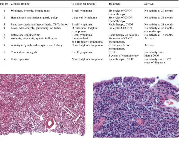
Table III. Localization and histological type of non-Hodgkin’s lymphoma in patients with hepatitis C virus.
Patient Clinical finding Histological finding Treatment Survival
1 Weakness, hypoxia, hepatic mass B cell lymphoma Six cycles of CHOP No activity at 29 months
chemotherapy
2 Hematemesis and melena, gastric polyp Large cell lymphoma Six cycles of CHOP No activity at 34 months
chemotherapy
3 Pain, paresthesia and hypoesthesia, T1-T6 lesion B cell lymphoma Radiotherapy, CHOP No activity at 38 months
4 Fever, adenomegaly, pulmonary infiltrates Diffuse non-Hodgkin’ Six cycles CHOP of No activity at 45 months
s lymphoma chemotherapy
5 Refractory conjunctivitis B cell lymphoma Radiotherapy-23 sessions No activity at 17 months
6 Asthenia, adynamia, splenic infiltration Immunoblastic Six monts of CHOP Activity
non-Hodgkin’s lymphoma chemotherapy
7 Activity in lymph nodes, spleen and kidney Non-Hodgkin’s lymphoma CHOP 4 cycles of Activity
chemotherapy
8 Cervical adenomegaly B cell lymphoma CHOP No activity since
4 cycles of chemotherapy March 2006
9 Fever, epistaxis Non-Hodgkin’s lymphoma Radiotherapy, CHOP No activity since 1997
(year of diagnosis)
Figure 1. Primary non-Hodgkin’s lymphoma of the liver. The total ar-chitecture of the liver is infiltrated by lymphocytic cells. An artery (arrow) and a central vein (double arrow) are shown.
edigraphic.com
SUSTRAÍDODE-M.E.D.I.G.R.A.P.H.I.C
:ROP ODAROBALE FDP
VC ED AS, CIDEMIHPARG
ARAP
ACIDÉMOIB ARUTARETIL :CIHPARGIDEM
2001, with tumor activity detected in the neck, pelvis, spleen and kidney. She received four cycles of chemo-therapy with cyclophosphamide, vincristine, mitoxantro-ne and prednisomitoxantro-ne. In April 2002, she had abnormal liver function tests. A percutaneous hepatic biopsy was done, and the result was indeterminate, suggesting drug liver in-jury. Therefore, chemotherapy was suspended.
Patient 8: A 67-year-old woman diagnosed with HCV infection by a screening test in February 2003. She re-ceived a blood transfusion in 1968 because of upper gas-trointestinal bleeding secondary to a gastric ulcer. She re-ported fatigue, and her aminotransferase activities were slightly elevated. A liver biopsy showed chronic hepatitis with mild activity. Unfortunately, she had no response to PEG-Interferon and ribavirin treatment, and HCV RNA was positive at the 24th week. Therefore, antiviral treat-ment was discontinued. In January 2005, three submaxil-lary lymph nodes were detected and a biopsy showed B cell NHL. She received four cycles of CHOP chemothera-py. The patient was asymptomatic until March 2006.
Patient 9: A 64-year-old woman diagnosed with nasal NHL in February 1997. She received CHOP chemothera-py and radiotherachemothera-py until October 1997. She has been in remission since then. In 2004, she was diagnosed with chronic hepatitis C after a screening test suggested by a friend because of easy bruising. She had a history of blood transfusions in 1973 and 1975 because of epistaxis. Aminotransferase activities were elevated, and a liver bi-opsy showed chronic hepatitis with a Knodell activity score of 8/22. She began PEG–Interferon and ribavirin treatment in 2005 with adequate tolerance.
Discussion
The viruses are the most important infectious agents associated with cancer development. The prevalence of viral markers in patients with cancer is always higher than the presence of cancer in patients with viral infections. It is well known that the expression of viral proteins can be associated with cancer development. There are other fac-tors (genetics, race, hormonal and immunologic facfac-tors) required for malignant transformation. Some oncogenic viruses, such as Epstein Barr virus, type 6 and 8 herpes virus, and type 1 and 2 associated with human T lympho-ma virus (HTLV-1, HTLV-2), begin a complex process of carcinogenesis by the expression of viral proteins that block some host proteins that are protective against cancer development. Conformational changes occur many years after the infection is acquired. During this latent period, the metabolic and immunologic factors that act as induc-ers of malignant transformation are added.3
The association between HCV and lymphoproliferative diseases has previously been documented. Its prevalence has been reported in Italy and other countries (Table IV). In addition, an association between HCV infection and mixed cryoglobulinemia, a proliferative disease
consid-ered an expression of low-grade NHL, has been shown. HCV RNA can be detected in patients with chronic hepa-titis C infection. This persistent viremia represents a chronic stimulation of B lymphocytes, causing clonal ex-pansion of these immunoglobulin-producing cells and originating malignant B-cell lymphoproliferative disease. Based on this hypothesis, and as was stated above, chron-ic infection by HCV alone or in combination with other factors can promote the development of B cell NHL.6
This article reports the association between HCV infec-tion and NHL in nine patients: one with primary liver lym-phoma, one with gastric lymlym-phoma, one with central ner-vous system lymphoma, two with systemic lymphomas, one with lymphoma of the conjunctiva, two with splenic lymphomas, and one with nasal lymphoma. In our popula-tion, the frequency of HCV infection in patients with lym-phoma is unknown. However, Italian studies have reported a high prevalence of between 9% and 32% of HCV infec-tion in patients with B cell NHL.7 Studies from different
authors have reported a 28% prevalence of HCV infection in patients with NHL compared with 2.9% in the general population and 3.1% in a group of patients with other ma-lignancies (Table II). It has been proved that the prevalence of low-grade lymphomas is particularly high (15.2%). It seems that HCV plays an important role in the development of low-grade NHL.8 In spite of the strong association
be-tween HCV infection and NHL reported in Italy, Asia and the United States, other European studies have not con-firmed this association, probably because of a particular geographic distribution, as it can be observed in some Jap-anese studies with high prevalence of HCV infection in pa-tients with NHL. Duberg et al. reported the association be-tween HCV infection and NHL in a Swedish cohort of 27,150 HCV-infected persons from 1990 to 2000, and there were 50 NHL cases diagnosed. The risk of NHL is signifi-cantly increased among patients with more than 15 years of infection (SIR 1.89 (95% CI, 1.10-3.03)).21
As previously published, in our study the frequency was higher in older women.6,9 It has been proposed that
the association between age and the development of NHL can be explained by an accumulative risk of HCV infec-tion and the long period that the virus needs to cause pro-liferation of lymphoid cells.
Lymphoplasmacytoid lymphoma/immunocytoma is the most frequent histological type among HCV-related lymphomas, with the infection being found in 26% to 49% of cases according to reports of more than 100 cas-es.11 Other types of lymphoma that have been associated
with HCV are the B cell monocytoid lymphoma and the diffuse large B cell lymphoma. Other histological types include the follicular central lymphoma, the marginal cell lymphoma and the immunoblastic NHL.6,12 The latter is a
infec-J Lizardi-Cervera et al. Hepatitis C virus infection and non-Hodgkin’s lymphoma
edigraphic.com
tion have been mostly detected in advanced stages of the disease, suggesting a direct viral promotion of tumor dis-semination and probable interference with immune sys-tem control. Both Hodgkin’s lymphoma and T-cell NHL consistently show no association with HCV.
Lymphomas associated with HCV infection more fre-quently present as primary extranodal lymphomas, especial-ly in liver, spleen and salivary glands. In our series of pa-tients, there was a primary liver lymphoma and two lympho-mas of the spleen. It is important to point out that other sites for NHL such as stomach (MALT),13 medulla and
conjuncti-va have also been reported, showing the ability of the virus to infect these structures. The gastric lymphoma reported in this series of patients was detected from a polypoid lesion that corresponded to a large B cell lymphoma. The search for
Helicobacter pylori was negative; thus, it is not known if this
lesion had an association with a mucosa lymphoma. Other studies have not found a relationship between MALT lym-phomas and HCV infection; thus, infection with hepatitis C is less common in these types of lymphoma. Finally, a new type of diffuse cell lymphoma known as hepatosplenic lym-phoma has been associated with HCV in 71.4% of cases in Japan and the United States.14
Treatment for patients with NHL and HCV infection is similar to the treatment of those without infection of HCV, with a very similar response in both groups. In
Table IV. Prevalence of HCV infection in series of patients with non-Hodgkin’s lymphoma22
Reference Country Number of patients Vhc (%)
Ferri, 1994 Italy 50 32
Silvestri, 1996 Italy 311 9
Luppi, 1998 Italy 157 22
Pioltelli, 1996 Italy 126 21
De Vita, 1997 Italy 162 22
De Rosa, 1997 Italy 263 22
Mazzaro, 1996 Italy 199 28
Vallisa, 1999 Italy 175 37
Pioltelli, 2000 Italy 300 16
Germanidis, 1999 France 201 2
Bauduer, 1999 France 136 8
Hausfater, 2000 France 1,485 2.5
Ellenrieder, 1998 Germany 69 4
Thalen, 1997 Netherlands 115 0
Cuculanu, 1999 Romania 68 30
Zucca, 2000 Switzerland 180 9
Paydas, 1999 Turkey 98 9
Timuraglu, 1999 Turkey 48 9
Hanley, 1996 England 38 0
Brind, 1996 England 63 0
McColl, 1997 England 72 0
Collier, 1999 Canada 100 0
Shariff, 1999 Canada 88 2
King, 1998 United States 73 1
Kashyap,1996 United States 312 7
Zuckerman, 1997 United States 120 22
Yoshikawa,1997 Japan 55 16
Izumi, 1997 Japan 25 16
Mizorogi, 2000 Japan 100 17
some studies, it was demonstrated that during treatment with chemotherapy for NHL, chronic HCV infected pa-tients had more adverse reactions than papa-tients without HCV. Commonly, there is a mild increase in liver en-zymes during treatment.6 However, cases of reported
hepatotoxicity are less than two among 110 patients. In our series, all patients received a multidrug chemo-therapy scheme based on cyclophosphamide, vincristine, prednisone, and doxorubicin (CHOP) without any com-plications, except for one patient who had abnormal liver function tests. A percutaneous hepatic biopsy done at that time suggested drug-induced liver injury; thus, chemo-therapy was suspended. The patients with conjunctival lymphoma and nasal NHL received radiotherapy, which was adequately tolerated.
Treatment in this group of patients has not been well es-tablished. Regression of mononuclear cell B expansion and elimination of the HCV has been demonstrated in patients with NHL treated with interferon.15,16 Regression has also
been achieved in MALT tumors in patients in whom
Heli-cobacter pylori was eradicated, although this has not been
demonstrated in patients with hepatitis C. Interferon may have direct antiproliferative activity against B cells, and it has been suggested that it can have an important role in the treatment of patients with NHL, mainly in low-grade lym-phomas through the elimination of HCV. In our series of patients, only two received treatment with interferon. One patient was given a reduced dose because of severe anemia and neutropenia associated with its administration. Interfer-on might be an attractive therapy for management of low-grade NHL, although more studies are required.
After a follow up of at least 30 months, there was no dif-ference in survival among patients with or without infec-tion with HCV, with similar results between low- and high-grade lymphomas. The three-year survival rate is 86% in patients without viral infection and 83% in patients with in-fection. In high-grade lymphomas, the three-year survival rate was 60% in patients without infection and 57% in pa-tients with viral infection. In this study, 100% of papa-tients were alive after 32 months. However, it is necessary to con-tinue with close follow-up to evaluate the real survival.
It is important to point out that survival shows no vari-ability in patients with lymphoma and HCV infection, but their quality of life is poorer.12,17,18
Coinfection of patients with HCV infection with the fla-vivirus known as GB type C virus or hepatitis G virus must be considered and ruled out. Recent studies have shown a high incidence of past infection with hepatitis G virus in patients with B cell lymphoma; however, this is controver-sial, and more studies are required to clarify the role of this virus in the development of lymphoma.19
Conclusion
al-edigraphic.com
SUSTRAÍDODE-M.E.D.I.G.R.A.P.H.I.C
:ROP ODAROBALE FDP
VC ED AS, CIDEMIHPARG
ARAP
ACIDÉMOIB ARUTARETIL :CIHPARGIDEM
though one hypothesis indicates that some HCV se-quences cannot be integrated into the host genome, and the virus acts as an external stimulus, inducing clonal proliferation of B cells.10 Although it was originally
as-sumed that perihepatic lymphadenopathy was a direct re-sult of liver inflammation, there is the possibility that viral infection may play a direct role in perihepatic lymph node hyperplasia, possibly contributing to HCV associated NHL and other B-cell lymphoproliferative disorders.23
New studies will help in understanding the physiopathol-ogy of the association between the hepatitis C virus and non-Hodgkin’s lymphoma as well as the most efficient treatment regimen for this lymphoproliferative disease as-sociated with HCV according to its natural history.
References
1. Morris J, Eddleston A, Crook T. Viral infection and cancer. Lancet 1995; 346 (8977): 754-58.
2. Alter MJ, Margolis HS, Krawezynsky K, Judson EN, Mares A, Alexander J, Ya Hu, P, Miller JK, Gerber MA, Sampliner RE The natural history of community acquired hepatitis C in the United States.
N Eng J Med 1992; 327(27): 1899-1905.
3. Zuckerman E, Zuckerman T, Levine AM, Douer D, Gutekjnst K, Mizokami M, Qian DG, Velankar M, Nathwanti BN, Fong TL. Hepa-titis C virus infection in patients with B-cell non-Hodgkin lymphoma.
Ann Intern Med 1997; 38: 423-428.
4. Vallisa D, Berte R, Rocca A, Civardi G, Giangregorio F, Ferrari B, Sbolli G, Cavanna L. Association between hepatitis C virus and non-Hodgkin’s lymphoma, and effects of viral infection on histologic subtype and clinical course. Am J Med 1999; 106(5): 556-560. 5. Karavattathayyil SJ, Kalkeri G, Liu HJ, et al. Detection of hepatitis C
virus RNA sequences in B-cell non-Hodgkin lymphoma. Am J Clin
Pathol 2000; 113: 391-398
6. Mazzaro C. Hepatitis C virus and non-Hodgkin’s lymphoma. Br J
Haematol 1996; 94(3): 544-50.
7. Mussini C, Ghini M, Mascia MT, Giovanardi P, Zanni G, Lattuada L, Morealli S, Longo G, Ferrari MG, Torelli G. Monoclonal gammopathies and hepatitis C virus infection. Blood 1995; 85: 1144-145.
8. Silvestri F, Barillari G, Fanin R, Salmaso F, Infanti L, Zaja F, Baccarani M. Hepatitis C virus infection (and additional neoplasms) among marginal zone lymphoma. Br J Haematol 1997; 96(2): 427-428.
9 . Izumi T, Sasaki R, Miura Y, Okamoto H. B cell malignancy and hepatitis C virus infection. Leukemia Research 1996; 20: 445. 10. King P, Wilkes J, Díaz-Arias A. Hepatitis C virus infection in
non-Hodgkin’s lymphoma. Clin Lab Haematol 1998; 20(2): 107-10. 11. Mazzaro C, Zagonel V, Monfardini S, Tulissi P, Pussini E, Fanni M,
Sorio R, Bortolus R, Crovatto M, Santini G, Tiribelli C, Sasso F, Masutti R, Pozzato G. Hepatitis C virus and non Hodgkin’s lympho-mas. Br J Haematol 1996; 94: 544-550.
12. Pozzato G, Mazzaro C, Santini G, Burrone O. Hepatitis C virus and non-Hodgkin’s lymphomas. Leuk Lymphoma 1996; 22(1-2): 53-60. 13. Sikuler E, Shnaider A, Zilberman D, Hilzenrat N, Shemer-Avni Y, Neumann L, Buskila D. Hepatitis C virus infection and extrahepatic malignancies. J Clin Gastroenterol 1997; 24(2): 87-9.
14. Page RD, Romaguera J, Osborne B, Cabanillas F. Primary hepatic lymphoma and association of hepatitis C viral infection. Blood 1996; 88(suppl 1): 223.
15. Vallisa D, Berte R, Rocca A, Civardi G, Giangregorio F, Ferrari B, Sbolli G, Cavanna L. Association between hepatitis C virus and non-Hodgkin’s lymphoma, and effect of viral infection on histologic sub-type and clinical course. Am J Med 1999; 106(5): 556-60. 16. Selva-O’Callaghan A, Rodriguez-Pardo D, Sánchez-Sitjes L,
Matas-Pericas L, Solans-Laque R, Bosch-Gil JA, Vilardell-Tarres M. Hepa-titis C virus infection, Sjogren’s syndrome, and non-Hodgkin’s lym-phoma. Arthritis Rheum 1999; 42(11): 2489-2491.
17. Silvestri F, Baccarani M. Hepatitis C-related lymphomas. Br J
Haematol 1997; 99(3): 475-480.
18. Satoh T, Yamada T, Nakano S, Tokunaga O, Kuramochi S, Kanai T, Ishikawa H, Ogihara T. The relationship between primary splenic malignant lymphoma and chronic liver disease associated with hepa-titis C virus infection. Cancer 1997; 15; 80(10): 1981-8.
19. Ellenrieder V, Weidenbach H, Frickhofen N, Michel D, Prummer O, Klatt S, Bernas O, Mertens T, Adler G, Beckh K. HCV and HGV in B-cell non-Hodgkin’s Lymphoma. J Hepatol 1998; 28: 34-39. 20. Imai Y, Ohsawa M, Tanaka H. High prevalence of HCV infection in
patients with B-cell non-Hodgkin’s lymphoma: Comparison with birth cohort- and sex-matched blood donors in a Japanese population.
Hepatology 2002; 35(4): 974-6.
21. Duberg AS, Nordström M, Törner A, Reichard O, Strauss R, et al. Non-Hodgkin’s lymphoma and other nonhepatic malignancies in Swedish patients with hepatitis C virus infection. Hepatology 2005; 41: 652-659.
22. Gisbert JP, García-Buey L, Pajares JM, et al. Prevalence of hepatitis C virus infection in B-cell non-Hodgkin´s lymphoma: systematic re-view and meta-analysis. Gastroenterology 2003; 125: 1723-1732. 23. Palm S, Sullivan DG, Kim S, Lai K, Kae J, Cotler SJ, Carithers RL,