TCR V
βββββ
Usage of Peripheral
Blood and Liver Infiltrating
Lymphocytes in Patients with Chronic Hepatitis B
Jianwei Zhou,* Cui Kong,** Bo Ban,*** Haixin Dong,* Chengqiang Jin*
* Clinical Laboratory, Affiliated Hospital of Jining Medical University, China. ** Nursing Department, Affiliated Hospital of Jining Medical University, China. *** Endocrinology Department, Affiliated Hospital of Jining Medical University, China.
March-April, Vol. 17 No. 2, 2018: 214-222
INTRODUCTION
Chronic hepatitis B (CHB) - defined as persistence of hepatitis B surface antigen (HBsAg) for six months or more - is a major public health problem. Worldwide, there are an estimated 240 million chronically infected persons worldwide, and it is estimated that about 650,000 people will die annually due to CHB.1 Universal hepatitis B
im-munization programmes that target infants have been high-ly effective in reducing the incidence and prevalence of hepatitis B (HB) in many endemic countries. However, these programmes will not have an impact on HBV-relat-ed deaths until several decades after their introduction.1
Therefore, it is still a long-time and arduous task for the researchers to prevent and therapy HB. Sadly, the exact mechanisms of the disease are yet unclear today; fortu-nately, a viewpoint has been determined that the occur-rence of liver injury is not caused by HBV itself but the cellular immune response.2 Among the process of immune
response, T cell plays the most important role.3,4 As to the
mechanism, several scholars considered that some cy-tokines secreted by HBV-activated T cells in the process of clearing HBV probably brought damage to liver cells.5
Presently, the relationship between the changes of T cell receptor (TCR) and HBV infection became a hot research topic.
Manuscript received: Manuscript received: Manuscript received: Manuscript received:
Manuscript received: January 08, 2016. Manuscript accepted:Manuscript accepted:Manuscript accepted:Manuscript accepted:Manuscript accepted: April 30, 2016.
DOI:10.5604/01.3001.0010.8636
A B S T R A C T A B S T R A C T A B S T R A C T A B S T R A C T A B S T R A C T
Introduction. Introduction. Introduction. Introduction.
Introduction. Chronic hepatitis B (CHB) is still a public health problem and its mechanism remains unclear. In this study, we de-tect the skewness of T cell receptor beta chain variable gene (TCR Vβ) in peripheral blood lymphocytes (PBL) and the liver infiltrat-ing lymphocytes (LIL) of patients with CHB; and hope to provide information for further research on the pathogenic mechanism of CHB. Material and methods.Material and methods.Material and methods.Material and methods.Material and methods. Fifteen patients with CHB, ten healthy volunteers and three patients with liver cysts were recruited as the subjects. The usage of TCR Vβ of PBL and LIL were measured and compared; the associations of the TCR Vβ usage of PBL with some hematological indices, including human leukocyte antigen (HLA) alleles, percents of CD4+ and CD8+ T cells, sera
levels of HBV-DNA and IFN-γ, were analyzed. Results. Results. Results. Results. Results. In PBL, Vβ12 and Vβ13.1 were the highest predominant usage genes which usage frequencies were all 46.7%; Vβ23 was the key limited usage gene (40.0%). In LIL, the mainly predominant and limited usage gene was Vβ13.1 (73.3%) and Vβ23 (46.7%), respectively. About half of the patients with CHB with HLA-DR9 or HLA-DR12 showed the predominant usage of Vβ5.2 or Vβ13.2. In patients with CHB, the percentage of CD4+ T cells was 33.41 ± 5.39 %, that
of CD8+ T cells was 28.67 ± 6.77 %; the concentration of IFN-γ was 182.52 ± 44.16 pg/mL. Compared to the healthy controls,
there were significant differences for these data (P < 0.05). Neither ALT nor HBV-DNA was relative to the usage of TCR Vβ. Con-Con-Con-Con- Con-clusions.
clusions. clusions. clusions.
clusions. PBL and LIL share the common sknewness of TCR Vβ genes which probably relates to some hematological indices. However, the roles of such similarities and associations in the development of CHB need further study.
Key words. Key words.Key words. Key words.
Key words. Complementarity determining region 3. Human leukocyte antigen. Liver infiltrating lymphocyte. Peripheral blood lym-phocyte. T cell receptor.
The Official Journal of the Mexican Association of Hepatology, the Latin-American Association for Study of the Liver and
As we known, T cells recognize the complex of anti-gens peptide and human leukocyte antigen (HLA) through TCR, which is composed mainly of receptor α and β
chains (more than 95%). The third complementarity de-termining region (CDR3) has been defined for the varia-ble regions of beta chains (Vβ). Functionally, Vβ genes associate with antigen-recognition process, and the part of the TCR Vβ mainly responsible for the specific interac-tion with the antigenic peptide is CDR3.6,7 T cells of
dif-ferent specificity express difdif-ferent CDR3 which vary in length or sequence.8,9 The specific recognition to antigen
peptides might result in clonal expansion of the T cells, and in which TCR Vβ CDR3 exhibit special changes. Therefore, measuring the frequency of specific CDR3 se-quences could reflect the degree of T cell clone. Accord-ingly, analysis of CDR3 size distribution has been used to define the degree of clonality of T cells in response to the special antigens.10 To date, the studies of the skewness of
TCR Vβ genes have involved some diseases, such as type 1 diabetes,11 colorectal carcinoma,12 tuberculosis,13 and so
on. The usage of TCR Vβ genes relates to HB has also been reported. Wu SQ, et al.14 reported that Vβ13.1, Vβ17
and Vβ22 were restrictedly used in some CHB patients, and suggested that the three predominant genes probably were the special clones to HB. In the study of Shi WJ, et al.,15 the expression levels of Vβ1, Vβ12 and Vβ20 of the
pa-tients with fulminant hepatitis B (FHB) were significantly higher than those of healthy controls, while the levels of Vβ5, Vβ7, Vβ13, Vβ14, Vβ15, Vβ22 and Vβ23 of the patients were lower than those of the controls. Accordingly, the authors thought that these Vβ genes probably related to the pathogenesis of the liver inflammation process of FHB. The two reports were different form another reference, in
which Vβ8, Vβ11, Vβ13, Vβ20 and Vβ24 were frequently used in the patients with chronic asymptomatic hepatitis B virus infection.16 Although the above studies exhibit some
significant findings, there are several limitations among them. For example, the skewed TCR Vβ genes are always different in different reports, and then, which is the real factor for the pathogenesis of HB still remains unclear; all the studies generally focus on the skewness of TCR V genes of the peripheral blood, how about those in liver tissue is rarely revolved. The existence of such limitations indicates that there will lots of work relate to TCR Vβ
gene usage to do in future.
In the previous reports,6,7,17 with real-time florescence
quantitative polymerase chain reaction (RQ-PCR) and melting curve analysis technique (MCAT), we successful-ly detected the skewness of several diseases. In this study, we would assay the TCR Vβ gene usage of peripheral blood lymphocyte (PBL) and liver infiltrating lym-phocyte (LIL) of patients with CHB with the same meth-od, and hope to provide information for the research on the pathogenesis of CHB through comparatively analyzing the clone features of the two specimens.
MATERIAL AND METHODS
Patients
Fifteen patients with CHB (as shown in Table 1) were diagnosed according to the Guideline on Prevention and Treatment of CHB (2010 version).18 Ten healthy
volun-teers and three patients with liver cysts were recruited as controls who provided peripheral blood and liver tissue samples, respectively. All subjects had not been treated
Table 1. The basic clinical information of all the patients with chronic hepatitis B.
Patients Age Sex HBsAg HBeAg HBeAb HBV-DNA ALT
(copies/mL) (U/L)
P1 61 M + + - 1.67 x 10⁶ 156
P2 42 F + + - 2.31 x 10⁴ 327
P3 55 M + + - 4.67 x 10⁵ 223
P4 62 M + + - 6.14 x 10⁴ 412
P5 57 M + + - 1.89 x 10⁷ 331
P6 39 M + - - 3.32 x 10³ 178
P7 57 M + - - 2.44 x 10⁴ 244
P8 55 M + - - 1.97 x 10³ 106
P9 43 M + - - 3.13 x 10³ 204
P10 46 F + - - 2.48 x 10⁴ 92
P11 54 M + - + 5.62 x 10³ 155
P12 50 M + - + 1.64 x 10³ 139
P13 44 M + - + 4.43 x 10³ 261
P14 57 F + - + 3.77 x 10³ 325
P15 49 M + - + 1.01 x 10⁴ 174
with immunomodulating drugs in the six months prior to the study and were seronegative for markers of the other hepatitis viruses (including hepatitis A, C, D and E virus), HIV and other pathogenic infections. Excluded from the study were patients with tumors and immunological dis-orders. Written informed consents were obtained from all the participants. This study protocol was approved by the Hospital Ethics Committee.
cDNA synthesis
PBL and LIL were isolated from peripheral blood and liver tissue samples by Ficoll-Hypaque density centrifuga-tion, respectively. Total RNA was extracted using an Omega RNA extraction kit according to the manufactur-er's instructions. 3 μg total RNA was reverse transcribed with 250 pm olig (dT), 200 U Moloney murine leukemia virus reverse transcriptase, and 5 μL of 10 mM dNTP mix (cDNA Synthesis Kit; MBI-Fermentas) in a eppendorf tube. The total volume was 50 μL. Six reactions were per-formed for each sample.
Detection of TCR Vβββββ usage
The sense primer and anti-sense primer for 24 TCR Vβ
genes families (both of Vβ5 and Vβ13 include two sub-families: Vβ5.1 and Vβ 5.2, Vβ13.1 and Vβ13.2) were previ-ously described.6,7,17 With RQ-PCR, 24 TCR Vβ genes
usage was detected, and the detail procedure was as fol-lowing: 2 μL sense primer and anti-sense primer, 2 μL MgCl2 (2.0 μM), 5 μL dNTP (10 mM), 5 μL 10x buffer, 2 μL cDNA template, and 1.4 U Taq-polymerase were mixed, followed by PCR under the conditions: 94°C for 5 min, 94°C melting for 1 min, primer annealing at 56°C for 1 min, and 72°C for 3 min, 35 cycles; then extension at 72°C for 12 min. Finally, PCR products of 24 TCR Vβ
genes were analyzed with MCAT.
HLA analysis
5 mL of blood was taken from each of the subjects, and with which HLA-A and HLA-DR were detected by polymerase chain reaction with sequences-specific prim-ers (PCR-SSP). The reagents were all bought from Shang-hai Shenggong Bioengineer Ltd., China.
Detection of homological indices
The percents of CD4+ and CD8+ T cells were de-tected with flow cytometer (BD FACSCaliber, USA). HBV-DNA was assayed with PCR instrument (ABI1500, USA), and the concentration of IFN-γ was detected with ELISA kit (Sigma, Singapore). All the experiments were
performed strictly according to the manufacturers in-structions.
Statistical analysis
TCR Vβ usage of PBL was compared with that of LIL. The comparison was performed according to two calcula-tion formulas.1
• Usage frequency (%) = N1/N0 x 100%. N1 meant the total number of certain a TCR Vβ gene family which exhibited advantage and/or limited usage; N0 repre-sented the total number of the same TCR Vβ genes. • Coincidence rate (%) = 2A/B x 100%.
A represented the total number of TCR Vβ genes which shared advantage (limited) usage in PBL and LIL; B represented the total number of all biased TCR Vβ genes in the two specimens. All the data were analyzed with the SPSS statistic software (version 15.0). χ2 test was used to
determine the difference of coincidence rates or the fre-quencies of HLA alleles. The level of 0.05 was taken as the criteria for significance.
RESULTS
TCR Vβββββ usages of PBL
Of all the patients with CHB, some TCR Vβ genes showed predominant and limited usage. The genes of highest predominant usage were Vβ12 and Vβ13.1, and the usage frequencies all were 46.7%. Vβ5.2 and Vβ7 were next to it with the same frequency of 33.3%. The most limited usage gene was Vβ23 (40.0%) which followed by Vβ16 (26.7%) (Table 2, Figure 1). In the ten healthy volunteers and three patients with liver cyst, there was no skewed TCR Vβ gene.
TCR Vβββββ usages of LIL
In the patients with CHB, there were some TCR Vβ
genes showed skewed. Vβ 13.1 was the highest predomi-nant usage gene (73.3%) which followed by Vβ 5.2 (66.7%). Vβ 23 was the limited usage gene with the highest fre-quency (46.7%); Vβ19 and Vβ 20 were next to it and the both frequencies were 20.0% (Table 2, Figure 1). In the three patients with liver cysts, no skewed TCR Vβ gene was found.
Comparison of TCR Vβββββ usages of PBL and LIL
(Table 2). The similarities shared in the two samples in-cluded three aspects:
• Vβ12 and Vβ 13.1 were the highest predominant usage genes.
• Vβ 10 and Vβ 23 genes were never predominantly used. • Vβ 19, Vβ 20 and Vβ 23 were the common limited
us-age genes.
The differences also contained three points:
• The number of preferential genes in LIL was larger than that in PBL.
• The predominant usage frequencies of many TCR Vβ
genes of LIL were significantly higher than those of PBL.
• The number of the restricted usage genes of PBL was more than that of LIL.
HLA alleles and TCR Vβββββ usage
As shown in table 3, HLA-A2 was the gene with the highest frequency (53.3%) in the patients with CHB; the corresponding data in the control group was 46.2%.
Table 2. The skewed TCR Vβ genes in PBL and LIL of the patients with CHB.
Patients TCR Vβ skewness in PBL TCR Vβ skewness in LIL Coin. Rate Predominant usage Limited usage Predominant usage Limited usage (%)
P1 Vβ 5.1,Vβ 8,Vβ 13.1 Vβ19, Vβ23 Vβ5.2,Vβ7,Vβ13.1,Vβ22 Vβ23 40.0
P2 Vβ 5.2,Vβ 12,Vβ 15 Vβ16 Vβ5.2,Vβ12,Vβ14,Vβ17,Vβ24 None 44.4
P3 Vβ 6,Vβ 9,Vβ 13.1,Vβ 18 Vβ20,Vβ23 Vβ7,Vβ13.1,Vβ14,Vβ18,Vβ21 Vβ20, Vβ23 61.5
P4 Vβ 1,Vβ 9,Vβ 21 Vβ14 Vβ4,Vβ7,Vβ13.1,Vβ14,Vβ17, None 0
Vβ 22, Vβ 24
P5 Vβ 3,Vβ 5.2,Vβ 12 Vβ16,Vβ19 Vβ3,Vβ5.2,Vβ12,Vβ13.1,Vβ22 Vβ 19, Vβ 23 66.7
P6 Vβ 5.2,Vβ 7,Vβ 12
Vβ 13.1,Vβ 15 None Vβ1,Vβ3,V 5.2,Vβ9,Vβ12, None 33.3
Vβ20, Vβ21
P7 Vβ 13.1,Vβ22 Vβ23 Vβ5.2,Vβ9,Vβ13.1,Vβ14, None 33.3
Vβ18, Vβ22
P8 Vβ 7,Vβ 11,Vβ 12 Vβ16,Vβ20 Vβ3,Vβ7,Vβ11,Vβ13.1,Vβ18, Vβ 20 50.0
Vβ24
P9 Vβ 6,Vβ 12,Vβ 13.1,Vβ 18 Vβ23 Vβ5.2,Vβ7,Vβ9,Vβ13.1,Vβ20, Vβ 23 33.3
Vβ21
P10 Vβ 5.2,Vβ 7,Vβ11 None Vβ9,Vβ13.1,Vβ17,Vβ19, Vβ21 Vβ23 0
P11 Vβ 12,Vβ 15,Vβ 18,Vβ19 Vβ2,Vβ16 Vβ3,Vβ5.2,Vβ7,Vβ6,Vβ9, Vβ 14, Vβ 21 23.5
Vβ12, Vβ13.1,Vβ19,Vβ20
P12 Vβ 7,Vβ 14,Vβ 21 Vβ23 Vβ5.2,Vβ12,Vβ13.1,Vβ16, Vβ19, Vβ23 18.2
Vβ22
P13 Vβ9,Vβ13.1 Vβ15,Vβ20 Vβ4,Vβ5.2,Vβ9, Vβ12, Vβ13.2, None 16.7
Vβ14,Vβ17,Vβ21
P14 Vβ5.2,Vβ13.1 Vβ17,Vβ19, Vβ5.2,Vβ13.1,Vβ13.2, Vβ18, Vβ17, Vβ23 66.7
Vβ23 Vβ20
P15 Vβ7,Vβ 9,Vβ12,Vβ18,Vβ24 Vβ1,Vβ5.1 Vβ7,Vβ 9,Vβ12,Vβ14,Vβ15, None 53.3
Vβ19, Vβ22, Vβ24
PBL: peripheral blood lymphocyte. LIL: liver infiltrating lymphocyte. UF: usage frequency. CR: coincidence rate.
In comparison, the difference was not significant (P > 0.05). The frequencies of HLA-DR9 and HLA-DR12 were 33.3% and 26.7% in the patients with CHB, respectively; both were significantly higher than those of the control group (P < 0.05). Combining these data with the results of the TCR Vβ usage, it is easy to find that more than half of cases which high expressed HLA-DR9 showed the pre-dominant usage of TCR Vβ5.2, such as P2, P6 and P14; al-most all the cases of HLA-DR12 positive predominantly expressed TCR Vβ13.1, e.g. P1, P6, P9 and P14.
Hematological indices and TCR Vβββββ usage
The percentage of CD4+ T cells was 33.41 ± 5.39 % in the patients with CHB, and that was 38.9 ± 6.17 % in the healthy volunteers. Comparatively, there was significant difference between the rates of the two groups of subjects (P < 0.05). The ratio of CD8+ T cells was 28.67 ± 6.77 %
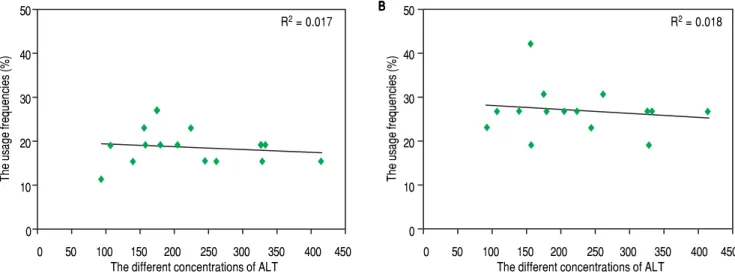
healthy controls (14.87 ± 9.95 pg/mL, P < 0.001). Not only in PBL but also in LIL, there was no correlation be-tween ALT and the skewness of TCR Vβ genes (Figure 3).
Relations between the
indices relative to HBV infection and TCR Vβββββ usage
As shown in table 1, according to the detection results of HBsAg, HBeAg and HBeAb, HBV infection was divid-ed into three serological patterns: HBsAg (+) & HBeAg (+) & HBeAb (-), HBsAg (+) & HBeAg (-) & HBeAb (-) and HBsAg (+) & HBeAg (-) & HBeAb (+). Not only in PBL but also in LIL, the predominant usage frequencies of TCR Vβ genes between the three patterns were similar (Figure 2). Besides, according to the comprehensive anal-ysis of TCR Vβ gene usage and HBV-DNA copies, there was no positive association between the skewed TCR Vβ
genes and HBV-DNA load.
DISCUSSION
The predominant usage of TCR Vβ in PBL from the patients with CHB has been ever described in a few re-ports. In Yao s study,19 Vβ8, Vβ12 and Vβ24 were the
pre-dominantly used genes. In another report, Vβ11 and Vβ12 highly expressed, and accordingly, the authors though that both genes probably associated with the occurrence of hepatitis B.20 In the present study, Vβ12 and Vβ13.1 were
found as the most predominant usage genes in PBL. Obvi-ously, Vβ12 was the common predominant usage gene in the three studies. This probably indicated that Vβ12 was the just predominant gene with high specificity to CHB. As to Vβ13.1, Wu, et al.14 also found that it was frequently
used in PBMC, and the usage frequency was high to 72.7% which was higher than the data of this study (46.7%). In another report, Vβ13 was also reported as the predominant usage gene,15 but because the researchers didn t identify it 50
40
30
20
10
0
,
The predominant
usage frequencies (%)
80
60
40
20
0
PBL
LIL
A
1 2 3 4 5.1 5.2 6 7 8 9 10 11 12 13.113.2 14 15 16 17 18 19 20 21 22 23 24
24 TCR Vβ genes
B
The limited
usage frequencies (%)
PBL LIL
Figure 1. Figure 1. Figure 1. Figure 1.
Figure 1. The comparison of the usage frequencies of 24 TCR Vβ genes of PBL with those of LIL in the patients with CHB. A. A. A. A. A. The comparison of the pre-dominant usage frequencies of 24 TCR Vβ genes of PBL with those of LIL. B.B.B.B.B. The comparison of the limited usage frequencies of 24 TCR Vβ genes of PBL with those of LIL.
1 2 3 4 5.1 5.2 6 7 8 9 10 11 12 13.113.2 14 15 16 17 18 19 20 21 22 23 24
Table 3. The frequencies of HLA-A and HLA-DR in CHB patient and control groups.
HLA CHB patient Control group HLA CHB patient Control group
group (n, %) (n, %) group (n, %) (n, %)
A1 1 (6.7) 0 (0) DR1 0 (0) 0 (0)
A2 8 (53.3) 6 (46.2) DR4 2 (13.3) 2 (15.4)
A3 0 (0) 1 (7.7) DR7 5 (33.3) 4 (30.8)
A11 3 (20.0) 3 (23.1) DR8 2 (13.3) 2 (15.4)
A23 0 (0) 0 (0) DR9 5 (33.3)* 3 (23.1)
A24 4 (26.7) 3 (23.1) DR10 0 (0) 0 (0)
A26 1 (6.7) 0 (0) DR11 0 (0) 1 (7.7)
A29 0 (0) 0 (0) DR12 4 (26.7)* 2 (15.4)
A30 4 (30.0) 3 (23.1) DR13 0 (0) 1 (7.7)
A31 2 (13.3) 1 (7.7) DR14 1 (6.7) 2 (15.4)
A32 0 (0) 0 (0) DR15 3 (20.0) 3 (23.1)
A33 4 (26.7) 3 (23.1) DR16 0 (0) 0 (0)
A68 0 (0) 0 (0) DR17 2 (13.3) 1 (7.7)
* Significant difference between CHB patient and control groups, P < 0.05.
A AA AA
The predominant usage frequencies (%)
100 90 80 70 60 50 40 30 20 10 0
The predominant usage frequencies (%)
B BB BB
100 90 80 70 60 50 40 30 20 10 0
Figure 2. Figure 2. Figure 2.
Figure 2. Figure 2. The comparison of the predominant usage frequencies of 24 TCR Vβ genes of PBL (LIL) in the patients with CHB between the three serological patterns of infection, which including HBsAg (+) & HBeAg (+) & HBeAb (-), HBsAg (+) & HBeAg (-) & HBeAb (-) and HBsAg (+) & HBeAg (-) & HBeAb (+). A.A.A.A.A. The comparison of the predominant usage frequencies of 24 TCR Vβ genes of PBL in the patients with CHB between the three serological patterns of infec-tion. B.B.B.B. The comparison of the predominant usage frequencies of 24 TCR Vβ genes of LIL in the patients with CHB between the three serological patterns ofB. infection.
,
1 2 3 4 5.1 5.2 6 7 8 9 10 11 12 13.1 13.2 14 15 16 17 18 19 20 21 22 23 24
24 TCR Vβ genes
1 2 3 4 5.1 5.2 6 7 8 9 10 11 12 13.1 13.2 14 15 16 17 18 19 20 21 22 23 24
24 TCR Vβ genes
P1-P5
P6-P10
P11-P15
P1-P5
P6-P10
as Vβ 13.1 or Vβ 13.2, so we could not determine whether their results were consistent with ours. Compared to PBL, fewer studies focused on the dominant usage of TCR Vβ of LIL. In this study, most of Vβ genes showed predominant usage including Vβ 13.1 and Vβ 5.2. This was similar to Zhang s study,21 in which Vβ 13.1 was reported
as the predominant usage gene with the highest frequency; Vβ 5.2 and Vβ 12 were next to it.
As to the limited usage Vβ genes, not only in PBL but also in LIL Vβ23 was always the highest frequent gene in this study. Dramatically, Vβ23 was the predominant usage gene in other several reports.14,16 According to our
knowl-edge, such a difference probably reflected the individuali-zation feature of the limited usage of TCR Vβ genes in patients with CHB.
To combine and analyze the skewness of TCR Vβ
genes of PBL with that of LIL, it was easy to found that there were similarities and differences between the two specimens. In our opinions, the mechanisms for the sim-ilarities and differences probably focused on two as-pects:
• Liver was the main place at where HBV located and proliferated, so the HBV antigen peptides in liver were much more than in peripheral blood. Naturally, as a key place for the cellular immune, there were more TCR Vβ genes skewed. This was the possible reason for what the total number of the skewed TCR Vβ
genes in LIL was higher than that in PBL.
• Some T cells containing the skewed TCR Vβ genes of peripheral blood probably migrated from liver,22 so
PBL and LIL shared the common skewed TCR Vβ
genes. However, these reasoning needed further
verifi-cation with more and powerful immunological or pathological experiments in future.
Recently, the association between HLA genes and the chronicity of HBV infection had been reported;23,24 but
few studies focused on the relativity between HLA alle-les and skewness of TCR Vβ genes of patients with CHB. In the present study, HLA alleles (HLA-A and HLA-DR) and TCR Vβ usage of the patients with CHB were simultaneously detected. In the results, there was no significant difference between the expression fre-quencies of HLA-A alleles of the patients with CHB and those of control group; while the frequencies of HLA-DR9 and HLA-DR12 of the CHB group were signifi-cantly higher than the data of the control group. This indicated that HLA-A gene probably played a small part in the chronicity of HBV infection; while HLA-DR was more important in such a progress. Besides, there was an interestingly phenomenon that the cases of HLA-DR9 or HLA-DR12 positive always showed the predominant us-age of Vβ 5.2 or Vβ 13.1. This further strongly suggested that HLA-DR9 or HLA-DR12 were very likely to associ-ate with the predominant usage of TCR Vβ genes. This finding was different from Sing's reports22, in which
HLA-B13, Bw4 and Cw3 associated with the oligoclonal use of Vβ 5.2, Vβ 11 and Vβ 17 gene families. However, both of studies showed that some HLA genes probably associated with the skewness of TCR Vβ genes for pa-tients with CHB.
In the results of hematological indices, the percentage of CD4+ T cells of the patients was obviously lower than
that of the healthy volunteers; while the rate of CD8+ T
cells of the patients was significantly higher than the
con-Figure 3. Figure 3. Figure 3. Figure 3.
Figure 3. The relation between the serum levels of ALT and the usage frequencies of TCR Vβ of PBL (LIL) in patients with CHB. A.A.A.A. The relation betweenA. the serum levels of ALT and the usage frequencies of TCR Vβ of PBL. B.B.B.B.B. The relation between the serum levels of ALT and the usage frequencies of TCR Vβ of LIL.
A A A A A
The usage frequencies (%)
50
40
30
20
10
0
0 50 100 150 200 250 300 350 400 450 The different concentrations of ALT
R2 = 0.017
B BB BB
The usage frequencies (%)
50
40
30
20
10
0
R2 = 0.018
trols. Companying these results with the data relate to the skewness of TCR Vβ genes, we considered that the im-balance of T cell subpopulation probably was one of the reasons for the skewness of TCR Vβ genes. This was con-sistent with other scholars reports that TCR Vβ genes of the CD8+ T cells of patients with HBV infection
exhibit-ed a greater number of biasexhibit-ed clones than CD4+ T
cells.25,26 Besides, the average IFN-γ concentration of the
patients with CHB was much higher than that of healthy volunteers. This finding indicated that the level of IFN-γ
probably associated with the skewness of TCR Vβ genes of patients with CHB.27,28 But the high IFN-γ
concentra-tion was the reason or the result for the skewness of TCR Vβ genes needed further study.
As shown in figure 2, the predominant usages of TCR Vβ of the patients with CHB were similar in the three se-rological patterns of infection. This probably because that the skewness of TCR Vβ genes kept a rather stable status under the long term of stimulation by HBV antigens; and moreover, such an antigen stimulation was uncorrelated with HBV-DNA load which can be known from the rela-tionship between HBV-DNA copies and TCR Vβ gene usage. Besides, no relation could be found between ALT and TCR Vβ gene usage in the study. This was consistent with another reports,29 and both studies gave a suggestion
that there was no obvious correlation between the TCR Vβ skewness and liver injury.
In conclusion, this study showed two important find-ings. One was that there were common and different fea-tures for TCR Vβ usage in PBL and LIL of patients with CHB; the other was that some hematological indices probably associated with the skewness of TCR Vβ genes, including HLA-DR9, HLA-DR12, CD8+ T cell and IFN-γ. However, due to the small size of cases, there were still some questions need to be cleared up in future, such as the sequences of the skewed TCR Vβ genes, the relation-ship between TCR Vβ genes and each of HLA alleles, the mechanism for the effects of imbalance of CD4+ and CD8+ T on TCR Vβ gene usage, and so on.
ACKNOWLEDGEMENTS
All the authors thank Dr. Xinsheng Yao for the guid-ance to the study.
GRANT SUPPORT
The work is granted by the Provincial Science and Technology Development Project (2012YD18054), the Provincial Nature Science Foundation (ZR2012HL29), the High School Science and Technology Plan Project (J11LF18), the Population and Family Planning Commis-sion ([2011]13), and the Development Plan Project of
Jin-ing Science and Technology Bureau of Shandong Prov-ince ([2011] 57, 2014jnjc12 and 2014jnyx14 ).
ABBREVIATIONS
• CDR3: the third complementarity-determining re-gion.
• CHB: chronic hepatitis B. • FHB: fulminant hepatitis B. • HB: hepatitis B.
• HBsAg: hepatitis B surface antigen. • HLA: human leukocyte antigen. • LIL: liver infiltrating lymphocytes. • MCAT: melting curve analysis technique. • PBL: peripheral blood lymphocytes.
• RQ-PCR: real-time florescence quantitative polymerase chain reaction.
• TCR: T cell receptor. • Vβββββ: beta chain variable gene.
REFERENCES
1. WHO. Guidelines for the prevention, care and treatment of persons with chronic hepatitis B infection. Executive sum-mary 2015; http://www.who.int/iris/handle/10665/154590. 2. Wei L. Natural history of chronic hepatitis B virus infection:
what determines prognosis after cirrhotic decompensation. J Gastroenterol Hepatol 2008; 23: 1631-2.
3. Chisari FV, Isogawa M, Wieland SF. Pathogeniesis of hepati-tis B infection. Pathol Biol 2010; 58: 258-66.
4. Grimm D, Heeg M, Thimme R. Hepatitis B virus: from immunol-ogy to immunotherapy. Clin Sci 2013; 19: 859-68.
5. Elgouhari HM, Abu-Rajib Tamimi II, Carey WD. Hepatitis B vi-rus infection: understanding its epidemiology, course, and di-agnosis. Clevel Clin J Med 2008; 75: 881-9.
6. Zhou J, Ma R., Luo R, He X, Sun W, Tang W, Yao X. Prima-ry exploration of molecular and spectratyping features of CDR3 of TCR β chain in the peripheral blood and tissue of patients with colorectal carcinoma. Cancer Epidemol 2010; 34: 33-40.
7. Zhou J, Kong C, Wang X, Jia Y, Wang L, Chang H, Sun L. In silico analysis of TCR Vβ 7 of two patients with type 1 dia-betes mellitus. J Lab Physicians 2013; 5: 79-82.
8. Miqueu P, Guillet M, Degauque N, Dore JC, Soulillou JP, Br-ouard S. Statistical analysis of CDR3 length distributions for the assessment of T and B cell repertoire biases. Mol Immu-nol 2007; 44: 1057-64.
9. Melenhourst JJ, Lay MDH, Price DS, Adams SD, Zeilah J, Sosa E, Hensel NF, et al. Contribution of TCR- locus and HLA to the shape of the mature human V repertoire. J Im-munol 2008; 180: 6484-9.
10. Attaf M, Huseby E, Sewell AK. αβ T cell receptors as pre-dictors of health and disease. Cell Mol Immunol 2015; 12(4): 391-9.
11. Fozza C, Contini S, Corda G, Virdis P, Galleu A, Bonfigli S, Pacifico A, et al. T-cell receptor repertoire analysis in monozygotic twins concordant and discordant for type 1 di-abetes. Immunobiology 2012 ; 217(9): 920-5.
12. Zhou JW, Ma R, Tang WT, Luo R, Yao XS. Primary explora-tion of the third complementarity determining region spectra-typing and molecular features of T cell receptor alpha chain
in the peripheral blood and tissue of patients with colorectal carcinoma. ACTA Medica Mediterranea 2011; 27: 97-104. 13. Yang J, He J, Huang H, Ji Z, Wei L, Ye P, Xu K, et al.
Molecu-lar characterization of T cell receptor beta variable in the pe-ripheral blood T cell repertoire in subjects with active tuberculosis or latent tuberculosis infection. BMC Infect Dis 2013; 13: 423.
14. Wu SQ, Yao XS, Qiu LM, Ma R, Bi XY, Chen Y. Analysis of the T lymphocyte receptor beta chain complementarity re-gion 3 spectratyping in the peripheral and hepatic tissue of patients with chronic hepatitis B. Chin J Infec Dis 2010; 28: 348-53.
15. Shi WJ, Wan H, Zhou J, Wei L, Yang ZM, Wang ZX. The study of the expression of TCR BV CDR3 family in fulminant hepatitis B patients. Zhonghua Shi Yan He Lin Chuang Bing Du Xue Za Zhi 2013; 27: 241-3.
16. Zhang GW, Yao XS, Ma SW, Yu LC, Wang ZH, Hou JL. Anal-ysis of T cell receptor repertoire in patients with chronic asymptomatic hepatitis B virus carriers. Jie Fang Jun Yi Xue Za Zhi 2006; 31: 246-9.
17. Zhou J, Kong C, Luo J, Cao J, Shi Y. Comparaing TCR beta chain variable gene skewness between children with tuber-culosis and BCG-vaccinated children. Arch Iran Med 2013; 16: 104-8.
18. Chinese Society of Hepatology, Society of Infectious Diseas-es. Guideline on Prevention and Treatment of Chronic Hepa-titis B (2010 version). Chin J Exp Clin Infect Dis 2011; 5: 79-100.
19. Yao XS, Diao Y, Sun WB, Luo JM, Qin M, Tang XY. Analysis of the CDR3 length repertoire and the diversity of TCR alpha chain in human peripheral blood T lymphocytes. Cell Mol Im-munol 2007; 4: 215-20.
20. Xiong Y, Song Y, Bi S, Tan Y. Study of clonaliy of TCR V gene subfamilies on peripheral blood CD8+ T lymphocyte in patient with chronic hepatitis B. Chin J Immunol 2011; 27: 751-6.
21. Zhang GW, Yao XS,Ma SW, Yang CG, Yu YC, Hou JL. Anal-ysis of T cell receptor BV dominant usage and CDR3 se-quences during acute exacerbation in patients with chronic hepatitis B. Zhonghua Gan Zang Bing Za Zhi 2006; 14: 23-8. 22. Sing GK, Li D, Chen X, Macnaughton T, Lichanska AM, But-terworth L, Ladhams A, et al. A molecular comparison of T lymphocyte populations infiltrating the liver and circulating in
the blood of patients with chronic hepatitis B: evidence for antigen-driven selection of a public complementarity-deter-mining region 3 (CDR3) motif. Hepatology 2001; 33: 1288-98. 23. Mbarek H, Ochi H, Urabe Y, Kumar V, Kubo M, Hosono N, Takahashi A, et al. A genome-wide association study of chronic hepatitis B identified novel risk locus in a Japanese population. Hum Mol Genet 2011; 20: 3884-92.
24. Doganay L, Fejzullahu A, Katrinli S, Yilmaz Enc F, Ozturk O, Colak Y, Ulasoglu C, et al. Association of human leukocyte antigen DQB1 and DRB1 alleles with chronic hepatitis B. World J Gastoenterol 2014; 20: 8179-86.
25. Yang J, He J, Lu H, Wei L, Li S, Wang B, Diao H, et al. Molec-ular features of the complementarity determining region 3 motif of the T cell population and subsets in the blood of pa-tients with chronic severe hepatitis B. J Transl Med 2011; 9: 210-8.
26. Das A, Hoare M, Davies N, Lopes AR, Dunn C, Kennedy PT, Alexander G, et al. Functional skewing of the global CD8 T cell population in chronic hepatitis B virus infection. J Exp Med 2008; 205: 2111-24.
27. Banu N, Chia A, Ho ZZ, Garcia AT, Paravasivam K, Groten-breg GM, Bertoletti A, et al. Building and Optimizing a Virus-specific T Cell Receptor Library for Targeted Immunotherapy in Viral Infections. Sci Rep 2014; 4: 4166.
28. Zhang YP, Yang R, Zhang GW. Characteristic of T lym-phocyte cell receptor repertoire in patients with chronic hep-atitis B and the relationship of it with early curative effect of interferon antiviral. Journal of Xinxiang Medical University 2014; 31: 195-201.
29. Ma SW, Li YY, Zhang GW, Huang X, Sun J, Li C, Abbott WG, et al. Complementarity-determing region 3 size spectratypes of T cell receptor β chains in CD8+ T cells following antiviral
treatment of chronic hepatitis B. Antimicrob Agents Chem-other 2011; 55: 888-94.
Correspondence and reprint request:
Jianwei Zhou, M.D.
Clinical Laboratory, Affiliated Hospital of
Jining Medical University, Jining, Shandong Province, China. Tel.: 86 537 2903223.