Otras secciones de este sitio:
☞ ☞ ☞ ☞
☞ Índice de este número
☞ ☞ ☞ ☞
☞ Más revistas
☞ ☞ ☞ ☞
☞ Búsqueda
Others sections in this web site:
☞ ☞ ☞ ☞
☞ Contents of this number ☞
☞ ☞ ☞
☞ More journals ☞
☞ ☞ ☞ ☞ Search
Article:
Obesity-related non-alcoholic steatohepatitis and TGF-β1 serum levels in relation to morbid obesity
Copyright © 2002: Mexican Association of Hepatology
ANNALS OF HEPATOLOGY
Number 1 January-March 2002
Volume 1
Annals of hepatology
Original Article
Obesity-related non-alcoholic steatohepatitis and
TGF-
β
1 serum levels in relation to morbid obesity
Ricardo N Sepúlveda-Flores MD,1 Lucio Vera-Cabrera MD,2 Juan P Flores-Gutiérrez MD,3 Héctor
Maldonado-Garza MD,1 Ricardo Salinas-Garza MD,1 Pablo Zorrilla-Blanco MD,1 Francisco J Bosques-Padilla MD1
Abstract
Non-alcoholic steatohepatitis (NASH) can vary from mild hepatic inflammation and steatosis to cirrhosis, and is most frequently associated with obesity, Type 2 diabetes mellitus, hypertension, and the female der. The prevalence of fatty liver and NASH in the gen-eral population is 20% and 3%, respectively. In Wes-tern countries, 15–20% of the population is obese and 74–90% of them exhibit fatty changes in liver biopsies. We assessed the prevalence of NASH in morbidly o-bese patients and evaluated serum TGF-βββββ1 concentra-tions in different stages of liver fibrosis. Thirty-five obese patients were evaluated, nine male and 26 female. Their mean body mass index (BMI) was 43.62 ± 7.92 kg/m2. Liver biopsies were evaluated by light
micros-copy; graded and staged according to Brunt’s system. Serum obtained from patients was used to detect TGF-βββββ1 concentrations by an ELISA method. Serum ala-nine transaminase (ALT) levels were elevated in four of the patients and the mean level was 49.98 ± 94.7 (8–65 IU/L). NASH was diagnosed in 32 (91%) of the biop-sies, and the most common pattern seen was mixed, predominantly macrovesicular steatosis. Some degree of fibrosis was seen in 34 (97%) of the biopsies and 22 (63%) were at stage 2 (range 1–3). Serum concentra-tions of TGF-βββββ1 had no relationship with the stages of fibrosis. In conclusion, NASH and fibrosis are com-mon in our obese patients, as observed in other stud-ies. TGF-βββββ1 may play a key role in liver fibrogenesis.
Key words: Obesity, Non-alcoholic steatohepatitis, Hepatic fibrosis, Steatosis.
Introducción
Obesity is a worldwide epidemic that is concerning public health organizations. In Latin America, 20–25% of the
popu-lation is obese1 and in the United States of America 40% of
the Mexican-American population is obese.2 Obesity alone
may not be a problem; however, the associated morbidity has a negative effect on health care. Hypertension, diabetes me-llitus, coronary diseases, and cerebrovascular accidents are just some examples of the diseases associated with obesity.
Non-alcoholic steatohepatitis (NASH) is a disease of the liver characterized by the presence of fat, inflamma-tion, necrosis, Mallory hyaline bodies and fibrosis seen in
liver biopsies.3 NASH has been associated with obesity,
diabetes, jejuno-ileal bypass, rapid weight loss, poor nu-trition, and with patients who have been put on parenteral
nutrition.3-6 The metabolic syndrome X is characterized by
insulin resistance, dyslipidemias, hypertension, and
male-type distribution of fat.7 There is a high association
be-tween metabolic syndrome X and NASH. In Spain,
García-Monzón et al.8 biopsied 46 obese patients and observed
that 69 % had NASH and 41 % had some stage of fibrosis.
Ratziu et al.9 studied 93 obese non-alcoholic subjects with
similar characteristics and found that 30% had septal fi-brosis and 11% had cirrhosis.
During hepatic fibrogenesis, stellate cells in the liver
produce Transforming Growth Factor-β (TGF-β), which
controls the extracellular matrix deposition of wound heal-ing proteins such as collagen, fibronectin, proteoglycans,
and hyaluronic acid.10, 11 This cytokine is also produced by
mesangial cells in the kidney, lung cells, and bone. Its main function is to induce wound healing, cell prolifera-tion, and apoptosis. It is also involved in the chemotaxis of neutrophils, monocytes, T lymphocytes, and
fibro-blasts.12 Tumor necrosis factor (TNF-α), interleukin-6
(IL-6) and TGF-β play important roles in hepatic wound
healing and must work all together.13 TNF-α and IL-6 are
both pro-inflammatory cytokines, except that the latter is
also responsible for hepatocyte development.14 TGF-β
was first isolated 10 years ago from platelets.15 There are
three isoforms: β1, β2, and β3.16,17 TGF-β1 is the most
abundant form secreted during fibrogenesis.16 It has three
cell membrane receptors.18,19 Binding to receptor βRI
in-duces synthesis and deposition of extracellular matrix;
βRII activation leads to cellular proliferation and βRIII
presents TGF-β to the other two receptors.12 It is possible
1 Departamento de Gastroenterología.
2 Laboratorio de Dermatología.
3 Departamento de Anatomía Patológica.
Hospital Universitario “Dr. José E. González”, Universidad Autó-noma de Nuevo León. Monterrey, N.L., México
Abbreviations: TGF-β1, transforming growth factor β1; LFT, liver function tests.
Address for correspondence: Francisco J Bosques-Padilla MD
RN Sepúlveda-Flores et al. / Obesity-related non-alcoholic steatohepatitis 37
edigraphic.com
to detect TGF-β1 in serum by enzyme-linked
immunosorb-ent assay (ELISA) with the help of specific microplates
pre-coated with TGF-βRII.20
The aim of this study was to assess the prevalence of hepatic injury in morbidly obese patients and to evaluate
serum TGF-β1 concentrations in different stages of liver
fibrosis in an attempt to validate its use as an indicator of liver disease progression.
Patients and methods
The study took place between September 2001 and Feb-ruary 2002 at the University Hospital “Dr. José E. Gonza-lez” of the University of Nuevo León in Monterrey, Nuevo León, Mexico. Candidates were chosen to participate in the study based on the following inclusion criteria: body mass index (BMI) >35%, alcohol consumption <30 g/day in men and < 20 g/day in women, age > 18 y, and a negative his-tory of viral, alcoholic, autoimmune or drug-related hepati-tis and tumors. Subjects who had been previously treated with amiodarone, corticosteroids, tamoxifen, methotrexate, or high-dose estrogens, or who had received a jejuno-ileal bypass or small bowel resection, or who had received total parenteral nutrition (TPN) were excluded from the study.
Thirty-five subjects who underwent gastric bypass sur-gery for weight reduction were included. Liver function tests (LFT), basic metabolic panel (BMP), a complete blood count (CBC), lipid profile, and measurements of blood levels of anti-hepatitis B virus (HBV) and C virus (HCV) were ordered for each patient. An abdominal ultra-sound was used to rule out any abscesses, tumors, or cysts in the liver before surgery.
This study conformed of Helsinki, and was approved by Hospital´s Research and Ethics Committe. Written in-formed consent was obtained from all patients.
Liver Biopsy
Immediately after the abdominal cavity was opened, a
3 × 3 × 1-cm liver biopsy was taken by excision. Each
biopsy was fixed in formaldehyde, embedded in paraffin wax, and stained with hematoxylin and eosin (H & E) and Masson’s Trichrome for the assessment of liver fibrosis. Brunt’s system of the histopathological lesions of NASH was used for the grading and staging of NASH and liver
fibrosis.21 The specimens were analyzed by one
patholo-gist with experience in liver pathology.
Serum TGF-βββββ1 Measurement
Before surgery, a 6-mL sample of venous blood was col-lected from each patient using an SST tube with EDTA as anticoagulant, and incubated in a 2–8 °C refrigerator
over-night to ensure complete release of TGF-β1. Each blood
sample was centrifuged at 100 g for 30 min at 4 °C. The obtained serum samples were then stored at –70 °C before
the assay. A Quantikine TGF-β1 immunoassay kit (R&D
Systems, Inc. Minneapolis, MN) was used. For the
activ-ation of latent TGF-β1 to immunoreactive TGF-β1, 0.1 mL
of a solution containing 2.5 N acetic acid and 10 M urea was added to 0.1 mL of serum and incubated for 10 minutes at room temperature. The acidified samples were neutral-ized by adding 0.1 mL of 2.7 NaOH and 1 M HEPES. The activated serum samples were then diluted tenfold. Each sample and standard were pipetted into a previously coated
microplate well containing recombinant human TGF-β
sol-uble receptor type II and assayed in duplicate. Each well was washed three times with a buffer solution to wash away any unbound substances, and polyclonal antibodies specific
for TGF-β1 conjugated to horseradish peroxide were added
to each well. Following a last wash, a substrate solution containing stabilized hydrogen peroxide and tetramethyl-benzidine was added to the wells. A bluish color developed
in proportion to the amount of bound TGF-β. Sulfuric acid
(2 N) was added to stop the reaction and optical density was read using a spectrophotometer at 450 nm corrected to 540 nm. The concentration of the samples was determined based on a standard curve prepared with samples of known concentrations.
Statistical Analysis
Data were collected and analyzed using Microsoft Ex-cel 2000 and Primer of Biostatistics software. Averages, medians, modes, and standard deviations of the variables were calculated. Analysis of variance (ANOVA) was used to test for any significance of differences, and a Bonferro-ni test to calculate the comparisons between groups.
Results
Demography
A total of 35 biopsies and serum samples were obtain-ed from a group of morbidly obese patients with a mean
BMI of 43.62 ± 7.92 kg/m2. Nine (26%) were men, 26
(74%) were women and their mean age was 33 ± 10 y. Their mean weights and heights were 120.3 ± 28.9 kg and 1.66 ± 0.09 m respectively. In this study, 24% of the pat-ients were known to have hypertension, 22% had fasting glucose levels >110 mg/dL and two were taking oral hy-poglycemic drugs at the time of the study.
Laboratory Analysis
239.3 ± 128.29 IU/L (range 100–225) and 99.4 ± 40.7 IU/ L (range 30–140), respectively. Only one patient had signif-icant elevation of all the liver enzymes measured.
Histopathological Analysis
NASH
Thirty-five biopsies were analyzed and classified accord-ing to Brunt’s gradaccord-ing and stagaccord-ing system. The most com-mon pattern of hepatic steatosis found in the biopsies was a predominantly macrovesicular steatosis mixed with micro-vesicular steatosis (43%). A mixed predominantly microvesi-cular steatosis pattern was present in 20% of the specimens. Chronic lobular inflammation was present in 16 (46%), and 31 (89%) presented acute lobular inflammation. Mild portal in-flammation was present in 18 (51%), moderate portal inflam-mation in 13 (37%) and one (3%) had no portal inflaminflam-mation. Grade 1 NASH was present in eight (23%), grade 2 in 16 (46%) grade 2 and grade 3 in seven (20%). Three biopsies (9%) could not be classified into a group (Figure 1).
Fibrosis
Thirty-four patients (97%) had some degree of fibrosis. Stage 1 fibrosis was present in six patients (17%), stage 2
in 22 (63%) and stage 3 in five (14%). In this category, only one patient (3%) did not have fibrosis (Figure 2).
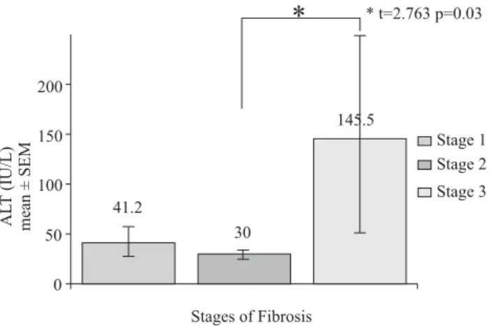
ALT levels and Stages of Fibrosis
The ALT levels were compared between the three stages of fibrosis using a Bonferroni t test (Figure 3). Significant statistical difference was found when comparing ALT lev-els between Stage 2 and Stage 3 of fibrosis (p = 0.03, t = 2.763). Not all subjects had elevation of ALT and AST lev-els (12% and 9% were elevated, respectively).
Serum TGF-βββββ1 concentration
Serum concentrations of TGF-β1 were grouped
accord-ing to the stage of fibrosis, calculataccord-ing the mean concen-tration (ng/mL) of each group, and a comparison was made between them to search for any difference between the groups (Figure 4). The concentration for stage 1 fibro-sis was 36.77 ± 6.16 ng/mL. For stage 2 fibrofibro-sis the con-centration was 41.57 ± 10.45 ng/mL, and it was 37.45 ± 7.83 ng/mL for stage 3. No statistically significant differ-ences were seen between groups.
Figure 1. Shows the percentage of patients with NASH in its three grades.
Figure 2. Percentage of patients for each stage of fibrosis.
Figure 3. Serum ALT levels (mean Standard Error of Means) for each
stage of fibrosis.
RN Sepúlveda-Flores et al. / Obesity-related non-alcoholic steatohepatitis 39
edigraphic.com
Discussion
TGF-β1 is just one of the many serum markers that
could potentially be used for a non-invasive diagnosis of hepatic fibrosis. Matrix deposition proteins, matrix pro-duction and degradation enzymes, and other fibrogenic cytokines could be used to assess this process in the liver. Among the members of the matrix molecule family, lami-nin, hyaluronic acid, and collagen type I and IV have been the most studied and have the most potential clinical utili-ty.22,23 Levels of other fibrogenic cytokines such as
plate-let-derived growth factor (PDGF) and connective tissue growth factor (CTGF) might also be clinically useful.
The results in the obese population we studied were sim-ilar to most of the published literature. A high prevalence of NASH was found in this group of patients. In our study, 32 (90.4%) had some grade of NASH and some degree of fibrosis was present in nearly all of them (97%). NASH is not always intentionally looked for when LFTs are within normal values in obese patients. As a high proportion of our population studied was diagnosed with NASH and yet the ALT levels were normal in 83%, routine LFT and ALT val-ues evidently lack sensitivity for this condition.
We do not have any doubts as to the importance
TGF-β has in the mechanism of wound healing and hepatic
fi-brosis. TGF-β1 serum concentrations were compared
be-tween fibrosis stage and grade groups, and no significant differences were found among them (Figure 4). Our
resul-ts show that even though TGF-β1 is not distributed evenly
among the three stages of fibrosis, it is possible to detect it in serum analysis. Therefore, our study suggests that
TGF-β1 is not the best serum marker for this particular
subgroup of patients. A well-selected control group should be included to further complement our results.
The diagnosis of NASH currently represents a clinical challenge and we must take into account several parameters such as anthropometric measurements, insulin resistance, blood pressure, and dyslipidemias to find it when studying
obese patients and assessing the presence of this condition.24
Genetic factors may contribute to insulin resistance,
and TNF-α promoter polymorphisms may influence it,
leading to non-alcoholic fatty livers and steatohepatitis.25
We conclude that liver biopsy remains the “gold-stand-ard” for diagnosing, grading, and staging liver diseases and a more sensitive serological marker is necessary to help us identify these patients before they develop termi-nal liver disease.
References
1. Organización Mundial de la Salud: http://www.who.int/whosis. 2. De la Mora G, Olivera M, de la Cerda R, Arista J, Kershenobich D,
Uribe M. Esteatohepatitis no alcohólica: Experiencia de 10 años en el Instituto Nacional de la Nutrición Salvador Zubirán. Rev Inv Clin 1994; 46:85–92.
3. Williams EJ, Gaá MDA, Brigstock DR, Arthur MJP, Benyon RC. Increased expression of connective tissue growth factor in fibrotic
human liver and in activated hepatic stellate cells. J Hepatol 2000; 32:754–61.
4. Fong DG, Nehra V, Lindor KD, Buchman AL. Metabolic and Nutritional Considerations in Nonalcoholic Fatty Liver. Hepatology 2000; 32:310. 5. Friedman SL, Maher JJ, Bissel DM. Mechanisms and Therapy of Hepatic Fibrosis: Report of the AASLD single Topic Basic Research Conference. Hepatology 2000; 32:1403–8.
6. Matteoni CA, Younoussi ZM, Gramlich T, Boparai N, Hu YC, McCullough AJ. Nonalcoholic Fatty Liver Disease: A spectrum of clinical and pathological severity. Gastroenterology 1999; 116: 1413–9. 7. Marceu P, Biron S, Hould FS, Marceu S, Simard S, Thung SN, Kral
JG. Liver Pathology and the Metabolic Syndrome X in Severe Obesity.
J Clin Endocrinol & Metab 1999; 84: 1513-7.
8. García-Monzón C, Martín-Pérez E, Lacono OL, Fernández-Bermejo M, Majano PL, Apolinario A, Larráñaga E, et al. Characterization of Pathogenic and Prognostic Factors of Nonalcoholic Steatohepatitis Associated with Obesity. J Hepatol 2000; 33:716–24.
9. Ratziu V, Giral P, Charlotte F, Bruckert E, Thibault V, Theodoru I, Khalil L, et al. Liver Fibrosis in Overweight Patients. Gastroenterology 2000; 118:1117–23.
10. Rockey DC. Pathophysiology of Disease: The Cell and Molecular Biology of Hepatic Fibrogenesis. Clin Liver Dis 2000; 4: 1-36. 11. Vera-Cabrera L, Handzel V, Laszlo A. Development of an Enzyme
Linked Immunosorbent Assay (ELISA) Combines With a Streptavidin Biotin and Enzyme Amplification Method to Detect Anti-2, 3-di-oacyltrehalose (DAT) Antibodies in Patients with Tuberculosis. J
Immun Met 1994; 177:69–77.
12. Border NA, Noble NA. Mechanisms of disease: Transforming Growth Facto (beta) in Tissue Fibrosis. N Eng J Med 1994; 331: 1286-1292. 13. Tilg H, Diehl AM. Cytokines in Alcoholic and Nonalcoholic
steatohepatitis. N Engl J Med 2000; 343:1467–76.
14. Streetz KL, Luedde T, Manus MP, Trautwein C. Interleukins 6 and Liver Regeneration. Gut 2000; 47:303–12.
15. Tsai JF, Chuang LY, Jang JE, Yang ML, Chang WY, Hsich MY, et al. Clinical Relevance of Transforming Growth Factor β1 as an indicator of Hepatic Function Impairment in Liver Cirrhosis. Medicine 1997; 76: 213-226.
16. Blobe GC, Scieman NP, Lodis HF. Role of Transforming Growth Factor β in Human Disease. N Engl J Med 2000; 342:1350–8. 17. George J, Roulot D, Kotelianski VE, Bissel DM. In Vivo inhibition of
rat stellate cell activation by soluble transforming growth factor β type II receptor: A potential new therapy for hepatic fibrosis. Proc
Natl Acad Sci U S A 1999; 96;12719–24.
18. Roulot D, Seucsik AM, Coste T, Strosberg AD, Marullo S. Role of Transforming Growth Factor β type II receptor in Hepatic Fibrosis: Studies of Human Chronic Hepatitis C and Experimental Fibrosis in Rats. Hepatology 1999; 29:1730–8.
19. Flisiak R, Pytel-Krolezuk B, Prokopowicz D. Circulating Transforming Growth Factor β1 as an indicator of Hepatic Function impairment in Liver Cirrhosis. Cytokine 2000; 12: 677–81. 20. Flisiak R, Pytel-Krolezuk B, Prokopowicz D. Circulating
Transforming Growth Factor β1 as an indicator of Hepatic Function impairment in Liver Cirrhosis. Cytokine 2000; 12: 677–81. 21. Murawaki Y. Plasma transforming growth factor beta 1 concentrations
in patients with chronic viral hepatitis. J Gastroenterol Hepatol 1998; 13:680–4.
22. Brunt EM. Nonalcoholic Steatohepatitis: Definition and Pathology.
Sem Liv Dis 2001; 21:3–16.
23. Oh S, Afdhal NH. Hepatic Fibrosis: Are any of the serum markers useful? Current Gastroenterology Reports 2001; 3:12–18. 24. Friedman SL. Seminars in Medicine of the Beth Israel Hospital Boston:
The Cellular Basis of Hepatic Fibrosis-Mechanisms and Treatment Strategies. N Engl J Med 1993; 328: 1828-1835.
25. Pagano G, Pacini G, Musso G, Gambino R, Mecca F, Depetris N, Cassader M et al. Nonalcoholic Steatohepatitis, Insulin Resistance, and Metabolic Syndrome: Further Evidence for an Etiologic Association. Hepatology 2002; 35:367–72.
26. Valenti L, Fracanzani AL, Dongiovanni P, Santorelli G, Branchi A, Taioli E, Fiorelli G et al. Tumor necrosis factor a promoter polymor-phisms and insulin resistance in nonalcoholic fatty liver disease.