Papillary in situ and intramucosal adenocarcinoma of the lower
third of common bile duct. A report and review of the literature
Eduardo E. Montalvo-Javé,*,†,§ Fredy Chablé-Montero,‡ Liz N. Toapanta-Yanchapaxi,*
Fernando Rojas-Mendoza,* Carlos López-Caballero,|| Mario L. Roca-Cabrera,¶ Germán E. Mendoza-Barrrera§
*Departamento de Gastroenterología, Fundación Clínica Médica Sur, Mexico City, Mexico. † Servicio de Cirugía General, Unidad 304, Hospital General de México. Mexico City, Mexico.
‡ Departamento de Patología, Fundación Clínica Médica Sur, Mexico City, Mexico. §
§ § §
§ Departamento de Cirugía, Facultad de Medicina, Universidad Nacional Autónoma de México (UNAM), Mexico City, Mexico. ||
|| ||
|| || Facultad Mexicana de Medicina, Universidad la Salle (ULSA), Mexico City, Mexico.
¶ Departamento de Anestesiología y Terapia Respiratoria. Fundación y Clínica Médica Sur, Mexico City, Mexico
ABSTRACT
We report the case of a 37-year-old woman with no relevant medical history. She was admitted to the hospital for epigastric pain related with food intake for 4 days; the pain did not improve with symptomatic management. A laparoscopic cholecystectomy due to acute lithiasic cholecystitis was performed. However, after 4 days, postoperative painless jaundice was evident; thus, endoscopic retrograde cholangiopancrea-tography was performed, which revealed an amputation of intrapancreatic common bile duct, as well as secondary intra- and extrahepatic bile duct dilatation. Brushing of the distal portion of the common bile duct revealed a well-differentiated adenocarcinoma. Therefore, a Whipple procedure with pylorus preser-vation was performed. Pathologic diagnosis of a papillary in situ adenocarcinoma with two microscopic foci of microinvasion was established. The pathologic Tumor-Node-Metastasis (TNM) stage was pT1, pN0, pM0, R0. The patient is asymptomatic and disease-free 24 months after surgery. In general, adenocarcinomas of the extrahepatic bile ducts are uncommon and have a poor prognosis. However, symptomatic patients with early disease stages are even rarer and can be cured surgically.
Key words. Papillary in situ. Intramucosal. Whipple. Surgery. Adenocarcinoma. Distal common bile duct.
Correspondence and reprint request: Eduardo E. Montalvo-Javé, MD, PhD,
FACS
Puente de Piedra, Núm. 150, Torre de Hospitalización, Piso 1, Servicio de Gastroenterología, Col. Toriello Guerra, 14050 México, D.F., México. Phone and Fax: (+52) (55) 5424-6892
E-mail: [email protected]
Manuscript received: October 20, 2014.
Manuscript accepted: November 26, 2014.
INTRODUCTION
Carcinomas of the extrahepatic bile ducts (EHBD) are uncommon. They represent 0.16% of all invasive carcinomas in males and 0.15% in females in the United States (U.S.) and constitute the third lead-ing cause of EHBD obstruction.1,2 Nearly 2,500 new cases are reported each year in the U.S., and their incidence is 0.88 per 100,000 inhabitants.1,2
Carcinomas have a major incidence in males, with a 1.3:1 ratio.1 Mean age at diagnosis is 63.3
years (range, 31-81 years). On evaluation of a specific population, Brugen, et al. reported on 43 patients: 93% were Caucasian; 5% Afro-American, and 2%, Hispanic.3 Carcinomas can arise in any part of the EHBD. For prognostic and therapeutic purposes, it has been useful to divide the extrahepatic ductal system into three parts: upper (hepatic ducts and the common hepatic duct); mid-dle (common hepatic duct and proximal common bile duct), and lower (intra- or peripancreatic zone of the common bile duct).1,4 Over 50% are located in the upper third, 18% in the middle third, and 22%, in the lower third.1,4,5
Because these tumors are rarely diagnosed in their early stages, we present a case of an in situ and intramu-cosal carcinoma of the lower third of common bile duct.
CLINICAL CASE
epigas-tric abdominal pain associated with consumption of food during 4 days. The patient did not have rele-vant medical history and was previously healthy. A clinical diagnosis of acute lithiasic cholecystitis was established; therefore, a laparoscopic cholecystecto-my was performed. Intraoperatively, the gallbladder was enlarged, had an edematous wall, and contained multiple gallstones. Moreover, external compression of the common hepatic duct by an impacted stone in cystic duct was observed.
During the postoperative period, the patient evolved favorably, but at 48 h, jaundice was docu-mented. The liver tests showed hyperbilirubinemia at the expense of direct bilirubin. After a 24-h obser-vation period, the following new changes in liver function tests were reported: Total bilirubin, 5.8 (previously 3.9); Direct bilirubin, 3.97; indirect bi-lirubin, 1.88; SGPT, 197; SGOT, 120; FA, 220, and GGT, 271.
Cholangiopancreatographic magnetic resonance (MR) was performed without relevant findings. En-doscopic retrograde cholangiopancreatography (ERCP) revealed stenosis and amputation of the in-trapancreatic common bile duct (Figure 1). A sphincterotomy with brushing of the lower third of the common bile duct was performed. Because of the pathological report, a pyloric-preserving Whipple surgery was performed.
GROSS AND MICROSCOPIC FINDINGS
Brush cytology exhibited groups and papillary structures of biliary epithelial columnar cells with
moderate and high-grade atypia (Figure 2). Diagno-sis of well-differentiated adenocarcinoma was estab-lished.
The Whipple specimen demonstrated fibrosis of the wall of the intrapancreatic portion of the com-mon bile duct. A mucosal lesion with micropapil-lary projections that extends into lumen and ampulla of Vater was observed. This mucosal lesion measured 1.3 cm at its greatest dimension (Figure 3). Microscopically, the lesion was composed of papillary structures lined predominantly with bil-iary type epithelium with moderate- and high-grade dysplasia/in situ adenocarcinoma, characterized by nuclear pseudostratification, prominent nucleoli, and mitotic figures (Figures 4 and 5). The tumor
Figure 1. Endoscopic retrograde cholangiopancreatography that shows stenosis and amputation of the lower third of the common bile duct. Proximal dilatation is also observed.
Figure 3. Macroscopic pathology. The distal portion of common bile duct showed fibrosis of the wall. Mucosal micro-papillary projections extend into the lumen and ampulla of Vater.
showed two foci of cribiform structures with strom-al microinvasion (Figure 6); these areas were strongly positive for MUC1. Moreover, foci of intestinal differentiation composed of a few goblet cells that expressed CDX2 were also observed. The proliferative index measured by Ki-67 was 95% (Figure 7). Surgical margins and 14 lymph nodes were negative. Perineural, lymphatic, and vascular invasion were not identified. A diagnosis of papillary in situ adenocarcinoma with two foci of intramucosal carcinoma was established. Tumor-Node-Metastasis (TNM) stage was pT1, pN0, pMX, R0.
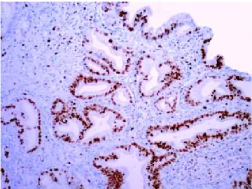
Figure 7. The proliferative index’’’ measured by Ki-67 was 95% in biliary-type in situ adenocarcinoma.
Figure 4. Whole mount section of the distal portion of the common bile duct that shows mucosal papillary structures that project into the lumen. Fibrosis of the wall is also observed.
Figure 5. Higher magnification of biliary-type epithelium with moderate- and high-grade dysplasia in situ adenocarcino-ma, characterized by nuclear pseudostratification, prominent nucleoli, and mitotic figures.
Figure 6. Intramucosal adenocarcinoma. A cribriform structure with stromal microinvasion is present. The adjacent glands showed biliary-type, high-grade dysplasia/in situ ade-nocarcinoma.
Follow-up
The patient is asymptomatic and disease-free 24 months after Whipple surgery.
DISCUSSION
These neoplasms have been associated with differ-ent risk factors. The most important predisposing factors for dysplasia and invasive carcinomas in-clude Primary sclerosing cholangitis (PSC), abnor-mal choledochopancreatic junction and fluke infestation.1,6-8 The presence of gallstones is a well-known risk factor for gallbladder cancer but the role of this entity in cancers of the extrahepatic bile duct is less established. Chronic biliary diseases may lead to long-term irritation and inflammation with re-sultant fibrosis and dysplasia, and may have con-tributed to the development of cancer in this patient with no other risk factor.9,10
Invasive adenocarcinomas are observed in 7-14%
of patients with PSC.1,6,7 But on thinking of
infectious parasites, Clonorchis sinensis and
Opisthorchis viverrini should be considered. In the Orient, infestations by Clonorchis sinensis and
Opisthorchis viverrini are the most common cause of carcinoma of the EHBD.11
The main symptoms are abdominal pain (right upper quadrant pain), pruritus, and weight loss dur-ing from days to weeks, but this can be variable. On physical examination, jaundice (93%), acholia, choluria, and hepatomegaly are observed.1,3
Laboratory tests will show an obstructive pat-tern, with prominent elevation of bilirubins and alkaline phosphatase, mild elevation of transami-nases, and prolongation of prothrombin time in some patients.
If ERCP is performed, cytology and biopsy for early diagnosis of the disease should be performed.
The use of other modalities, such as SpyGlass®, increase sensitivity.15
Histologically, dysplasia and carcinoma in situ have been recognized adjacent to invasive carcino-mas of the EHBD (10-75%), and can be multicen-tric in the majority of patients.1,16 High-grade dysplasia/carcinoma in situ of the EHBD have also been described in association with PSC and less frequently with ulcerative colitis.1,17-19 Incidence varies according to the series from about 0 to 1.8%.1,18,19 The incidence of high-grade dysplasia/ carcinoma in situ in the bile duct mucosa adjacent to carcinomas arising on a background of primary sclerosing cholangitis is higher (60%).19 Carcino-mas of the EHBD probably evolve through a dys-plasia-carcinoma sequence.
Well and moderately differentiated adenocarcino-mas are the most common invasive type of the EHBD. The morphology of these carcinomas is het-erogeneous.20,21 The most common variants of aden-ocarcinoma are biliary and intestinal.1
Total resection is possible in only 25-30% of le-sions in distal origin. In the case of a distal lesion, a Whipple procedure should be performed.22
The overall 5-year survival rate for these neo-plasms is 28%. Bile duct tumors confined to the wall have a 10-year survival of 19%, but if there is re-gional invasion or metastasis, survival can be worse (9 and 1%, respectively). If pancreatic invasion is documented, prognosis is poor.23 Prognosis of carci-nomas that arise in the upper third of the EHBH is worse than for those localized in the remaining por-tions.1,23
Another point for consideration is that the major-ity of tumors are multifocal. Up to 5% of patients with EHBD carcinomas may have a synchronic neo-plasm of the gallbladder, and an examination of the entire biliary tree is recommended.25,26
Finally, perineural and lymphatic invasion is common in this type of carcinoma, but at early stag-es, it is less frequent, up to 11%, and should be eval-uated specifically.23
In conclusion, we present the case of a woman with a papillary in situ and intramucosal carcinoma of the EHBD that was completely resected, and at this time the woman is disease-free 24 months after surgery.
REFERENCES
1. Albores-Saavedra J, Henson DE, Klimstra DS. Tumors of the gallbladder, extrahepatic bile ducts and ampulla of Vater. Atlas of Tumor Pathology. Third series. Fascicle 27. Was-hington, D.C., USA: Armed Forces Institute of Pathology. 2000.
2. Argani P. Pathology of the Gallbladder and Extrahepatic Bile Ducts. In: Gastrointestinal and Liver Pathology. 2nd Ed. New York: Elsevier, Inc.; 2012, p. 490-513.
3 . Bruggen JT, McPhee MS, Bhatia PS, Richter JM. Prima-ry adenocarcinoma of the bile ducts. Clinical charac-teristics and natural history. Dig Dis Sci 1986; 31: 840-6.
4. Launois B, Reding R, Lebeau G, Buard JL. Surgery for hilar cholangiocarcinoma: French experience in a collective survey of 552 extrahepatic bile duct cancers. J
Hepatobi-liary Pancreat Surg 2000; 7: 128-34.
5. Akamatsu N, Sugawara Y, Hashimoto D. Surgical strategy for bile duct cancer: advances and current limitations. J
Clin Oncol 2001; 2: 94-107.
6. Katabi N, Albores-Saavedra J. The extrahepatic bile duct lesions in end-stage primary sclerosing cholangitis. Am J
Surg Pathol 2003; 27: 349-55.
7. Khan SA, Toledano MB, Taylor-Robinson SD. Epidemiology, risk factors, and pathogenesis of cholangiocarcinoma. HPB 2008; 10: 77-82.
8. Lewis JT, Talwalkar JA, Rosen CB, Smyrk TC, Abraham SC. Prevalence and risk factors for gallbladder neoplasia in pa-tients with primary sclerosing cholangitis: evidence for a metaplasia-dysplasia carcinoma sequence. Am J Surg
9. Hsing AW, Gao YT, Han TQ, Rashid A, Sakoda LC, Wang BS, Shen MC. Gallstones and the risk of biliary tract cancer: a population-based study in China. Br J Cancer 2007; 97: 1577-82.
10. Jae MC, Myung HK, Se JJ. Early bile duct cancer. World J
Gastroenterol 2007; 13: 3409-16.
11. Sripa B. Pathobiology of opisthorchiasis: an update. Acta
Trop 2003; 88: 209-20.
12. Choi SH, Han JK, Lee JM, Lee KH, Kim SH, Lee JY, Choi BI. Differentiating malignant from benign common bile duct stricture with multiphasic helical CT. Radiology 2005; 236: 178-83.
13. Zech CJ, Schoenberg SO, Reiser M, Helmberger T. Cross-sectional imaging of biliary tumors: current clinical status and future developments. Eur Radiol 2004; 14: 1174-87. 14. Rosch T, Hofrichter K, Frimberger E, Meining A, Born P,
Weigert N, Allescher HD, et al. ERCP or EUS for tissue diagnosis of biliary strictures? A prospective comparative study. Gastrointest Endosc 2004; 60: 390-6.
15. Fukuda Y, Tsuyuguchi T, Sakai Y, Tsuchiya S, Saisyo H. Diagnostic utility of peroral cholangioscopy for various bile-duct lesions. Gastrointest Endosc 2005; 62: 374-82. 16. Gilloteaux J, Combetta J. Carcinoma in situ of the cystic
duct. Ultrastruct Pathol 2005; 29: 79-84.
17. Fleming KA, Boberg KM, Glaumann H, Bergguist A, Smith D, Clausen OP. Biliary dysplasia as a marker of cholangiocar-cinoma in primary sclerosing cholangitis. J Hepatol 2001; 34: 360-5.
18. Hoang MP, Murakata, LA, Padilla-Rodriguez AL, Albores-Saavedra J. Metaplastic lesions of the extrahepatic bile ducts. A morphologic and immunohistochemical study. Mod
Pathol 2001; 14: 1119-25.
19. Lewis JT, Talwalkar JA, Rosen CB, Smyrk TC, Abraham SC. Precancerous bile duct pathology in end stage primary
sclerosing cholangitis, with and without cholangiocarcino-ma. Am J Surg Pathol 2010; 34: 27-34.
20. Washington MK, Berlin J, Branton PA, Burgart LJ, Carter DK, Compton CC, Fitzgibbons PL, et al. Protocol for the examination of specimens from patients with carcinoma of the distal extrahepatic bile ducts. Arch Pathol Lab Med 2012; 134: e8-e13.
21. Washington MK, Berlin J, Branton PA, Burgart LJ, Carter DK, Compton CC, Fitzgibbons PL, et al. Protocol for the examina-tion of specimens from patients with carcinoma of the peri-hilar bile ducts. Arch Pathol Lab Med 2010; 134: e19-e24. 22. DeOliveira ML, Cunningham SC, Cameron JL, Kamangar F,
Winter JM, Lilemoe KD, Choti MA, et al. Cholangiocarcino-ma: thirty-one-year experience with 564 patients at a single institution. Ann Surg 2007; 245: 755-62.
23. Henson DE, Schwartz AM, Nsouli H, Albores-Saavedra J. Carcinomas of the pancreas, gallbladder, extrahepatic bile ducts, and ampulla of Vater share a field for carcinogene-sis: a population-based study. Arch Pathol Lab Med 2009; 133: 67-71.
24. Bortolasi L, Burgart LJ, Tsiotos GG, Luque-De León E, Sarr MG. Adenocarcinoma of the distal bile duct. A clinicopa-thologic outcome analysis after curative resection. Dig
Surg 2000; 17: 36-41.
25. Sakamoto Y, Kosuge T, Shimada K, Sano T, Ojima H, Yamamoto J, Yamasaki S, et al. Prognostic factors of surgical resection in middle and distal bile duct can-cer: an analysis of 55 patients concerning the signi-ficance of ductal and radial margins. Surgery 2005; 137: 396-402.