Original Article
ped_2884113..117Pyuria is not always sterile in children with Kawasaki disease
Sheng-Ling Jan,1,2
Meng-Che Wu,1,4
Ming-Chih Lin,1
Yun-Ching Fu,1,2
Sheng-Ching Chan3
and Shing-Jong Lin2 1
Division of Cardiology, Department of Pediatrics, Taichung Veterans General Hospital,2
Institute of Clinical Medicine, National Yang-Ming University,3
Department of Nursing, Ta-Jen University and4
Department of Pediatrics, Chang Bing Show Chwan Memorial Hospital, Taiwan
Abstract Background: Although Kawasaki disease (KD) often presents with sterile pyuria, bacterial pyuria (urinary tract infection [UTI]) occasionally occurs.
Methods: This was a retrospective cohort study of 285 children with KD diagnosed between 1995 and 2005. Among these patients, a total of 210 patients underwent routine urine tests and 75 children underwent urine culture tests. This study was conducted to investigate the incidence, clinical manifestations, management and outcome of KD with pyuria. Results: The incidence of pyuria was 29.5% (62/210). Among the 75 children undergoing urine culture tests, 34 had sterile pyuria (45.3%), eight had bacterial pyuria (10.7%), two had UTI without pyuria (2.7%) and 31 had neither pyuria nor UTI (41.3%). When pyuria was used as a predictor of KD with UTI, the positive and negative predictive values were 19% and 93.9%, respectively. There were no significant differences in demographic data, clinical presentations, laboratory results, duration of fever, ratio of resistant KD or risk level, except in the nitrite test, between both groups. Conclusions: Pyuria was not always sterile in patients with KD. Although there was no different clinical phenotype or coronary outcome in KD patients with or without UTI, we suggest that UTI should be considered and evaluated in KD patients with pyuria, a positive nitrite test or a positive result of urine culture. If UTI is definitively diagnosed, the patient should be treated for a UTI as well as for KD and complete post-UTI work-up is recommended.
Key words Kawasaki disease, outcome, pyuria, urinary tract infection.
Introduction
Kawasaki disease (KD) is a systemic vasculitis that most fre-quently affects infants and young children under 5 years of age. It has been reported as the leading cause of acquired heart disease in children around the world. KD often presents with abnormal urinary findings, such as pyuria, proteinuria, and microscopic hematuria.1Pyuria in patients with KD is always considered as
sterile. It originates from the urethra and/or kidneys as a result of mild and subclinical renal injury or vasculitis. Clinically, fever with pyuria is routinely considered as a urinary tract infection (UTI) and antibiotics for treatment of UTI are started soon after urine collection.2If urine culture has no bacterial growth or signs
of KD develop, UTI will be excluded. In other words, when KD is definitively diagnosed, no urine culture will be done or the result of urine culture will be neglected because “Pyuria is always sterile”. However, bacterial pyuria in patients with KD occasion-ally occurs.3,4For antimicrobial therapy considerations, the
pos-sibility of UTI has to be assessed. The diagnosis of UTI, continuation of antibiotics and cardiac or renal outcome should be considered when KD is definitively diagnosed. We have briefly reported the preliminary result of our study of KD with
pyuria in the past.5In this study, we further discuss the topic of
whether KD with pyuria is really sterile, and investigate the incidence, clinical manifestations, management and outcome of KD with or without UTI.
Methods
This was a retrospective cohort study of 285 children who were treated for clinically diagnosed KD at Taichung Veterans General Hospital, a tertiary medical center in central Taiwan, between January 1995 and December 2005. The diagnosis of KD was based on the American Heart Association criteria.5Atypical or
incomplete KD was diagnosed and recorded on the surveillance form when the patient had three or four of the principal criteria plus coronary artery lesion (CAL) detected by echocardiography. The CAL determination was based on: (i) coronary ectasia, if the internal diameter of the coronary artery was>3 mm in children younger than 5 years or>4 mm in children at least 5 years old but a segmental aneurysm was not apparent; and (ii) coronary aneu-rysm, if the internal diameter of a segment was>1.5 times that of an adjacent segment, and segmental coronary aneurysms were classified as small (3 to 5 mm internal diameter), medium (5 to 8 mm internal diameter), or giant (>8 mm internal diameter).6,7
Echocardiography was performed at the time of diagnosis during the acute phase, at 1 month and 6 months later. The degree of coronary artery involvement and risk levels were based on the guidelines for long-term management of children with KD.6
Correspondence: Meng-Che Wu, MD, 160, Sec 3, Chung-Kang Road, Taichung 40705, Taiwan. Email: [email protected]
Address for reprint requests to Dr Meng-Che Wu.
The definition of pyuria was based on urinary white blood cell (WBC) count>5 cells/high power field. The definitive diagnosis of UTI was based on culture of a single organism from a cath-eterized urine culture with a colony count greater than 10 000 (100 000 colonies in uncircumcised boys), from a suprapubic culture with greater than 1000 colonies, or from a midstream urine culture with greater than 100 000 colonies. The midstream urine cultures were collected from patients 3 and 8 when, during attempted catheterization, the patients urinated after the foreskin had been retracted and the urethra was sterilized with aqua beta iodine, and a clean container was placed in line with the urine stream. Urines with microscopic examination of the sediment revealing epithelial cells or those in which two or more bacterial species were isolated by culture were excluded. Common bacte-rial contaminants of urine cultures includingStaphylococcus epi-dermidisand anaerobic bacteria were also excluded. All of the routine urine tests and cultures were carried out before antibiotic and intravenous immunoglobulin (IVIG) administration.
Clinical data, including age, gender, fever course, WBC, hemoglobin (Hb), platelet count, differential count, C-reactive protein (CRP), erythrocyte sedimentation rate (ESR), routine urine test and/or culture, blood culture, the usage of IVIG or methylprednisolone (MTP) pulse therapy, echocardiographic results and the risk level of KD, were compared between KD patients with and without UTI. Parametric data were expressed as median with range. Statistical analysis was performed with SPSS, Version 10.0 for Windows (SPSS, Chicago, IL, USA). A comparison of characteristics between patients with and without UTI was carried out using Pearson’sc2-test, Fisher’s exact test or
the Mann–WhitneyU-test. A probability less than 0.05 was con-sidered as statistically significant.
Results
A total of 285 patients were diagnosed as having KD during the study period, including typical KD in 249 and atypical KD in 36. Boys outnumbered girls by a ratio of 1.77 : 1. The onset median age was 16 months (range from 1 to 108 months). A total of 232 (81.4%) children received one course of IVIG and 37 (13%) received more than one course of IVIG. Sixteen children (5.6%) did not receive IVIG treatment due to other reasons such as late presentation or misdiagnosis. The clinical parameters and follow-up echocardiographic results are listed in Tables 1 and 2, respectively.
A total of 210 patients underwent a routine urine test, and the incidence of pyuria was 29.5% (62/210). Most pediatricians thought that was just sterile pyuria in KD, so many patients did not undergo urine culture tests. So, only 75 patients underwent urine culture tests during the acute stage of KD in this study. Among the 75 patients undergoing urine culture tests, eight patients (10.7%) had bacterial pyuria, two (2.7%) had UTI (bac-terial growth on urine culture) without pyuria, 34 (45.3%) had sterile pyuria and 31 (41.3%) had neither pyuria nor UTI. The patients with KD who underwent urine culture tests were divided into two groups: KD with UTI and KD without UTI (10 and 65 patients, respectively). Although the incidence of pyuria is higher in KD with UTI (80% vs 52.3%), there was no significant
difference (P=0.17). When pyuria was used as a predictor of KD with UTI, there was a low positive predictive value of 19% (8/42) but a high negative predictive value of 93.9% (31/33). In our study, among the 10 KD patients with UTI, eight were diagnosed as typical and two as atypical. All of the patients with UTI received at least one course of IVIG and three of them received two courses of IVIG. Seven of 10 patients took antibiotics for UTI. The rate of IVIG resistance was 30% vs 15% and the total duration of fever was 9.5 vs 8.0 days in patients with UTI vs those without UTI, respectively, although the difference was not statistically significant. There were no significant differences in demographic data, clinical presentations, laboratory results, or risk level between the two groups, except in the nitrite test (30% in the UTI group vs 1.4% in the group without UTI,P=0.007). The detailed results are shown in Tables 2 and 3. The pathogens and UTI survey results are listed in Table 4. The UTI survey, including renal sonography, Tc 99m-dimercaptosuccinic acid (DMSA) renal scan, and voiding cystourethrogram (VCUG), was incomplete in most KD patients with UTI. The renal sonography revealed right acute pyelonephritis in one case and mild left hydronephrosis in two cases. Two patients underwent VCUG and three underwent DMSA renal scan and the results were all nega-tive. All of the results of blood culture were aseptic except one, which was contaminated byStaphylococcus epidermidis. It could exclude the UTI caused by blood stream infection.
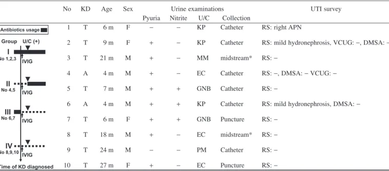
Regarding the usage of antibiotics for treatment of UTI, patients 1 to 3 were classified into group I: antibiotics were prescribed before KD diagnosis and complete antibiotic treat-ment was given for UTI. Patients 4 and 5 were classified into group II: antibiotics were not prescribed before KD diagnosis but used after positive result of urine culture and definitive diagnosis
Table 1 Clinical features and treatment of the 285 KD patients
during acute stage
Variables n(%) or median
(range) Typical KD/Atypical KD 249/36 (87.4%/12.6%)
Female/male 103/182 (36.1%/63.9%)
Age (months) 16 (1–108)
Total duration of fever (days) 7 (4–30) Duration of fever before KD diagnosis
(days)
6 (3–30)
Duration of fever after KD diagnosis (days)
1 (0–13)
Complete blood count
WBC (/mm3) 14 900 (1230–52 200)
Hb (g/dl) 10.8 (6.9–14.8)
Platelet (¥103/cumm3) 385 (16–1345)
CRP (mg/dl) 7.8 (0.01–51.6)
ESR (mm/hr) 78 (1–150)
IVIG usage (times)
0 16 (5.6%)
1 232 (81.4%)
32 37 (13%)
of UTI. Patients 6 and 7 were classified into group III: antibiotics were prescribed initially but discontinued after KD diagnosis. Patients 8 to 10 were classified into group IV: antibiotics were never used even after definitive diagnosis of UTI. Groups I and II were considered as completely treated UTI cases, and groups III and IV as incompletely treated UTI cases, as shown in Table 4.
Discussion
The incidence of KD with pyuria has been reported to be from 10% to 63%,1,2,8,9and was 29.5% in this study. KD should be
considered in young children with prolonged fever, pyuria, no response to antibiotic therapy, no bacterial growth in urine culture and/or other signs of KD development.3,8Although sterile
pyuria with fever is often found in KD, 10.7% of KD patients had bacterial pyuria in our series. Among these, about 19% (positive predictive value) of KD patients had bacterial pyuria with UTI.
Even in KD patients without pyuria, the possibility of UTI was 2.7%. The incidence of KD with UTI may be underestimated.
It is reasonable that UTI will be diagnosed according to the clinical presentations of fever with bacterial pyuria. However, even when KD is definitively diagnosed, UTI is not often con-sidered. Sometimes the urine sample is contaminated, or urinary leukocytes in KD patients are mononuclear cells but not neutro-phils, or there is just a misdiagnosis.1,2,8,9In most cases of pyuria
with the non-specific finding of severe inflammation in KD, a possible diagnosis of UTI is not automatically considered. Shouldn’t the possibility that KD is associated with UTI and renal sequelae be considered? Wanget al.10reported that 46% of
KD with renal scarring was detected with a DMSA renal scan at 6 months follow up, even though it did not necessarily relate to UTI. We believe that KD can coexist with UTI. Therefore, we were especially interested in the clinical presentations and
out-Table 2 Coronary artery outcomes of the KD patients by follow-up echocardiography
Total (n=285)
KD with UTI (n=10)
KD without UTI (n=65)
P-value
Echocardiographic results
CAL (+) initially 89 (31.3%) 3 (30%) 22 (33.8%) 1.000‡
CAL (+) at 1 month f/u 48 (18.2%) 3 (37.5%) 17 (28.3%) 0.685‡
CAL (+) at 6 months f/u 25 (9.9%) 2 (25%) 7 (12.3%) 0.305‡
Risk level 0.832†
I 180 (63.4%) 6 (60%) 36 (55.4%)
II 75 (26.4%) 2 (20%) 19 (29.2%)
III 26 (9.2%) 2 (20%) 9 (13.8%)
IV 2 (0.7%) 0 (0%) 1 (1.5%)
V 1 (0.4%) 0 (0%) 0 (0%)
Data are presented as case number (percentages).
A comparison of characteristics between patients with and without UTI was performed by using†Pearson’sc2-test or‡Fisher’s exact test.
CAL, coronary artery lesion; KD, Kawasaki disease; UTI, urinary tract infection.
Table 3 Clinical characteristics of acute stage of KD patients who had undergone urine culture tests
Variables KD with UTI
(n=10)
KD without UTI (n=65)
P-value
Pyuria 8 (80%) 34 (52.3%) 0.170†
Urine nitrite test (+) 3 (30%) 1 (1.4%) 0.007†
Typical KD 8 (80%) 52 (80%) 1.000†
Gender (F/M) 4/6 (40%/60%) 23/42 (35%/65%) 1.000†
IVIG usage (times) 0.364†
21 7 (70%) 55 (85%)
32 3 (30%) 10 (15.4%)
Antibiotics usage 7 (70%) 42 (65.6%) 1.000†
Age (months) 8 (4–27) 11 (2–108) 0.685‡
Total duration of fever (days) 9.5 (4–16) 8.0 (4–22) 0.464‡
Duration of fever before KD diagnosis (days) 5.5 (3–14) 6.0 (3–20) 0.747‡
Duration of fever after KD diagnosis (days) 2 (0–8) 2 (0–11) 0.388‡
WBC (cumm) 14 450 (10 030–29 600) 15 600 (3900–52 200) 0.827‡
Hb (g/dl) 10.5 (8.2–12.1) 10.4 (7.4–13.5) 0.785‡
Platelet count (¥103cumm) 369.5 (160–936) 365.0 (31–1345) 0.919‡
CRP (mg/dL) 9.5 (1.2–36.8) 8.3 (0.2–44.5) 0.575‡
ESR (mm/h) 60 (6–180) 97 (2–150) 0.104‡
Data are presented as case number, percentages or median (range).
Statistics were obtained using†Fisher’s exact test or the‡Mann–WhitneyU-test.
comes of KD with or without UTI, the relationship between KD and UTI, and the treatment for UTI in patients with KD.
Although there was no statistically significant difference in age, patients in the KD with UTI group were younger than those in the KD without UTI group (median age of 8 months in the group with UTI vs 11 months in the group without UTI). Sixty percent (6/10) of patients with UTI were younger than 12 months. Similarly, Wirojanan et al.8 found that KD patients
younger than 1 year were more likely to have pyuria than those older than 1 year. There may be a trend in which KD with UTI more commonly occurs in the infant stage. However, further large studies are needed for confirmation.
After a presumptive diagnosis of KD, antibiotics are usually discontinued. However, if KD coexists with UTI or other bacte-rial infections, should antibiotics be stopped? Benseler et al.4
reported that 33% of KD patients had concurrent confirmed infections that could be a bacterial or viral infection, and that infections at diagnosis of KD did not affect the patients’ response to IVIG treatment and coronary artery outcome when compared with those patients without infections. Wuet al.9reported that
incomplete KD with UTI showed a poor response to antibiotics treatment, but fever subsided after IVIG. Different attending phy-sicians have different managements for UTI in KD patients, and there are no available standard guidelines for this patient group. Among 10 KD patients with UTI in this study, all received IVIG treatment. Five patients (groups I +II) underwent a complete treatment for UTI and the others (groups III+IV) underwent an incomplete treatment (Table 4). In this study, we found the pres-ence of UTI did not alter the clinical phenotype of KD or the
coronary artery outcome. Although we were concerned about the correlation between complete or incomplete UTI treatment and genitourinary tract outcome in KD patients with UTI, the geni-tourinary tract outcome was unclear in this study because most of the patients underwent incomplete post-UTI work-up. Because of the possible beneficial effect of intravenous hydration and IVIG in KD patients with UTI, we speculated that there would be no significant difference between patients with and without com-plete UTI treatment. There might be two underlying disorders, inflammation and infection, in KD patients with UTI. This could also explain why KD with UTI showed poor response to the antibiotics treatment but fever subsided after intravenous hydra-tion and IVIG. Because pediatricians did not pay special atten-tion to the presence of UTI in KD patients in the past and sometimes performed incomplete study of post-UTI work-up, we suggest that reassurance of UTI and complete post-UTI work-up such as renal sonography, DMSA renal scan, and VCUG should be done selectively in future research on KD patients with UTI to clarify its effect.
We concluded that pyuria was not always sterile in KD patients in this study. There was no different clinical phenotype or coronary artery outcome in KD patients with or without UTI. We suggest that urine culture should be completed before KD is diagnosed or at least in those of KD with pyuria and/or positive nitrite test. If a urine culture is reported to be positive in a child with KD, the culture should be repeated by catheterization or suprapubic punc-ture prior to starting antibiotics, and if that repeated culpunc-ture is positive, the patient should be treated for a UTI as well as for KD and complete post-UTI work-up is recommended.
Table 4 Clinical features and the usage of antibiotics (graph) for treatment of UTI in 10 patients with KD. Groups I, II underwent complete UTI
treatment and groups III, IV underwent incomplete UTI treatment
No KD Age Sex Urine examinations UTI survey
Pyuria Nitrite U/C Collection
IVIG I
II
III
IV
Group U/C (+)
Time of KD diagnosed IVIG
IVIG
IVIG Antibiotics usage
No 1,2,3
No 4,5
No 6,7
No 8,9,10
1 T 6 m F - - KP Catheter RS: right APN
2 T 9 m F + - KP Catheter RS: mild hydronephrosis, VCUG:-, DMSA:
-3 T 21 m M + - MM midstream* RS:
-4 A 4 m M + - EC Catheter RS:-, DMSA:-VCUG:
-5 T 7 m M + + GNB Catheter RS:
-6 A 4 m M + + KP Catheter RS: mild hydronephrosis, DMSA:
-7 T 6 m F + + GNB Puncture RS:
-8 T 18 m M + - EC midstream* RS:
-9 T 24 m M - - PM Catheter RS:
-10 T 27 m F + - EC Puncture RS:
--, negative results; *, mid-stream clean-catch urine method; A, atypical KD; APN, acute pyelonephritis; DMSA, Tc 99m-dimercaptosuccinic acid (DMSA) renal scan; EC,Escherichia coli; GNB, Gram-negative; KP,Klebsiella pneumoniae; MM, Morganella morganii; PM,Proteus
mirabilis; RS, renal sonography; T, typical KD; U/C(+), the time of positive result of urine culture; UTI, urinary tract infection; VCUG, voiding
References
1 Ristoska-Bojkovska N, Stavric K, Tasic V. Kawasaki disease mis-diagnosed as acute pyelonephritis. Pediatr. Nephrol. 2003; 18: 851–2.
2 Watanabe T, Abe Y, Sato S, Uehara Y, Ikeno K, Abe T. Sterile pyuria in patients with Kawasaki disease originates from both the urethra and the kidney.Pediatr. Nephrol.2007;22: 987–91. 3 Shino N, Koga Y, Ito H, Egawa K, Ono S, Itami N. Really
sterile pyuria with Kawasaki disease?Pediatr. Nephrol.2004;19: 124.
4 Benseler SM, McCrindle BW, Silverman ED et al. Infections and Kawasaki disease: Implication for coronary artery outcome.
Pediatrics2005;116: 760–6.
5 Wu MC, Jan SL, Lin MC, Fu YC, Lin SC. Is Kawasaki disease with pyuria always sterile? Pediatr. Infect. Dis. J. 2008; 7: 1121.
6 Newburger JW, Takahashi M, Gerber MA et al. Diagnosis, treatment, and long-term management of Kawasaki disease:
A statement for health professionals from the Committee on Rheu-matic Fever, Endocarditis and Kawasaki Disease, Council on Car-diovascular Disease in the Young, American Heart Association.
Circulation2004;110: 2747–71.
7 Research committee on Kawasaki disease, Ministry of Health and Welfare. Report of the subcommittee on standardization of diag-nostic criteria and reporting of coronary artery lesions in Kawasaki disease. Tokyo, Japan: Research committee on Kawasaki disease, Ministry of Health and Welfare, 1984.
8 Wirojanan J, Sopontammarak S, Vachvanichsanong P. Sterile pyuria in Kawasaki disease.Pediatr. Nephrol.2004;19: 363. 9 Wu CY, Hsieh KS, Chiou YHet al.Prolonged fever and pyuria: A
urinary tract infection presentation of incomplete Kawasaki disease.Acta Paediatr.2005;94: 375–7.
10 Wang JN, Chiou YY, Chiu NT, Chen MJ, Lee BF, Wu JM. Renal scarring sequelae in childhood Kawasaki disease.Pediatr. Nephrol.