,1 ,
,1
*Departamento de Bioquı´mica y Biologı´a Molecular y Fisiologı´a-IBGM, Universidad de Valladolid-CSIC, Valladolid, Spain
Departamento de Gene´tica, Universidad de Sevilla, Sevilla, Spain
àCIBER de enfermedades respiratorias, Instituto de Salud Carlos III, Madrid, Spain.
§De´partement des Sciences Biologiques, Centre BioMed, Universite´ du Que´bec a` Montre´al, Montre´al, Canada
Cells in the nervous system (NS) are exposed to a strong and constant production of reactive oxygen and nitrogen species (RS) because of their highly demanding metabolism. This oxidative load is tightly regulated to sustainable limits by an effective array of antioxidant proteins and compounds. However, physiological aging and a number of genetic and environmentally-induced degenerative diseases affect both the production and the clearance of RS. The subsequent oxidative stress (OS) clearly contributes to the pathogenic mechanisms underlying these conditions.
As a way of studying the antioxidant mechanisms participating in NS homeostatic regulation, several methods of RS induction have been used. Some of these methods, involving treatments with drugs such as MPTP, maneb and paraquat (PQ; 1,1¢-dimethyl-4,4¢-bipyridinium), in model organisms are able to totally or partially mimic the signs and symptoms of a devastating neurodegenerative process such as Parkinson’s disease (Drechsel and Patel 2008).
The extensive work of several groups with these com-pounds has uncovered the detailed process of the response of the NS to the experimentally-induced OS. The treatment with chronic sublethal doses of these compounds elicits NS specific responses that are generated by the different cell types involved. Early responses to PQ in gene expression
Received January 31, 2011; revised manuscript received March 16, 2011; accepted March 31, 2011.
Address correspondence and reprint requests to Maria D. Ganfornina and D. Sanchez, Instituto de Biologı´a y Gene´tica Molecular, c/Sanz y Fore´s 3, Universidad de Valladolid-CSIC, 47003 Valladolid, Spain. E-mail: [email protected]
1These authors contributed equally to this study.
Abbreviations used: ApoD, Apolipoprotein D; FC, fold change; FDR, false discovery rate; GC, GeneChip; GO, gene ontology; hApoD, human ApoD; KO, knock-out; NS, nervous system; OS, oxidative stress; Pkcd, Protein kinase Cd; PQ, paraquat; RMA, robust multiarray average algorithm; RS, reactive species; Tg, transgenic; WT, wild-type. Abstract
The lipocalin Apolipoprotein D (ApoD), known to protect the nervous system against oxidative stress (OS) in model organisms, is up-regulated early in the mouse brain in response to the ROS generator paraquat. However, the pro-cesses triggered by this up-regulation have not been explored. We present here a study of the effect ofApoDon the early transcriptional changes upon OS in the mouse cerebellum using microarray profiling. ApoD-KO and transgenic mice over-expressingApoDin neurons are compared to wild-type controls. In control conditions,ApoDaffects the transcriptional profile of neuron and oligodendrocyte-specific genes involved in neuronal excitability, synaptic function, and myelin homeostasis. When challenged with paraquat, the absence of
ApoDmodifies the response of genes mainly related to OS management and myelination. Interestingly, the over-expres-sion ofApoDin neurons almost completely abolishes the early transcriptional response to OS. We independently evaluate the expression of protein kinase Cd, a gene up-regulated by OS only in the ApoD-KO cerebellum, and find it over-expressed in cultured ApoD-KO primary astrocytes, which points to a role forApoDin astrocyte-microglia signaling. Our results support the hypothesis thatApoDis necessary for a proper response of the nervous system against physiological and pathological OS.
Keywords: astrocytes, lazarillo, lipocalin, oligodendrocytes, paraquat, Pkcd.
have been reported for non-neural tissues (Edwards et al. 2004; Tomita et al. 2006, 2007), but no study has been performed in the NS.
Our laboratory studies the role in the nervous system of the gene Apolipoprotein D (ApoD) and its homologs in Drosophila melanogaster. Using experimentally-induced OS by PQ treatment, we have demonstrated that ApoD has protective effects over the organism survival both in mouse and flies, and that it helps to maintain the NS tissue homeostasis by maintaining low levels of lipid peroxidation (Sanchez et al.2006; Ganfornina et al. 2008; Hull-Thomp-son et al. 2009). ApoD mRNA expression is transiently induced in the mouse brain upon PQ treatment with an early peak at 3 h, and this up-regulation is specific for the neural tissue (Ganforninaet al.2008).
We have previously analyzed how the lack of ApoD generates specific imbalances in the transcriptional response of peripheral nerves upon injury, indicating that at least part of the complex response to injury is modulated by ApoD (Ganfornina et al. 2010). However, whether ApoD is also an important contributor shaping the early transcriptional response of the CNS to OS is still unknown. In this work, we analyze the transcriptional profile of PQ-challenged cere-bellum of wild-type (WT), ApoD loss-of-function [ApoD-knock-out (KO)] and transgenic mice over-expressing human ApoDin neurons (hApoD-Tg) using oligonucleotide micro-array technology. Besides its function in motor coordination and learning, the cerebellum is a OS-sensitive brain region found to be altered in aging and many NS pathologies (Apps and Garwicz 2005).
The alteration ofApoDexpression results in transcriptional changes of genes involved in neuron electrical activity and synaptic function, and in myelin homeostasis. In addition, ApoDregulates the expression of several genes that control the cellular response to environmental stimuli such as OS. On the other hand, the expression profile of the OS-challenged cerebellum shows a number of genes with ApoD-dependent expression that, aside of OS management, are related to nervous system development, cell differenti-ation and the myelindifferenti-ation process. Our results support the hypothesis that the presence ofApoDin the nervous system is necessary for a proper response against physiological and pathological OS.
Experimental procedures
Animals and cell cultures
In this study, we used adult (80 ± 5 days old) male mice of three genotypes: ApoD-KO, hApoD-Tg and their WT littermates. The loss-of-function mutant ApoD-KO mice were generated by homo-logous recombination, and the mutation is evidenced by PCR-genotyping with two different primer pairs as described previously (Ganfornina et al.2008). The gain-of-function mutant hApoD-Tg mice over-express the humanApoD gene under the control of the
neuron-specific Thy-1 promoter, and their characterization and genotyping procedures have been already reported (Ganfornina et al. 2008; Do Carmo et al. 2009). In order to avoid potential maternal effects ofApoDand to generate WT and ApoD-KO cohorts of homogeneous genetic background, the experimental cohorts used in this study are the F1 generation of homozygous crosses of each genotype. The parental generation was composed ofApoD)/)and ApoD+/+ littermates from heterozygous crosses of the ApoD-KO line. The hApoD-Tg animals used in this study were heterozygous mutants. Both mutations have been backcrossed > 11 generations into the C57Bl/6J genetic background.
All mice were housed in positive pressure-ventilated racks at 25 ± 1C with a 12 h light/dark cycle, fedad libitumwith a standard rodent pellet diet (Global Diet 2014; Harlan Inc., Indianapolis, IN, USA), and allowed free access to filtered and UV-irradiated water. Experimental procedures were approved by the Animal Care and Use Committees of the University of Valladolid (UVa) and Universite´ du Que´bec a` Montre´al (UQAM) and were in accordance with the Guidelines for the Care and Use of Mammals in Research (European Commission Directive 86/609/CEE and Spanish Royal Decree 1201/2005).
Primary glial cultures were prepared from the cortices of neonatal (P0) mice, treated with 10 mg/mL trypsin for 15 min at 37C, mechanically dissociated, and incubated in Dulbecco’s Modified Eagle’s medium supplemented with 10% fetal bovine serum (FBS), 1% L-Glutamine and 1% Penicillin (10 U/lL) – Streptomycin (10lg/lL) – Amphotericyn B (25lg/mL) at 37C in 5% CO2with 90–95% humidity. The medium was weekly replaced, and after 2–3 subculture steps, over 95% of type 1 astrocytes were present, as estimated by glial fibrillary acidic protein (GFAP) labeling and by morphological criteria. The cultures had a minor contribution of microglial cells (Cd11b marker). Oligodendrocytes were not detected (pi-GST marker).
Experimental oxidative stress treatments and tissue collection Nine mice of each genotype were either treated with a single intraperitoneal injection of PQ (30 mg/kg) in 200lL sterile saline (Experimental group), or a similar volume of sterile saline (Control group). Six hours after injection, each mouse was killed with CO2 and the cerebellum was immediately removed and frozen.
In the chronically treated cohort, male mice (n= 6/genotype for PQ andn= 4/genotype for control) were injected intraperitoneally with 10 mg/kg PQ or phosphate-buffered saline for a total of seven injections (two per week for the first 2 weeks, one per week for three additional weeks). Tissue collection was carried out 7 days after last injection.
Paraquat injections were performed by the same experimenter to minimize differences in animal stress. The brain samples were extracted at the same time of the day in order to avoid gene expression variations due to circadian rhythms.
amounts of total RNA from three randomly selected mice for each genotype and experimental condition were pooled, rendering three biological replicates to hybridize with the arrays.
cDNA was synthesized and purified from 5lg of each RNA sample with the One Cycle cDNA synthesis (Affymetrix, Santa Clara, CA, USA). The generation, labeling and purification of cRNA was performed using the IVT kit (Affymetrix).
Ten micrograms of the biotinylated and fragmented probes were hybridized to Affymetrix GeneChip Mouse Genome 430A 2.0 arrays (Lot # 4029603) at 45C for 16 h, following the manufac-turer’s protocols. After washes, the arrays were incubated with anti-biotin streptavidin-phycoeritrin antibody and scanned with an Affymetrix GeneChip Scanner 7G. Probe synthesis and hybridiza-tions were performed at the Genomics facility of the Centro de Investigacion del Cancer (Salamanca, Spain).
Microarray data analysis
The analysis of gene expression and the comparative expression between genotype and experimental conditions were performed using the Affymetrix CEL files and both, the GEPAS platform (Tarragaet al.2008) and the FlexArray v1.4.1 program (Blazejczyk et al. 2007). The original CEL files are available at the GEO Database (Accession number GSE28643).
Robust normalization using MAS 5.0 (Affymetrix) was per-formed to estimate a changep-value and its associated change call in gene expression for each probeset. Data pre-processing was carried out with FlexArray using the robust multiarray average algorithm (RMA) and GeneChip RMA (GC-RMA) algorithms with back-ground corrections and normalization, and with GEPAS using RMA-quantiles for background correction and normalization. Only perfect-match probesets were considered in both analyses.
Differentially expressed genes were evaluated with FlexArray by two sample comparisons with the cyberT-test (Baldi and Long 2001), using a threshold of 2-fold change (FC) and ap-value < 0.05. False discovery rate (FDR) correction was performed using the Benjamini-Hochberg method. ANOVA was performed on the GC-RMA processed probes with FDR = 1% to further select candidate genes specifically affected by PQ treatment and/or genotype. ANOVA was also selected in GEPAS to study FDR-corrected differentially expressed genes with a FC‡±2 cut-off value and an adjustedp-value < 0.05. Genes that showed consensus expression changes by ANOVA and cyberT-test were selected for further study.
As a final filter for analyzing genes whose expression is affected by the levels ofApoD, we compared the list of genes generated with the RMA/GC-RMA/cyberT/ANOVAlists generated by FlexArray and the GEPAS analysis platform. From a consensus analysis we selected the genes for further exploration. Probe sets derived from uncharacterized genes were not considered for the final discussion of differentially expressed genes.
The genes selected from our microarray analysis were subjected to gene ontology (GO) and pathway analyses. Results coming from the two background correction and normalization procedures were compared, and genes that showed expression changes under both methods were considered for discussion and future experimental analysis.
Data mining with GO classification of the selected transcripts and GO comparisons between datasets were carried out with the
GOEAST (http://omicslab.genetics.ac.cn/GOEAST/) and DAVID 6.7 platforms (http://david.abcc.ncifcrf.gov/home.jsp) (Zheng and Wang 2008; Huang daet al.2009). Pathway analysis was performed using MouseNet (http://avis.princeton.edu/mouseNET/index.php).
A meta-analysis of microarray studies reporting transcriptional changes induced by OS was performed by using the LOLA database and analysis software (http://lola.gwu.edu/) (Cahanet al.2005) to compare gene transcriptional changes with a statistical assessment of the congruencies or differences.
We also performed a comparison of the gene sets obtained in our study with the genes reported to be cell-type enriched in the nervous system (2618 astrocyte-enriched genes, 2036 neuron-enriched genes, and 2228 oligodendrocyte-enriched genes) by Cahoyet al.(2008).
Quantitative real-time RT-PCR
RNAs for qRT-PCR experiments were extracted with TRIzol (Invitrogen) either from the pooled samples of mouse cerebella described above, from homogenized mouse diencephalons, or from cultured astroglial cells. Total RNA (1lg) was reverse-transcribed with PrimeScriptTM(Takara Bio Inc., Otsu, Japan) and treated with DNaseI. The cDNA obtained was used as template for qRT-PCR using SybrGreen (SYBRPremix Ex Taqkit, Takara) amplifications. The oligonucleotide primers used in our amplifications are shown in Table S2. The geneRpl18was used as a reference because neither genotype nor treatment gives a significant fold change for this gene. Amplifications were performed in quadruplicate in an ABI Prism 7900HT or a Rotor-Gene RG-3000 (Corbett-Qiagen Iberia) thermal cycler. Standard cycling conditions were: 95C, 5 min; 40 cycles (95C, 30 s; 60C, 1 min).
Changes in transcriptional expression were estimated with the
DDCTmethod (Livak and Schmittgen 2001). The following criteria were applied to our amplifications: (i) Replicates with variation coefficient > 2.5% were excluded; (ii) Undetermined CT values (gene expression below detection levels) were assignedCT= 35; (iii) Pairwise comparisons where the gene averageCT> 35 cycles in both conditions were excluded from the analysis; (iv) Only transcriptional changes greater than or equal to twofold were included in the analysis. Significant differences of gene transcrip-tional changes were evaluated with a Mann–WhitneyU-test (Yuan et al.2006), using theDCTof each replica. Values are expressed as mean Log2)DDCt± SD, and the level of significance was set at p< 0.05. Only statistically significant differences of expression are presented in results and discussed in the text.
Immunoblot experiments
Brain tissue was homogenized in lysis buffer [1% Nonidet P-40 (Calbiochem, Merck KGaA, Darmstadt, Germany), 0.1% sodium dodecyl sulfate, 0.5% sodium deoxycholate, and 10% Complete Protease Inhibitors (Roche Molecular Biochemicals, Indianapolis, IN, USA) in phosphate-buffered saline], cleared by centrifugation, and the supernatant was stored at)80C.
Goat anti-Rabbit or Donkey anti-goat IgG (Santa Cruz) were used. Protein loads were normalized with the signal obtained with a horseradish peroxidase-conjugated anti-bactin antibody (Sigma, St Louis, MO, USA). Membranes were developed with ECL (Milli-pore, Billerica, MA, USA). The integrated optical density of the immunoreactive protein bands was measured in images taken within the linear range of the digital camera (VersaDoc, Bio-Rad Laboratories, Hercules, CA, USA). The mean ± SD of arbitrary density units was calculated from at least duplicate blots.
Statistical analysis
Statistical analyses were performed with Statgraphics plus (v 5.0) (Statpoint Technologies Inc., Warrenton, VA, USA) and SPSS (v 18) (IBM, New York, NY, USA) softwares.p< 0.05 was defined as a threshold for significant changes.
Results and discussion
The gene expression profiles of several tissues subjected to experimental oxidative stress (OS) have been studied by other authors using microarray analysis in model organisms such as Drosophila and mouse. In the nervous system, several brain regions showing selective vulnerability to OS, such as hippocampus, substantia nigra and striatum, have been studied (Chung et al. 2005; Wang et al. 2007; Chin et al. 2008). However, the transcript profile of the challenged cerebellum, home of a massive number of OS-sensitive granule cells (Gonzalez-Polo et al. 2004; Wang et al.2009), has not been experimentally assessed.
Besides, ApoD is consistently expressed in the rodent cerebellum, mainly in oligodendrocytes and astrocytes (Provostet al.1991; Ong et al.1999; Navarroet al. 2004; Ganfornina et al. 2005), and the ApoD-KO mouse shows behavioral defects in cerebellar-related motor coordination (Ganforninaet al.2008).
Therefore, we selected the cerebellum to assay the effect of altering the expression ofApoDon the early response to an acute experimental OS produced by a single dose of PQ. At the time point selected, 6 h after PQ exposure, ApoD transcript up-regulation has taken place and elevated levels of ApoD protein are present in the tissue, but neither brain lipid peroxidation nor neuronal cell death have yet increased over the basal levels (McCormacket al.2005; Prasadet al.2007; Ganfornina et al. 2008). Using this protocol we expect to isolate the direct transcriptional response to PQ from responses derived as secondary consequences of cell death occurring in the tissue, or other slow-paced cellular events that are also modified by ApoD, like lipid peroxidation. Therefore, only transcriptional changes underlying functional responses of neurons and glia are expected, and their dependence onApoDfunction can be discerned.
Quality controls and validation of microarray results The quality of hybridization signals in our arrays was assessed according to standard Affymetrix guidelines. The
percent of present (P) vs. absent (A) calls (average P: 64.1 ± 2.3%) is in the accepted range, as it is also the number of concordant calls in the triplicates, that averages 87.3 ± 2.1%. The reliability index (the Cron-bach’s a coefficient estimated from multiple regression analysis) of the triplicate hybridization values averages 0.99 ± 0.01 and indicates an adequate level of reproduc-ibility (Table S1).
Differential gene expression upon constitutive loss-of-function and over-expression ofApoD
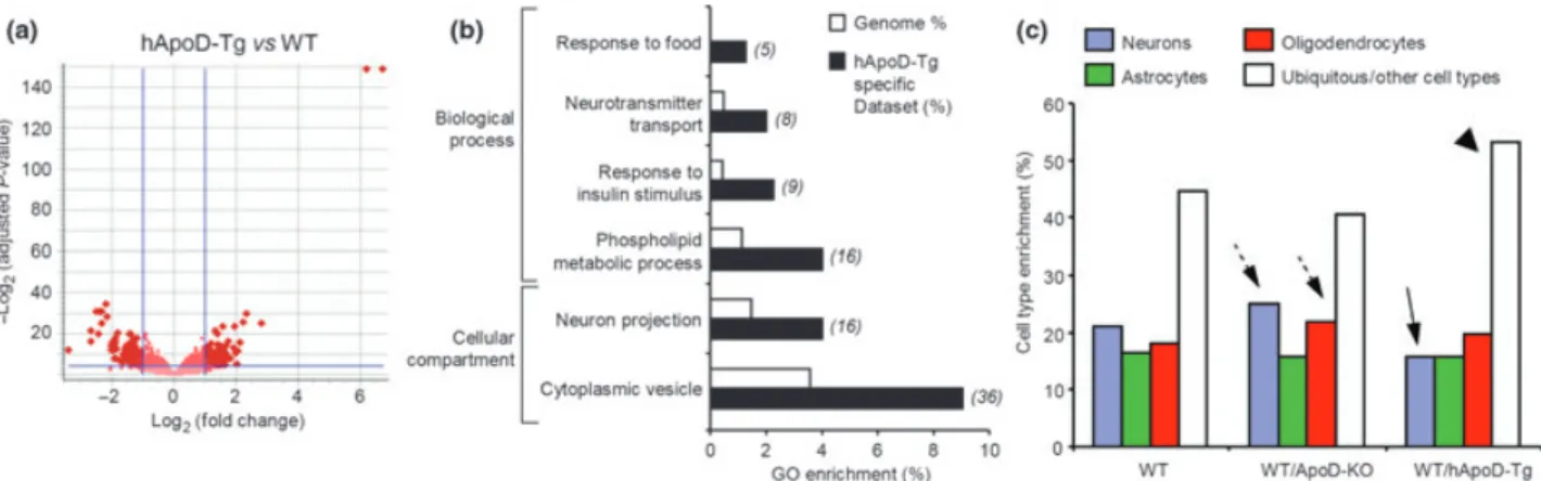
Our first inquiry was to assay the effects on transcription because of the constitutive absence ofApoDin the cerebellum of young adult mice. Twenty eight genes passed our selection criteria (adjustedp-value < 0.05 after FDR correction, and a FC‡±2 threshold) for robust changes in expression in the ApoD-KO mice under control conditions (Fig. 1a; Table S3). In this set, 70% of the genes are down-regulated. The fact that ApoD is the gene most down-regulated in the ApoD-KO samples (arrow in Fig. 1a) was an expected outcome and supports the array results. Despite the reduced number of differentially expressed genes obtained, a significant enrich-ment occurs in Gene Ontology (GO) terms related to transcrip-tional regulation and to neuron excitability (Fig. 1b; Table S9). Several genes related to the transmission of neuronal action potentials appear down-regulated in the ApoD-KO neural tissue. One of them is Mbp, a myelin-associated protein that contributes to the formation of compact myelin (Simons and Trotter 2007) and thus improves axonal conduction velocity. We have found a similar down-regula-tion of Mbp in ApoD-KO peripheral nerves (Ganfornina et al. 2010), stressing the link between ApoD and the myelination process. Also, the modulation of synaptic transmission and neuronal firing patterns by Ca2+-activated K+ channels is expected to be altered as theKcnma1 gene (Salkoffet al.2006) is down-regulated in ApoD-KO mice. In relation to neurotransmission as well, the synaptic machinery appears to react with an increased transcription of ionotropic glutamate receptor GluR4 [a -amino-3-hydroxy-5-methyl-isoxazole-4-propionate (AMPA) receptors] and the neuro-transmitter vesicle-related genes (Kf1bandVapb) to increase a possibly reduced synaptic efficacy in ApoD-KO brain. In relation to this, we have reported a significant decrease of functional glutamate receptors in the brain of ApoD-KO mice (Boer et al. 2009) that could cause a compensatory transcriptional up-regulation of some glutamate receptors.
transcription levels ofMbpin the diencephalon of ApoD-KO mice (Fig. 1c), and immunoblot analysis of WT and ApoD-KO whole brains confirms that lower amounts of Mbp protein (Fig. 1d) is a general effect ofApoDloss.
A different set of ApoD-dependent genes, such as the Map3K7, the nuclear factorNf1a and the chemokineCcl21 are related to stress responses. The absence of ApoD up-regulates Map3k7, a kinase involved in cell responses to environmental stresses through activation of c-Jun N-terminal kinase (MAPK8/JNK) and mitogen-activated pro-tein kinase kinase 4 (MAP2K4/MKK4) signaling cascades. Map3k7 is activated by arachidonic acid, a candidate physiological ligand for ApoD (Vogt and Skerra 2001).Nfia, also up-regulated in the ApoD-KO, regulates the expression of stress-response glial proteins such as glial fibrillary acidic protein (GFAP) (Gopalan et al. 2006). Finally, the chemo-kine Ccl21, involved in the neuronal response to ischemia (de Jong et al. 2005), is down-regulated in the ApoD-KO mice. We confirmed that transcriptional levels ofCcl21and Nfiaalso show similar differences in the ApoD-KO cerebel-lum when studied by qRT-PCR (Fig. 1e).
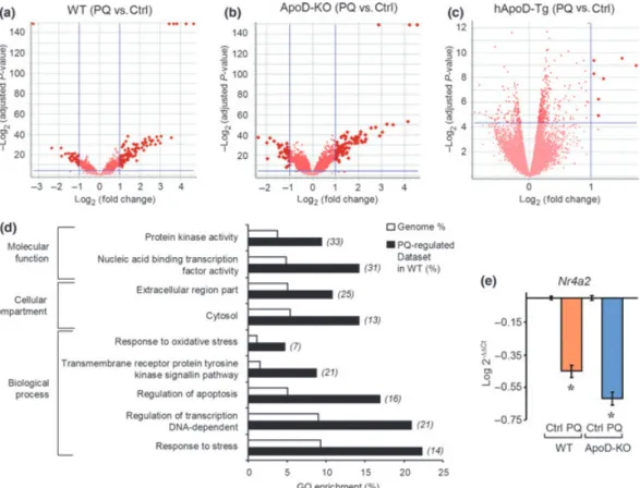
Our second query was to assay the effects on transcription of over-expressing hApoD in neurons. The microarray analysis for the hApoD-Tg mice revealed a substantial number of genes (388) with significant changes in expression levels in the cerebellum (Fig. 2a). The pool of genes fulfilling our criteria for selection (adjustedp-values < 0.05 after FDR correction, and FC‡±2) are shown in Table S4. In terms of enrichment of GO terms (Table S10, Fig. 2b), hApoD-Tg mice show significant changes in hormone receptor binding, transporter activity for neurotransmitters, phospholipid metabolism and response to metabolic stimuli. An interesting observation is the numerous up-regulated genes evidenced in the hApoD-Tg cerebellum related to vesicle dynamics in axonal and synaptic function ( synapto-tagmins I,II,XIII; syntaxin 6; rabphilin 3A; kinesins Ia,Va,Vc; dynein light chain,asynuclein, dynamin 2), possibly related to the ectopic secretion in neurons of hApoD itself and the subsequent secretory vesicle load. These changes could in turn affect synaptic functions.
In order to test whether the transcriptome of a particular NS cell type is modified by the ApoD genotype, we Fig. 1Transcriptional profile of the ApoD-KO cerebellum in adult mice
in control conditions. (a) Volcano plot showing the relationship be-tweenp-value and log2(fold change) for genes differentially expressed in the ApoD-KO compared to the WT tissue. The statistically signifi-cant genes (FC‡2; FDR-adjustedp-value < 0.05) are pointed by dark red dots. Arrow marks the down-regulation ofApoD. (b) Gene ontology terms significantly enriched in genes differentially expressed in ApoD-KO cerebellum. The number of genes in the experimental dataset is
shown in italics. (c) qRT-PCR analysis of Mbp expression in the diencephalon (WT tissue is used as the calibrator sample). (d) Immunoblot analysis of Mbp protein expression in WT and ApoD-KO brains. Protein levels were quantified by band densitometry normal-ized tob-actin signal. (e) qRT-PCR analysis of the expression of the
compared our lists of genes with those reported by Cahoy et al. (2008) to be enriched in different cell types of the mouse brain. Under control conditions, the genes differen-tially regulated in the cerebellum of ApoD-KO mice are distributed in a cell type pattern similar to that of WT brain. Only a moderate increase in the representation of neuronal and oligodendroglial genes is apparent (dashed arrows, Fig. 2c) at the expense of ubiquitous genes, whereas astrocyte genes are equally abundant. However, the hApoD-Tg cerebellar arrays show a marked decrease in the transcription of neuron-specific genes (arrow, Fig. 2c) accompanied by an increase in ubiquitous genes (possibly those with housekeeping functions; arrowhead in Fig. 2c). This might reflect the response of neurons to the ectopic expression of hApoD.
In summary, both a constitutive absence of ApoD expression and an over-expression of ApoD in mouse neurons result in transcriptional changes in the adult mouse cerebellum of genes related to neuronal function, mainly affecting action potential conduction and synaptic function, as well as genes related to myelin management. Moreover, several genes that control the cellular response to environmental stimuli appear regulated by ApoD expression, suggesting that ApoD-KO brains are suffering from constitutive stress such as OS or inflammation. This result is supported by their elevated basal levels of lipid peroxides (Ganfornina et al. 2008) and is in agreement with our findings in the PNS, where the transcriptional profile of injury-regulated genes in the intact ApoD-KO nerves resembles the profile of a damaged WT sciatic nerve (Ganfornina et al. 2010). Appropriate levels of ApoD are thus essential for a proper nervous system homeostasis.
Comparative gene expression profile analysis of wild type cerebellar tissue exposed to experimental oxidative stress To identify gene networks participating in the early cellular response of the cerebellum to OS, we studied the gene expression changes in WT samples 6 h after PQ treatment.
We found 118 genes that showed regulation in the WT cerebellum by the experimental treatment, most of them (71%) presenting up-regulations (Fig. 3a). Table S5 lists only the genes with FC‡±3. The GO analysis identifies gene functional groups related to the cellular response to OS, the regulation of cell death and proliferation, and the regulation of transcription (Fig. 3d). It is also apparent the enrichment in cytosolic and extracellularly secreted proteins. Furthermore, genes related to kinase signaling pathways, critically involved in the cell response to OS, are also enriched in this dataset.
To corroborate our results, as well as to detect potentially important genes common to the response to OS in different tissues, we performed a meta-analysis of microarray studies that explored transcriptional changes upon OS (Edwards et al. 2004; Tomita et al. 2006, 2007; Wang et al. 2007, 2009; Chinet al.2008; Olesenet al.2008; Patelet al.2008; Sforza 2008) using the LOLA database and analysis software (http://lola.gwu.edu/) (Cahanet al.2005).
Thirty genes that appear differentially expressed in our analysis of the WT cerebellum upon PQ treatment (some of them highlighted in bold in Tables S5 and S6) showed concordant regulations in other microarray reports (see above) using diverse OS experimental paradigms. It is important to mention that genes such asFkbp5, Zfp36, Ctgf, Sgk3, Cebpd, Gadd45g, Nr4a1, S3-12, Cdkn1a, Pdk4, Mt2 and Map3k6, present in our PQ-regulated dataset, have been reported as early response genes to PQ treatment in other Fig. 2 Effects of hApoD over-expression in cerebellar gene
expres-sion (a and b) and modifications in cell-type enrichment distributions in theApoD-dependent gene pools (c). (a) Volcano plot for genes dif-ferentially expressed in the hApoD-Tg compared to the WT tissue in control conditions. Genes statistically significant (FC‡2; FDR-adjustedp-value < 0.05) are shown by dark red dots. (b) GO terms
tissues. Therefore, they must be part of a common set of PQ-responding genes.
Thus, our array analysis identifies genes that organize the early response of cells to OS in the WT cerebellum. The fact that we find a significant portion of genes common to the response to OS in many tissues contributes to validate our results and supports the subsequent hypotheses on ApoD effects on the OS-responsive tran-scription network.
Effect ofApoD expression on the early response to experimental oxidative stress in cerebellar tissue
To reveal the biological processes triggered as a consequence of theApoDup-regulation in response to OS, we compared the gene expression profiles of ApoD-KO, hApoD-Tg and WT cerebellum upon PQ treatment.
From the genes that respond to PQ in the WT and ApoD-KO cerebellar tissue, we selected first those that show a common regulation by PQ. This set contains 72 genes with genotype-independent changes, that is, they respond to PQ in a similar way regardless of the presence or absence ofApoD (Fig. 4a, red dots: Fig. 4b, diagram intersection). This pool
represents 61% of the genes that organize the early response to PQ in the WT (see above).
We randomly selected a gene moderately regulated by PQ treatment that, in addition, showed slight differences in expression in WT and ApoD-KO cerebellum, so that we can test whether small expression differences detected in the array are validated by an independent quantification method. The Nuclear receptor subfamily 4, group A, member 2 (Nr4a2), involved in dopaminergic neurons development and function (Maguire-Zeiss and Federoff 2010) appears down-regulated by PQ in both the WT and ApoD-KO cerebellum ()3.22 FC in WT; )3.91 FC in ApoD-KO). Comparable down-regulations ()2.81 FC in WT;)4.15 FC in ApoD-KO) were obtained by qRT-PCR experiments using the same RNA samples used for the arrays (Fig. 3e).
Next, we analyzed the genes showing a differential response to PQ in WT and ApoD-KO cerebellum. Seventy seven genes showed ApoD-dependent changes in their transcriptional levels (Tables S6 and S7, and Fig. 4b). Thirty one transcripts change their expression specifically in ApoD-KO cerebellum (Table S7), and can thus be considered genes that respond to the OS generated by PQ only whenApoDis Fig. 3Transcriptome profile of oxidative stress-challenged
cerebel-lum. (a–c) Volcano plots for the genes constituting the early response to a single dose of PQ in WT (a), ApoD-KO (b) and hApoD-Tg (c) cerebellum. (d) Enrichment plot of GO terms for the PQ-challenged WT tissue. The number of genes in the experimental dataset is shown
not being expressed by cerebellar astrocytes and oligoden-drocytes. In contrast, 46 transcripts were specifically regu-lated in WT, i.e., they are genes that require the presence of ApoDto respond to OS (Table S6).
The GO analysis of the 77 genes (46 WT specific and 31 ApoD-KO specific) differentially regulated by PQ in a genotype-dependent manner, uncovers a significant enrich-ment in terms related to the regulation of transcription, nervous system development, and the response to stress (Fig. 4c). Similarly, an enriched set of these genotype-dependent genes code for membrane-related proteins, which suggests a role of ApoD in the effect of OS on cell membranes.
In addition to the genes that show all-or-none ApoD -dependent responses, there are others, among the 72 common genes (intersection in Fig. 4b), that differ in the magnitude of the response to PQ. Our analysis identifies five genes with |FC(KO) ) FC(WT)|‡1.5 in their response to PQ. The genes Rhoj, Cdkn1a, Polr3e and Pdk4 are found more
up-regulated in ApoD-KO, andFosis less down-regulated in ApoD-KO. Interestingly, three of these genes are part of the common pool of early-responders to OS that we have uncovered in our meta-analysis (bold type in Table S5).
Finally, we studied the cell type enrichment patterns of the groups of genes that respond to PQ in WT and ApoD-KO cerebellum (Fig. 4d). In these comparisons, the most obvious result is the enrichment of astrocyte-specific genes and the under-representation of neuronal genes (Fig. 4d, arrowhead) in the WT response to PQ. This is consistent with the known critical role of astrocytes in the OS-challenged brain (Rossi and Volterra 2009). A similar pattern is found for the common genes equally regulated by PQ in both WT and ApoD-KO cerebellum. However, superimposed to this common pattern in the response to PQ, the pool of genes that specifically respond to PQ either in the WT or in the KO show a marked increase in oligodendrocyte-specific genes at the expense of ubiquitous genes (arrow in Fig. 4d).This result supports our hypothesis that the absence of ApoD Fig. 4 Early transcriptional response to PQ in the ApoD-KO
cere-bellum. (a) Correlation plot of genes differentially expressed upon PQ treatment in WT and ApoD-KO mice. The gray box marks the boundaries for non-statistically significant changes. Red dots point to genes that show genotype-dependent regulation by PQ. Black dots are genes equally regulated in WT and ApoD-KO mice. (b) Venn diagram of PQ-regulated genes in WT and ApoD-KO
cerebella. (c) Plot showing the enrichment in GO terms of PQ-regulated genes that are dependent on ApoD genotype. The number of genes in the experimental dataset is shown in italics. (d) Cell type enrichment analysis in PQ-regulated genes, grouped according to their dependence onApoDgenotype and compared to the cell type representation in the WT mouse brain (Cahoy et al.
makes lipid-bearing cell compartments more susceptible to oxidative stress (Sanchez et al. 2006; Ganfornina et al. 2008). We can predict from this pattern thatApoDfunction is important for oligodendrocytes, with their lipid-enriched myelin, especially in pro-oxidant situations. Among the genes that would normally respond to the OS in oligoden-drocytes, they must either need ApoD to respond or their response is inhibited byApoD.
A second set of comparisons was performed of the genes regulated by PQ-treatment in WT and hApoD-Tg cerebellum. Surprisingly, only six hApoD-Tg genes appear regulated by PQ (Table S8). Three of them are well known OS-regulated genes (S3-12, Fkbp5and Xdh), but their FC are well below the levels attained in the WT tissue (see Table S5 for comparison). Other genes with expression levels related to brain pathologies (Ttr, Folr1and Sdc4) appear up-regulated in the brain of PQ-challenged hApoD-Tg mice. The lack of an early gene regulation in response to OS when ApoD is over-expressed in the brain further confirms the improved survival and the prevention of lipid peroxide brain accumu-lation previously reported (Ganforninaet al.2008).
In view of our GO and cell-type enrichment analyses, two main biological processes appear as clearly dependent on the
function ofApoDin OS conditions: myelin management and glial responses to stress.
Among the genes whose response to PQ is genotype-dependent, a significant number (Aspa1, Tnc, Cldn5, Cdh11, Elovl7, Eomes, Sox4, Sox10, Tyro3andUgt8a) are related to the myelination process in the CNS. Seven of these genes are down-regulated by PQ, but they belong to the WT-specific group, meaning that their OS-induced inhibition is absent in the ApoD-KO cerebellum. These genes are normally shut down upon OS, and are thus halting myelin synthesis when OS affects oligodendrocytes. The anomalous maintenance of their expression in the absence of ApoD might potentially enhance the already high vulnerability of oligodendrocytes. This effect can in turn be aggravated by the ApoD-KO specific down-regulation of genes such asAspa1andTncthat is correlated with demyelination processes (Zhaoet al.2009; Mattanet al.2010).
The cellular response to OS, and particularly the astroglial and microglial responses, is especially relevant to our proposal thatApoDis involved in the detoxification system against OS. A group of 18 genes (highlighted in italics in Tables S6 and S7) are related to such glial responses.
One of these OS-responsive genes is ApoD itself. We observed an up-regulation ofApoDmRNA in response to PQ in the ApoD-KO cerebellum (see Table S7). AlthoughApoD expression is up-regulated by OS (Ganfornina et al. 2008), this result is a priori unexpected in the ApoD-KO back-ground. We confirmed by qRT-PCR that an increased amount ofApoDmRNA also exists in a different sample of WT and ApoD-KO brains a week after a chronic treatment with PQ (Fig. 5a). However, we already reported that a truncated mRNA species is produced in the ApoD-KO brain (Gan-fornina et al. 2008), and a lack of translation was clear by immunoblot (Fig. 5b). Interestingly, no transcriptional regu-lation of mouse ApoD was observed in the PQ-treated hApoD-Tg tissue (Table S4). These results support that ApoDis required in the normal cell response to OS, and that the mechanisms regulating ApoD gene transcription, nor-mally part of an early response, can persist chronically under conditions of null protein expression.
A set of genes among those specifically down-regulated upon PQ in ApoD-KO cerebellum,Cd44, Phlda1andEfnb2, are also related to the molecular pathways of OS-responding genes. These genes are known to be abundantly transcribed in the OS-resistant mesencephalic A10 dopaminergic neu-rons (Chung et al. 2005). A10 neurons also produce high amounts of several neuropeptides, such as pituitary adenylate cyclase-activating polypeptide (PACAP), that confer resis-tance to MPTP-associated OS (Chunget al.2005). Interest-ingly, ApoD has been recently found to induce pituitary adenylate cyclase-activating polypeptide (PACAP) expres-sion from neuronal primary cultures (Kosackaet al. 2011). Together with our findings, these data suggest that ApoD must be required, at least in the less labile sets of Fig. 5Glial genes involved in the oxidative stress response of
dopaminergic neurons, to keep appropriate levels of protec-tors under OS conditions.
Finally, an interesting gene specifically up-regulated in PQ-challenged ApoD-KO cerebellar arrays isProtein kinase Cd(Pkcd). Its up-regulation is further confirmed in the same samples by qRT-PCR (Fig. 5c). Although expressed ubiqui-tously and involved in a wide range of cellular functions, Pkcdis among the genes significantly enriched in astrocytes in the Cahoyet al.(2008) analysis. Also, it has been recently linked to the PQ-induced OS generation and the astroglial response in the nervous system (Kimet al.2008). Given the major role of astrocytes in the PQ response, their expres-sion of ApoD upon stressful situations, and the specific vulnerability of ApoD-KO astrocytes to PQ-generated OS (our unpublished results; Bajo-Gran˜eraset al.) we tested by qRT-PCR the transcription of Pkcd in astrocyte-enriched primary glial cultures of WT and ApoD-KO mice upon 6 h of PQ treatment. This acute PQ treatment produced, how-ever, no significant regulation ofPkcdin astrocyte cultures of either genotype (Fig. 5d), suggesting that the specific early up-regulation ofPkcd we observe in the PQ-treated ApoD-KO cerebellum could occur in microglial cells, which are also known to express high levels ofPkcd. In microglia,Pkcdis in fact linked to PQ-dependent reactive oxygen species (ROS) production mediated by activation of NADPH oxidase (Milleret al.2007). On the other hand, in basal conditions a significant increase in the levels ofPkcdexpression is seen in ApoD-KO astrocytes (Fig. 5d), supporting the enhanced vulnerability of the ApoD-KO nervous system to either physiological or pathologically generated OS.
As glial cells are also implicated in the priming effects that occur in PQ-related neurodegeneration, and this process is dependent on signals exchanged among microglia, astrocytes and neurons (Purisai et al. 2007; Klintworth et al. 2009), studying the role of ApoD in neuron-glia and glia-glia interactions is of paramount importance, and is the logical next step in our research program aiming to understand the role of this lipocalin in nervous system development and function. In summary, the altered expression profiles in the ApoD-KO cerebellum, both in control conditions and after PQ treatment, along with the deficient transcriptional response to PQ observed in the hApoD-Tg tissue, strongly support that the presence ofApoDin the neural environment is necessary for a proper protection against oxidative damage.
Acknowledgements
We thank J.R. Acebes, E. Gonza´lez and E. Martı´n for technical assistance, and the Lazarillo Lab (M. Ruiz, N. Garcı´a-Mateo, M. del Can˜o & A. Pe´rez-Castellanos) for their helpful discussions and positive criticisms. We thank S. Sanz for help with some of the qRT-PCR experiments. Thanks also to Dr. E. Fermin˜an (Genomics facility at the Centro de Investigacion del Cancer) for performing the array hybridizations. This work was supported by grant CIHR MOP
15677 to E.R.; FRSQ and CRSNG studentships to S.D.C.; grants BFU2007-61848 (DGICYT) and CIBER CB06/06/0050 (FISS-ICiii) to C.G.; and grants MEC BFU2005-00522, JCyL VA049A05, and MICINN BFU2008-01170 to M.D.G. and D.S. Authors declare that no conflict of interest exists in relation to the content of this manuscript. Neither the author’s institutions nor the funding agencies had a role in the study design, data collection and analysis, decision to publish, or preparation of the manuscript.
Supporting information
Additional supporting information may be found in the online version of this article:
Table S1. Parameters used to evaluate the quality of the microarray hybridization signals.
Table S2.Oligonucleotide primers used for qRT-PCR.
Table S3. Genes differentially expressed in ApoD-KO cerebel-lum in control conditions (Fold change‡±2).
Table S4.Genes differentially expressed in hApoD-Tg cerebel-lum in control conditions (Fold change‡±2).
Table S5.PQ-regulated genes in WT cerebellum (Fold change‡
±2).
Table S6. PQ-regulated genes specific for WT (Fold change‡
±2).
Table S7. PQ-regulated genes specific for ApoD-KO (Fold change‡±2).
Table S8. PQ-regulated genes specific for hApoD-Tg (Fold change‡±2).
Table S9.GO Terms enriched in ApoD-KO vs. WT comparison. Table S10.GO Terms enriched in hApoD-Tg vs. WT compar-ison.
Table S11.GO terms enrichment in WT PQ-regulated genes. Table S12. GO terms enrichment in genotype-dependent PQ-regulated genes.
As a service to our authors and readers, this journal provides supporting information supplied by the authors. Such materials are peer-reviewed and may be re-organized for online delivery, but are not copy-edited or typeset. Technical support issues arising from supporting information (other than missing files) should be addressed to the authors.
References
Apps R. and Garwicz M. (2005) Anatomical and physiological foun-dations of cerebellar information processing.Nat. Rev. Neurosci.6, 297–311.
Baldi P. and Long A. D. (2001) A Bayesian framework for the analysis of microarray expression data: regularized t -test and statistical inferences of gene changes.Bioinformatics17, 509–519. Blazejczyk M., Miron M. and Nadon R. (2007)FlexArray: A Statistical
Data Analysis Software for Gene Expression Microarrays. Genome Quebec, Montreal, Canada.
Boer S., Sanchez D., Reinieren I., van den Boom T., Udawela M., Scarr E., Ganfornina M. D. and Dean B. (2009) Decreased kainate receptors in the hippocampus of apolipoprotein D knockout mice. Prog. Neuropsychopharmacol. Biol. Psychiatry34, 271–278. Cahan P., Ahmad A. M., Burke H.et al.(2005) List of lists-annotated
Cahoy J. D., Emery B., Kaushal A. et al. (2008) A transcriptome database for astrocytes, neurons, and oligodendrocytes: a new resource for understanding brain development and function. J. Neurosci.28, 264–278.
Chin M. H., Qian W. J., Wang H.et al. (2008) Mitochondrial dys-function, oxidative stress, and apoptosis revealed by proteomic and transcriptomic analyses of the striata in two mouse models of Parkinson’s disease.J. Proteome Res.7, 666–677.
Chung C. Y., Seo H., Sonntag K. C., Brooks A., Lin L. and Isacson O. (2005) Cell type-specific gene expression of midbrain dopaminergic neurons reveals molecules involved in their vulnerability and pro-tection.Hum. Mol. Genet.14, 1709–1725.
Do Carmo S., Fournier D., Mounier C. and Rassart E. (2009) Human apolipoprotein D overexpression in transgenic mice induces insulin resistance and alters lipid metabolism.Am. J. Physiol. Endocrinol. Metab.296, E802–E811.
Drechsel D. A. and Patel M. (2008) Role of reactive oxygen species in the neurotoxicity of environmental agents implicated in Parkin-son’s disease.Free Radic. Biol. Med.44, 1873–1886.
Edwards M. G., Sarkar D., Klopp R., Morrow J. D., Weindruch R. and Prolla T. A. (2004) Impairment of the transcriptional responses to oxidative stress in the heart of aged C57BL/6 mice.Ann. N Y Acad. Sci.1019, 85–95.
Ganfornina M. D., Sanchez D., Pagano A., Tonachini L., Descalzi-Cancedda F. and Martinez S. (2005) Molecular characterization and developmental expression pattern of the chicken apolipopro-tein D gene: implications for the evolution of vertebrate lipocalins. Dev. Dyn.232, 191–199.
Ganfornina M. D., Do Carmo S., Lora J. M.et al.(2008) Apolipoprotein D is involved in the mechanisms regulating protection from oxi-dative stress.Aging Cell7, 506–515.
Ganfornina M. D., Do Carmo S., Martinez E., Tolivia J., Navarro A., Rassart E. and Sanchez D. (2010) ApoD, a glia-derived apolipo-protein, is required for peripheral nerve functional integrity and a timely response to injury.Glia58, 1320–1334.
Gonzalez-Polo R. A., Rodriguez-Martin A., Moran J. M., Niso M., Soler G. and Fuentes J. M. (2004) Paraquat-induced apoptotic cell death in cerebellar granule cells.Brain Res.1011, 170–176.
Gopalan S. M., Wilczynska K. M., Konik B. S., Bryan L. and Kordula T. (2006) Nuclear factor-1-X regulates astrocyte-specific expression of the alpha1-antichymotrypsin and glial fibrillary acidic protein genes.J. Biol. Chem.281, 13126–13133.
Huang da W., Sherman B. T. and Lempicki R. A. (2009) Systematic and integrative analysis of large gene lists using DAVID bioinformatics resources.Nat. Protoc.4, 44–57.
Hull-Thompson J., Muffat J., Sanchez D., Walker D. W., Benzer S., Ganfornina M. D. and Jasper H. (2009) Control of metabolic homeostasis by stress signaling is mediated by the lipocalin NLaz. PLoS Genet.5, e1000460.
de Jong E. K., Dijkstra I. M., Hensens M., Brouwer N., van Amerongen M., Liem R. S., Boddeke H. W. and Biber K. (2005) Vesicle-mediated transport and release of CCL21 in endangered neurons: a possible explanation for microglia activation remote from a pri-mary lesion.J. Neurosci.25, 7548–7557.
Kim S., Hwang J., Lee W. H., Hwang D. Y. and Suk K. (2008) Role of protein kinase Cdelta in paraquat-induced glial cell death. J. Neurosci. Res.86, 2062–2070.
Klintworth H., Garden G. and Xia Z. (2009) Rotenone and paraquat do not directly activate microglia or induce inflammatory cytokine release.Neurosci. Lett.462, 1–5.
Kosacka J., Schroder T., Bechmann I., Kloting N., Nowicki M., Mittag A., Gericke M., Spanel-Borowski K. and Bluher M. (2011) PACAP up-regulates the expression of apolipoprotein D in 3T3-L1 adipo-cytes. DRG/3T3-L1 co-cultures study.Neurosci. Res.69, 8–16.
Livak K. J. and Schmittgen T. D. (2001) Analysis of relative gene expression data using real-time quantitative PCR and the 2(-Delta Delta C(T)) Method.Methods25, 402–408.
Lyons D. A., Naylor S. G., Scholze A. and Talbot W. S. (2009) Kif1b is essential for mRNA localization in oligodendrocytes and development of myelinated axons. Nat. Genet. 41, 854– 858.
Maguire-Zeiss K. A. and Federoff H. J. (2010) Future directions for immune modulation in neurodegenerative disorders: focus on Parkinson’s disease.J. Neural Transm.117, 1019–1025. Mattan N. S., Ghiani C. A., Lloyd M., Matalon R., Bok D., Casaccia P.
and de Vellis J. (2010) Aspartoacylase deficiency affects early postnatal development of oligodendrocytes and myelination. Neurobiol. Dis.40, 432–443.
McCormack A. L., Atienza J. G., Johnston L. C., Andersen J. K., Vu S. and Di Monte D. A. (2005) Role of oxidative stress in paraquat-induced dopaminergic cell degeneration.J. Neurochem.93, 1030– 1037.
Miller R. L., Sun G. Y. and Sun A. Y. (2007) Cytotoxicity of paraquat in microglial cells: involvement of PKCdelta- and ERK1/2-dependent NADPH oxidase.Brain Res.1167, 129–139.
Navarro A., Del Valle E. and Tolivia J. (2004) Differential expression of apolipoprotein d in human astroglial and oligodendroglial cells. J. Histochem. Cytochem.52, 1031–1036.
Olesen B. T., Clausen J. and Vang O. (2008) Characterization of the transcriptional profile in primary astrocytes after oxidative stress induced by Paraquat.Neurotoxicology29, 13–21.
Ong W. Y., Lau C. P., Leong S. K., Kumar U., Suresh S. and Patel S. C. (1999) Apolipoprotein D gene expression in the rat brain and light and electron microscopic immunocytochemistry of apolipoprotein D expression in the cerebellum of neonatal, immature and adult rats.Neuroscience90, 913–922.
Patel S., Singh K., Singh S. and Singh M. P. (2008) Gene expression profiles of mouse striatum in control and maneb + paraquat-in-duced Parkinson’s disease phenotype: validation of differentially expressed energy metabolizing transcripts.Mol. Biotechnol. 40, 59–68.
Prasad K., Winnik B., Thiruchelvam M. J., Buckley B., Mirochnitchenko O. and Richfield E. K. (2007) Prolonged toxic-okinetics and toxicodynamics of paraquat in mouse brain.Environ. Health Perspect.115, 1448–1453.
Provost P. R., Villeneuve L., Weech P. K., Milne R. W., Marcel Y. L. and Rassart E. (1991) Localization of the major sites of rabbit apoli-poprotein D gene transcription by in situ hybridization.J. Lipid Res.32, 1959–1970.
Purisai M. G., McCormack A. L., Cumine S., Li J., Isla M. Z. and Di Monte D. A. (2007) Microglial activation as a priming event leading to paraquat-induced dopaminergic cell degeneration. Neurobiol. Dis.25, 392–400.
Rossi D. and Volterra A. (2009) Astrocytic dysfunction: insights on the role in neurodegeneration.Brain Res. Bull.80, 224–232. Salkoff L., Butler A., Ferreira G., Santi C. and Wei A. (2006)
High-conductance potassium channels of the SLO family. Nat. Rev. Neurosci.7, 921–931.
Sanchez D., Lopez-Arias B., Torroja L., Canal I., Wang X., Bastiani M. J. and Ganfornina M. D. (2006) Loss of glial lazarillo, a homolog of apolipoprotein D, reduces lifespan and stress resistance in Drosophila.Curr. Biol.16, 680–686.
Sforza D. M. (2008) Gene expression changes in multiple brain regions of a mouse MPTP model of Parkinson’s disease. Gene Expression Omnibus, Vol. GSE7707. National Center for Biotechnology Information.
Tarraga J., Medina I., Carbonell J.et al.(2008) GEPAS, a web-based tool for microarray data analysis and interpretation.Nucleic Acids Res.36, W308–W314.
Tomita M., Okuyama T., Katsuyama H., Hidaka K., Otsuki T. and Ishikawa T. (2006) Gene expression in rat lungs during early response to paraquat-induced oxidative stress.Int. J. Mol. Med.17, 37–44.
Tomita M., Okuyama T., Katsuyama H., Miura Y., Nishimura Y., Hidaka K., Otsuki T. and Ishikawa T. (2007) Mouse model of paraquat-poisoned lungs and its gene expression profile. Toxi-cology231, 200–209.
Vogt M. and Skerra A. (2001) Bacterially produced apolipoprotein D binds progesterone and arachidonic acid, but not bilirubin or E-3M2H.J. Mol. Recognit.14, 79–86.
Wang X., Pal R., Chen X. W., Kumar K. N., Kim O. J. and Michaelis E. K. (2007) Genome-wide transcriptome profiling of region-specific
vulnerability to oxidative stress in the hippocampus.Genomics90, 201–212.
Wang X., Zaidi A., Pal R., Garrett A. S., Braceras R., Chen X. W., Michaelis M. L. and Michaelis E. K. (2009) Genomic and biochemical approaches in the discovery of mechanisms for selective neuronal vulnerability to oxidative stress.BMC Neurosci. 10, 12.
Yuan J. S., Reed A., Chen F. and Stewart C. N. Jr. (2006) Statistical analysis of real-time PCR data.BMC Bioinformatics7, 85. Zhao C., Fancy S. P., Franklin R. J. and ffrench-Constant C. (2009)
Up-regulation of oligodendrocyte precursor cell alphaV integrin and its extracellular ligands during central nervous system re-myelination.J. Neurosci. Res.87, 3447–3455.
Arrays (3/group) Scale factor Present Calls Concordant Calls Reliability index
WT-Ctrl 0.352±0.05 65.2±1.29% 88.13% 0.996
WT-PQ 0.349±0.02 64.5±0.66% 88.51% 0.997
ApoD KO-Ctrl 0.358±0.01 64.2±0.38% 89.16% 0.999
ApoD KO-PQ 0.347±0.02 61.4±4.29% 82.75% 0.997
hApoD Tg-Ctrl 0.337±0.05 65.8±0.81% 87.68% 0.991
Gene-primer Sequence Acc. Number Oligonucleotide sequence
Rpl18-Forward 5’-TTCCGTCTTTCCGGACCT
Rpl18-Reverse 5’- TCGGCTCATGAACAACCTCT
ApoD-Forward 5’- GAAGCCAAACAGAGCAACG
ApoD-Reverse 5’- TGTTTCTGGAGGGAGATAAGGA
Nr4a2-Forward 5’- AGTGCCTAGCTGTTGGGATGGT
Nr4a2-Reverse 5’- TAGTCAGGGTTTGCCTGGAA
Pkcd-Forward 5’- CACCAATAGCCGGGACACCATCT
Pkcd-Reverse 5’- TGGTTGATACCACACAGGTTG
Ccl21-Forward 5’- AGGCTGGGTGCAGAACCTGAT
Ccl21-Reverse 5’- TGAAGTTCGTGGGGGATCT
Nf1a-Forward 5’- TGGAGGTTGGACCTCGTCATGGT
Nf1a-Reverse 5’- CTGGCTGGGACTTTCAGATT
Mbp-Forward 5’- GCTGAGAAGGCCAGTAAGGA
Mbp-Reverse 5’- CCACGCTTCTCTTCTTTCCA
NM_011124.4
NM_001122952.1
NM_010777.3 NM_009077.2
NM_007470.2
NM_013613.2
UniGene ID Gene Title Gene Symbol Fold change P value
Mm.260456 Vesicle-associated membrane protein B and C Vapb 3.41 4.0E-10
Mm.31274 Nuclear factor I/A Nfia 3.39 4.1E-09
Mm.12145 Retinoblastoma binding protein 4 Rbbp4 3.14 5.0E-13
Mm.268548 Max protein Max 2.54 5.6E-07
Mm.402393 Kinesin family member 1B Kif1b 2.09 1.1E-07
Mm.209263 Glutamate receptor. ionotropic. AMPA4 (alpha 4) Gria4 2.07 3.3E-07 Mm.258589 Mitogen activated protein kinase kinase kinase 7 Map3k7 2.07 8.5E-08 Mm.21841 Splicing factor. arginine/serine-rich 2 (SC-35) Sfrs2 2.06 8.0E-06
Mm.253518 Bromodomain containing 4 Brd4 -2.00 1.5E-06
Mm.3360 Tyr-3/trp-5-monooxygenase activation protein Ywhaz -2.07 2.6E-07
Mm.5001 DNA methyltransferase 3A Dnmt3a -2.08 3.2E-05
Mm.280842 Heterogeneous nuclear ribonucleoprotein A/B Hnrpab -2.09 4.0E-07
Mm.383196 Nuclear receptor subfamily 2. group C. member 2 Nr2c2 -2.09 1.6E-04 Mm.8687 CAP. Adenylate cyclase-associated protein 1 (yeast) Cap1 -2.20 7.6E-08
Mm.3815 Syndecan 4 Sdc4 -2.28 6.1E-07
Mm.311912 Cys-rich transmembrane BMP regulator 1 (chordin like) Crim1 -2.29 9.9E-10
Mm.331626 Synaptic nuclear envelope 1 Syne1 -2.44 4.2E-07
Mm.439824 Similar to Protein tyrosine phosphatase type IVA protein 2 PRL-2 -2.48 5.3E-12
Mm.259197 RNA binding motif protein 5 Rbm5 -2.57 1.7E-08
Mm.12926 Mediator complex subunit 1 Med1 -2.58 6.7E-12
Mm.203921 OTU domain. ubiquitin aldehyde binding 1 Otub1 -2.59 5.3E-10
Mm.455873 Nuclear receptor interacting protein 1 Nrip1 -2.62 1.7E-05
Mm.259197 RNA binding motif protein 5 Rbm5 -2.68 2.4E-08
Mm.343607 K+ calcium-activated channel. subfamily M. member 1 Kcnma1 -3.55 4.0E-10
Mm.252063 Myelin basic protein Mbp -3.85 5.4E-11
Mm.450416 Chemokine (C-C motif) ligand 21 Ccl21 -4.17 4.2E-14
Mm.270999 GATA zinc finger domain containing 2B Gatad2b -7.22 9.0E-14
UniGene ID Gene Title Gene name Fold change P-value
Mm.17484 Synuclein, alpha Snca 107.15 0.0E+00
Mm.458208 Dynein light chain tctex-type 1 Dynlt1 5.05 9.8E-10
Mm.181166 Rabphilin 3a Rph3a 4.72 1.7E-08
Mm.131074 Btb (poz) domain containing 14a Btbd14a 4.36 2.0E-05
Mm.43081 Mitogen-activated protein kinase 8 interacting protein 3 Mapk8ip3 4.24 3.9E-04
Mm.218875 Target of myb1-like 2 (chicken) Tom1l2 4.07 3.1E-02
Mm.271898 Unc-51 like kinase 1 (c. Elegans) Ulk1 3.99 6.3E-04
Mm.254515 Digeorge syndrome critical region gene 2 Dgcr2 3.84 1.4E-04
Mm.103551 Toll interacting protein Tollip 3.52 4.3E-03
Mm.24044 Beta-site app cleaving enzyme 1 Bace1 3.38 1.4E-05
Mm.33490 Synaptotagmin xiii Syt13 3.28 2.2E-03
Mm.270278 Thyrotroph embryonic factor Tef 3.16 3.7E-02
Mm.250605 Sel-1 suppressor of lin-12-like (c. Elegans) Sel1l 3.13 1.6E-03
Mm.372314 Heat shock protein 1b Hspa1b 3.13 9.5E-04
Mm.1682 Signal-regulatory protein alpha Sirpa 3.08 3.1E-03
Mm.23047 Transmembrane and coiled coil domains 3 Tmcc3 3.06 3.1E-04
Mm.237099 Amylo-1,6-glucosidase, 4-alpha-glucanotransferase Agl 3.06 2.7E-03 Mm.44245 Adenylate cyclase activating polypeptide 1 receptor 1 Adcyap1r1 3.05 1.6E-02
Mm.10728 Myosin binding protein c, cardiac Mybpc3 3.04 1.5E-02
Mm.30837 N-myc downstream regulated gene 1 Ndrg1 2.88 2.2E-04
Mm.254144 Adrenergic receptor kinase, beta 1 Adrbk1 2.88 2.9E-04
Mm.256342 Kinesin family member 5 Kif5 2.88 3.2E-02
Mm.6645 Thymoma viral proto-oncogene 1 Akt1 2.88 2.6E-04
Mm.332295 Microtubule-associated protein, rp/eb family, member 3 Mapre3 2.86 5.4E-05
Mm.230249 Cathepsin e Ctse 2.85 8.9E-03
Mm.235194 Thymoma viral proto-oncogene 3 Akt3 2.83 2.5E-03
Mm.259295 Pbx/knotted 1 homeobox Pknox1 2.81 5.0E-05
Mm.402393 Kinesin family member 1b Kif1b 2.81 8.3E-03
Mm.196532 Splicing factor 3b, subunit 2 Sf3b2 2.81 1.1E-04
Mm.270484 Makorin, ring finger protein, 1 Mkrn1 2.79 1.7E-04
Mm.28017 F-box and wd-40 domain protein 11 Fbxw11 2.78 2.0E-02
Mm.340818 Atpase, h+ transporting, lysosomal v0 subunit a1 Atp6v0a1 2.74 1.6E-02
Mm.289702 Synaptotagmin i Syt1 2.73 1.6E-02
Mm.256342 Kinesin family member 5c Kif5c 2.73 4.0E-02
Mm.261168 Potassium inwardly-rectifying channel, subfamily j, member 9 Kcnj9 2.71 6.3E-04
Mm.275003 Melanoma cell adhesion molecule Mcam 2.71 1.1E-03
Mm.284503 Cdp-diacylglycerol synthase (phosphatidate cytidylyltransferase) 2 Cds2 2.68 5.6E-03
Mm.370185 Guanine nucleotide binding protein, alpha 12 Gna12 2.68 8.4E-04
Mm.39040 Myelin and lymphocyte protein, t-cell differentiation protein Mal 2.66 3.5E-05 Mm.41812 G protein-regulated inducer of neurite outgrowth 1 Gprin1 2.66 9.0E-04
Mm.265347 Annexin a6 Anxa6 2.63 5.1E-03
Mm.6379 Solute carrier family 1 (glutamate/neutral amino acid transporter), member 4 Slc1a4 2.61 1.3E-02 Mm.171484 Pyridoxal-dependent decarboxylase domain containing 1 Pdxdc1 2.61 2.0E-03 Mm.293120 Signal transducer and activator of transcription 2 Stat2 2.60 3.2E-03 Mm.24724 Protein phosphatase 1, regulatory (inhibitor) subunit 3c Ppp1r3c 2.58 1.5E-02 Mm.40331 Bromodomain adjacent to zinc finger domain, 1b Baz1b 2.53 2.2E-03
Mm.316628 Adenosine deaminase, rna-specific Adar 2.53 2.1E-02
Mm.271656 Rab and dnaj domain containing Rbj 2.52 7.9E-04
Mm.4364 Interleukin 6 signal transducer Il6st 2.52 4.5E-03
Mm.426936 Nuclear factor i/c Nfic 2.51 4.2E-03
Mm.197387 Bicaudal d homolog 2 (drosophila) Bicd2 2.50 8.9E-03
Mm.385012 Sodium channel, voltage-gated, type viii, alpha Scn8a 2.49 1.8E-03
Mm.130227 Flotillin 2 Flot2 2.48 6.1E-04
Mm.196067 Adenylate kinase 3 Ak3 2.47 5.7E-03
Mm.412319 Sphingosine phosphate lyase 1 Sgpl1 2.47 2.9E-03
Mm.37371 Procollagen-lysine, 2-oxoglutarate 5-dioxygenase 1 Plod1 2.46 3.9E-03
Mm.5137 Double c2, beta Doc2b 2.46 3.5E-03
Mm.147946 Myb binding protein (p160) 1a Mybbp1a 2.43 3.5E-03
Mm.303059 Ubiquilin 4 Ubqln4 2.43 4.3E-03
Mm.6904 Fibroblast growth factor receptor 3 Fgfr3 2.43 2.4E-02
Mm.44249 Nitric oxide synthase 1, neuronal Nos1 2.42 1.7E-02
Mm.329963 Stromal membrane-associated protein 1 Smap1 2.42 9.9E-03
Mm.207 Homeo box b5 Hoxb5 2.41 5.4E-03
Mm.5102 Synaptotagmin ii Syt2 2.41 1.9E-02
Mm.268548 Max protein Max 2.40 2.8E-02
Mm.252987 Solute carrier family 12, member 5 Slc12a5 2.40 3.7E-03
Mm.244549 Solute carrier family 6 (neurotransmitter transporter, glycine), member 9 Slc6a9 2.40 1.9E-02
Mm.318841 Erbb receptor feedback inhibitor 1 Errfi1 2.39 2.8E-03
Mm.466617 Ankyrin 1, erythroid Ank1 2.39 7.9E-02
Mm.28587 Mitogen activated protein kinase kinase kinase 4 Map3k4 2.37 1.5E-03 Mm.29855 Protein tyrosine phosphatase, receptor type, f Ptprf 2.35 3.0E-03
Mm.234912 Plectin 1 Plec1 2.35 1.6E-02
Mm.140761 Dnaj (hsp40) homolog, subfamily c, member 5 Dnajc5 2.35 2.8E-03
Mm.23739 Xpa binding protein 2 Xab2 2.32 6.9E-04
Mm.300594 Surfeit gene 4 Surf4 2.32 1.5E-03
Mm.258589 Mitogen activated protein kinase kinase kinase 7 Map3k7 2.31 7.9E-03
Mm.30602 Ubiquitin specific peptidase 22 Usp22 2.31 3.2E-02
Mm.458114 Tbc1 domain family, member 14 Tbc1d14 2.31 6.5E-03
Mm.265716 Fibroblast growth factor receptor 1 Fgfr1 2.29 2.6E-04
Mm.30012 High density lipoprotein (hdl) binding protein Hdlbp 2.29 2.6E-02
Mm.248096 Run and sh3 domain containing 2 Rusc2 2.29 2.9E-03
Mm.28347 Nad kinase Nadk 2.28 1.1E-02
Mm.29274 Rab31, member ras oncogene family Rab31 2.28 1.8E-02
Mm.4375 Fat mass and obesity associated Fto 2.28 6.4E-02
Mm.312893 Ctd phosphatase, subunit 1 Ctdp1 2.28 5.1E-03
Mm.268317 Pleckstrin homology domain containing, family h member 1 Plekhh1 2.28 6.0E-03
Mm.46401 Son cell proliferation protein Son 2.28 8.4E-03
Mm.333380 Atrophin 1 Atn1 2.26 1.1E-02
Mm.248353 Host cell factor c1 Hcfc1 2.26 6.7E-03
Mm.40989 Hyaluronic acid binding protein 4 Habp4 2.26 1.5E-02
Mm.102278 Secretory carrier membrane protein 5 Scamp5 2.25 3.4E-02
Mm.43871 Tripartite motif-containing 35 Trim35 2.25 2.2E-02
Mm.42047 Matrix metallopeptidase 17 Mmp17 2.25 3.5E-03
Mm.433257 Dynamin 2 Dnm2 2.25 6.1E-03
Mm.427626 Zinc finger, cchc domain containing 3 Zcchc3 2.25 4.7E-03
Mm.323901 Solute carrier family 20, member 2 Slc20a2 2.24 1.6E-02
Mm.28521 Arp1 actin-related protein 1 homolog b (yeast) Actr1b 2.24 4.4E-03 Mm.255858 Ctr9, paf1/rna polymerase ii complex component Ctr9 2.23 1.3E-03
Mm.103748 Exostoses (multiple)-like 3 Extl3 2.23 1.8E-02
Mm.157119 Sortilin 1 Sort1 2.22 1.1E-02
Mm.260504 Map/microtubule affinity-regulating kinase 4 Mark4 2.22 9.9E-03
Mm.206536 Syndecan 3 Sdc3 2.22 3.1E-03
Mm.103711 Chemokine (c-x3-c motif) ligand 1 Cx3cl1 2.22 4.2E-02
Mm.217318 Microtubule-associated protein 4 Mtap4 2.21 6.1E-05
Mm.3213 Low density lipoprotein receptor Ldlr 2.21 2.2E-03
Mm.149954 Huntingtin interacting protein 1 related Hip1r 2.21 2.2E-03
Mm.277409 Growth factor receptor bound protein 2-associated protein 1 Gab1 2.21 1.1E-03 Mm.233799 Insulin-like growth factor binding protein 4 Igfbp4 2.20 3.9E-03
Mm.66264 Syntaxin 6 Stx6 2.19 7.8E-03
Mm.57247 Adenomatosis polyposis coli 2 Apc2 2.18 5.5E-03
Mm.289707 Fascin homolog 1, actin bundling protein (strongylocentrotus purpuratus) Fscn1 2.18 1.1E-02
Mm.31274 Nuclear factor i/a Nfia 2.17 1.5E-02
Mm.29210 Vesicle amine transport protein 1 homolog (t californica) Vat1 2.17 1.8E-03
Mm.249364 Interferon gamma receptor 2 Ifngr2 2.16 9.5E-03
Mm.2969 A kinase (prka) anchor protein 1 Akap1 2.16 9.0E-04
Mm.63584 Sv2 related protein Svop 2.16 4.5E-03
Mm.274942 Progestin and adipoq receptor family member iv Paqr4 2.15 5.4E-04
Mm.21912 F-box protein 21 Fbxo21 2.15 2.0E-02
Mm.56930 Potassium voltage-gated channel, shaker-related subfamily, member 2 Kcna2 2.15 3.1E-02
Mm.328872 Rab40c, member ras oncogene family Rab40c 2.15 2.3E-02
Mm.39487 Sal-like 2 (drosophila) Sall2 2.14 1.5E-03
Mm.172947 Tuftelin interacting protein 11 Tfip11 2.14 3.9E-03
Mm.34650 Paralemmin Palm 2.14 1.0E-02
Mm.1775 Hematological and neurological expressed sequence 1 Hn1 2.13 4.7E-03
Mm.38016 Sterol regulatory element binding factor 2 Srebf2 2.13 9.6E-03
Mm.435 Potassium channel tetramerisation domain containing 20 Kctd20 2.13 5.1E-03
Mm.289106 Adducin 1 (alpha) Add1 2.13 9.1E-03
Mm.253280 Abelson helper integration site Ahi1 2.12 2.4E-02
Mm.22682 Phosphatidylinositol-5-phosphate 4-kinase, type ii, gamma Pip4k2c 2.12 1.2E-02
Mm.21198 Gap junction protein, beta 1 Gjb1 2.12 1.1E-02
Mm.260647 Efr3 homolog a (s. Cerevisiae) Efr3a 2.12 3.0E-03
Mm.311337 Mitogen activated protein kinase 14 Mapk14 2.11 1.2E-02
Mm.38993 Calsyntenin 1 Clstn1 2.11 1.1E-03
Mm.19133 Amyloid beta (a4) precursor-like protein 2 Aplp2 2.11 3.3E-02
Mm.393405 Coactosin-like 1 (dictyostelium) Cotl1 2.10 7.9E-03
Mm.1845 Pyruvate carboxylase Pcx 2.09 5.7E-03
Mm.285075 Opioid receptor-like 1 Oprl1 2.09 6.1E-02
Mm.253090 Adaptor protein complex ap-2, alpha 2 subunit Ap2a2 2.09 1.1E-02
Mm.172720 Leucine rich repeat containing 59 Lrrc59 2.09 3.6E-02
Mm.276155 Inositol hexaphosphate kinase 1 Ihpk1 2.08 4.6E-02
Mm.275393 Protein phosphatase 2a, regulatory subunit b (pr 53) Ppp2r4 2.07 4.4E-04 Mm.260256 Eukaryotic translation initiation factor 4, gamma 1 Eif4g1 2.07 1.4E-03
Mm.18526 Eh-domain containing 3 Ehd3 2.06 6.4E-03
Mm.9394 Nuclear factor i/x Nfix 2.06 1.8E-02
Mm.270044 Gata zinc finger domain containing 2a Gatad2a 2.06 1.3E-02
Mm.282039 Atp citrate lyase Acly 2.05 3.1E-03
Mm.274553 Solute carrier family 6 (neurotransmitter transporter, creatine), member 8 Slc6a8 2.05 4.0E-02
Mm.455813 Ubiquitin-conjugating enzyme e2h Ube2h 2.04 8.5E-03
Mm.235123 Inner membrane protein, mitochondrial Immt 2.04 2.3E-02
Mm.40546 Intersectin 1 (sh3 domain protein 1a) Itsn1 2.02 1.8E-02
Mm.20472 Lin-7 homolog b (c. Elegans) Lin7b 2.02 8.4E-03
Mm.347430 Glucose-6-phosphate dehydrogenase 2 G6pd2 2.02 2.6E-02
Mm.297109 Neurofibromatosis 2 Nf2 2.01 6.1E-03
Mm.1458 Putative phosphatase RP23-136K12.4 2.01 2.0E-02
Mm.100116 Zxd family zinc finger c Zxdc 2.01 1.5E-02
Mm.313977 Phosphatidylinositol-5-phosphate 4-kinase, type ii, alpha Pip4k2a 2.01 1.3E-02 Mm.10125 Intraflagellar transport 81 homolog (chlamydomonas) Ift81 -2.00 8.4E-03
Mm.254017 Notch gene homolog 2 (drosophila) Notch2 -2.01 1.7E-03
Mm.237064 Heterogeneous nuclear ribonucleoprotein a1 Hnrnpa1 -2.02 2.7E-03 Mm.458583 Solute carrier organic anion transporter family, member 1a4 Slco1a4 -2.03 1.0E-02
Mm.381170 Protein kinase, cgmp-dependent, type i Prkg1 -2.03 1.8E-03
Mm.124502 Ras p21 protein activator 2 Rasa2 -2.03 2.0E-02
Mm.271160 Upf3 regulator of nonsense transcripts homolog b (yeast) Upf3b -2.04 6.1E-03
Mm.427162 Traf and tnf receptor associated protein Ttrap -2.04 6.1E-03
Mm.273379 Sorting nexin 5 Snx5 -2.04 1.2E-02
Mm.4465 F-box and wd-40 domain protein 2 Fbxw2 -2.04 2.4E-03
Mm.209650 Helicase-like transcription factor Hltf -2.05 2.5E-02
Mm.336245 Brain expressed gene 4 Bex4 -2.05 9.0E-03
Mm.32886 Deleted in lymphocytic leukemia, 2 Dleu2 -2.05 2.9E-03
Mm.21686 Udp-galnac:betaglcnac beta 1,3-galactosaminyltransferase, polypeptide 2 B3galnt2 -2.06 7.7E-03
Mm.6766 G protein-coupled receptor 177 Gpr177 -2.06 3.1E-03
Mm.293321 Ubx domain containing 2 Ubxd2 -2.06 3.0E-02
Mm.332268 Myeloid/lymphoid or mixed-lineage leukemia 3 Mll3 -2.06 7.6E-02
Mm.274318 Las1-like (s. Cerevisiae) Las1l -2.07 4.3E-03
Mm.51049 Prp38 pre-mrna processing factor 38 (yeast) domain containing b Prpf38b -2.08 4.7E-03
Mm.175612 Cyclin l1 Ccnl1 -2.08 4.6E-02
Mm.426680 Heterogeneous nuclear ribonucleoprotein d-like Hnrpdl -2.08 2.5E-02 Mm.260545 Synaptotagmin binding, cytoplasmic rna interacting protein Syncrip -2.08 2.8E-02
Mm.3862 Insulin-like growth factor 2 Igf2 -2.08 5.2E-03
Mm.18742 Nuclear protein 1 Nupr1 -2.09 3.1E-04
Mm.203921 Otu domain, ubiquitin aldehyde binding 1 Otub1 -2.10 1.7E-02
Mm.245715 Synaptosomal-associated protein 23 Snap23 -2.10 6.6E-03
Mm.170103 Vacuolar protein sorting 54 (yeast) Vps54 -2.12 1.0E-02
Mm.155896 Heterogeneous nuclear ribonucleoprotein a2/b1 Hnrnpa2b1 -2.13 2.8E-03 Mm.213292 Translocase of outer mitochondrial membrane 70 homolog a (yeast) Tomm70a -2.14 1.9E-03
Mm.2454 Sh3 domain protein d19 Sh3d19 -2.14 6.2E-04
Mm.25059 Jumonji, at rich interactive domain 2 Jarid2 -2.14 1.2E-02
Mm.5011 Zinc finger protein 37 Zfp37 -2.14 1.5E-02
Mm.280920 Ring finger protein 25 Rnf25 -2.14 6.2E-02
Mm.3810 Phosphatidylinositol 3-kinase, c2 domain containing, alpha polypeptide Pik3c2a -2.14 5.7E-03 Mm.2863 Stt3, subunit of the oligosaccharyltransferase complex, homolog a (s. Cerevisiae) Stt3a -2.15 2.7E-02 Mm.281885 Solute carrier family 35 (cmp-sialic acid transporter), member 1 Slc35a1 -2.15 2.5E-03
Mm.52356 Zinc finger, cchc domain containing 9 Zcchc9 -2.16 3.8E-03
Mm.133293 Membrane protein, palmitoylated 7 (maguk p55 subfamily member 7) Mpp7 -2.16 3.0E-03 Mm.72753 Dead/h (asp-glu-ala-asp/his) box polypeptide 26b Ddx26b -2.17 1.9E-02
Mm.274784 Heterogeneous nuclear ribonucleoprotein h3 Hnrph3 -2.17 1.4E-03
Mm.219648 Tho complex 1 Thoc1 -2.17 5.6E-02
Mm.275281 Nmda receptor-regulated gene 1 Narg1 -2.17 4.8E-02
Mm.455873 Nuclear receptor interacting protein 1 Nrip1 -2.17 6.5E-02
Mm.148425 Solute carrier family 44, member 2 Slc44a2 -2.18 1.7E-02
Mm.45436 Lysozyme Lyzs -2.18 6.0E-03
Mm.260433 Protein tyrosine phosphatase, non-receptor type 2 Ptpn2 -2.18 2.0E-02
Mm.211131 Polyribonucleotide nucleotidyltransferase 1 Pnpt1 -2.19 1.6E-02
Mm.287146 Fibromodulin Fmod -2.20 4.9E-03
Mm.43152 Rap2c, member of ras oncogene family Rap2c -2.21 3.1E-03
Mm.327681 Glutamate receptor, ionotropic, ampa3 (alpha 3) Gria3 -2.21 3.6E-03
Mm.215745 Rearranged l-myc fusion sequence Rlf -2.21 3.2E-03
Mm.422826 Histone cluster 1, h2ao Hist1h2ao -2.22 3.7E-02
Mm.426956 Heterogeneous nuclear ribonucleoprotein u Hnrnpu -2.22 9.8E-03
Mm.1442 Brain derived neurotrophic factor Bdnf -2.22 4.3E-02
Mm.249232 Golgi integral membrane protein 4 Golim4 -2.22 7.5E-03
Mm.275608 Dystrophin, muscular dystrophy Dmd -2.23 1.9E-03
Mm.5548 Serine active site containing 1 Serac1 -2.24 2.7E-03
Mm.117068 Claudin 2 Cldn2 -2.26 3.8E-06
Mm.423324 Ribosomal protein l5 Rpl5 -2.27 2.1E-03
Mm.288645 Cleavage and polyadenylation factor subunit homolog (s. Cerevisiae) Pcf11 -2.29 4.9E-03 Mm.207354 Atp-binding cassette, sub-family b (mdr/tap), member 1a Abcb1a -2.29 8.6E-03
Mm.23335 Yme1-like 1 (s. Cerevisiae) Yme1l1 -2.29 4.7E-03
Mm.25656 Map3k12 binding inhibitory protein 1 Mbip -2.30 4.3E-03
Mm.279741 Retinol binding protein 1, cellular Rbp1 -2.31 4.0E-03
Mm.22661 Obg-like atpase 1 Ola1 -2.32 5.7E-03
Mm.245395 Phosphatase and tensin homolog Pten -2.32 2.5E-03
Mm.218637 Cd2-associated protein Cd2ap -2.34 4.9E-04
Mm.333574 Zinc finger protein 329 Zfp329 -2.34 1.8E-02
Mm.141936 Insulin-like growth factor binding protein 2 Igfbp2 -2.35 2.0E-03
Mm.252063 Myelin basic protein Mbp -2.36 9.1E-03
Mm.15793 T-cell specific gtpase Tgtp -2.36 2.7E-04
Mm.24738 Ribonucleotide reductase m2 b (tp53 inducible) Rrm2b -2.37 1.1E-02 Mm.392493 Mob1, mps one binder kinase activator-like 1a (yeast) Mobkl1a -2.37 3.5E-03