Epigenetics of Antimicrobial Resistance in Gram-
Negative Bacteria
Ester Fusté i Domínguez
Aquesta tesi doctoral està subjecta a la llicència Reconeixement 3.0. Espanya de Creative Commons.
Esta tesis doctoral está sujeta a la licencia Reconocimiento 3.0. España de Creative Commons.
PhD THESIS
EPIGENETICS OF ANTIMICROBIAL
RESISTANCE IN GRAM-NEGATIVE BACTERIA
DOCTORAL PROGRAM IN BIOMEDICINE
AUTHOR: ESTER FUSTÉ I DOMÍNGUEZ
DIRECTOR: MIQUEL VIÑAS CIORDIA
DEPARTMENT OF PATHOLOGY AND EXPERIMENTAL
THERAPEUTICS – IDIBELL
Faculty of Medicine and School of Nursery
UNIVERSITY OF BARCELONA
EPIGENETICS OF ANTIMICROBIAL
RESISTANCE IN GRAM-NEGATIVE BACTERIA
DOCTORAL PROGRAM IN BIOMEDICINE
AUTHOR: ESTER FUSTÉ I DOMÍNGUEZ
DIRECTOR: MIQUEL VIÑAS CIORDIA
Dept. De Patologia i Terapèutica Experimental
Facultat de Medicina
Campus de Ciències de la Salut de Bellvitge
Miquel Viñas Ciordia, Catedràtic de Microbiologia del Departament de Patologia i Terapèutica Experimental de la Facultat de Medicina de la Universitat de Barcelona
F A I G C O N S T A R,
Que la Tesi Doctoral presentada per Ester Fusté Domínguez i titulada “EPIGENETICS OF ANTIMICROBIAL RESISTANCE IN GRAM-NEGATIVE BACTERIA” ha estat desenvolupada per l’autora sota la meva supervisió.
Que el manuscrit compleix els requisits formals i conceptuals per a que pugui ser defensada davant del tribunal corresponent.
I per a que consti signa el present a Bellvitge, l’Hospitalet de Llobregat, el dia 24 de Juliol de 2012.
Faculty of Medicine
Department of Pathology and Experimental Therapy Doctoral Program in Biomedicine
Thesis submitted by Ester Fusté Domínguez for the degree of Doctor (Faculty of Medicine, University of Barcelona) including the mention of “European doctor” under the direction of Miquel Viñas Ciordia, Professor of Microbiology in the University of Barcelona.
“L'essentiel est invisible pour les yeux.”
“[…] dehors des grosses planètes comme la Terre,
Jupiter, Mars, Vénus, aux quelles on a donné des noms, il y en a des centaines d'autres qui sont quelque-fois si petites qu'on a beaucoup de mal à les apercevoir au télescope. Quand un astronome découvre l'une d'elles, il lui donne pour nom un numéro.
tesis no és el resultat d’un esforç individual, sinó
el fruit de la participació directa o indirecta de
totes aquelles persones que heu confiat en mi.
Aquestes paraules són per expressar el més sincer
agraïment als que m’heu despertat l’esperit crític,
als que m’heu transmès la il·lusió per l’apassionant
món de la ciència i la investigació, als que m’heu
inspirat, als que m’heu ajudat, motivat, corregit i
donat ànims en els moments de crisi i de felicitat.
Gràcies per la vostra paciència!
Al Dr. Miquel Viñas, director d’aquesta tesi, per
haver confiat en mi donant-me l’oportunitat de formar
part del seu grup de recerca i fer realitat un dels
meus grans somnis. A vegades no som prou conscients de
lo afortunats que som d’estar envoltats de persones
que et brinden l’oportunitat d’aconseguir metes
inimaginables. Tu n’ets la principal. Mai et podré
agrair prou la teva amistat, ajuda, consells i
il·lusió per aprendre els grans misteris que amaguen
els minúsculs microbis. I què dir dels viatges cap a
Pobla… a canvi d’una dotzena d’ous! Gràcies a tu
també, Anna!
A l’amiga i estimada Dra. Teresa Vinuesa, per la seva
disposició a donar sempre un cop de mà i a ajudar en
tot moment. Per les seves ensenyances, comprensió i
afecte. Pels bons moments viscuts a dins i fora del
Al Dr. Rogelio Martín i al Dr. José Pedro Martínez,
membres de la comissió de seguiment del programa de
doctorat de biomedicina, pels seus suggeriments,
comentaris i cordialitat. José Pedro, gràcies també
per la teva amistat i viatges en Euromed cap a la
Ciutat Comtal!
I would like to thank Prof. Roland Benz, for accepting
me to his laboratory in Bremen to learn bilayer with
his wonderful team (I miss you my friends!).
Thanks also for providing me insightful discussions
about the research. I will never forget you and
Janelore. You are so lovely!
Professor Winterhalter, I hope that I could be as
lively, enthusiastic, and energetic as you in my
scientific career. Thanks for your patience and
helpful advices.
Specially thanks to Dr. Iwona Beech, for accepting me
to her team in the Applied Microbiology Laboratory at
the University of Portsmouth (UK) and support my work.
Martin, thanks a lot for helping me to establish my
scientific life in Portsmouth and be my English
family. I will never forget the big lobsters…
A l’Elisabet Grenzner del Laboratori de diagnòstic
clínic de l’Hospitalet-Cornellà per haver-me cedit les
ajuda en la realització dels experiments de PFGE.
Al Departament d’Infermeria de Salut Pública, Salut
Mental i Maternoinfantil, especialment a l’Assumpta
Rigol i a la Carmen López per haver-me donat
l’oportunitat de formar part del seu equip docent.
Milions de gràcies també als meus companys i companyes
de laboratori, als d’abans i als d’ara! Per la vostra
excel·lent i immillorable simpatia, amabilitat,
recolzament i ganes de “disfrutar” fent ciència i
vivint el dia a dia. Especialment a la Lidieta i a la
Lupe (més val que no contem les hores que hem passat
juntes, que veurem “lo velles” que ens fem!). Sou
genials!
Eva, sempre disposada a ajudar en tot moment; César,
Lídia, Jana, Anna, Àlex, Ira, Eulàlia, Sergi, Cate,
Toni, Carolina, Erica, Clara, Elena, Betty, Rida,
Indira, Josep, Pepe: gràcies pels bons moments viscuts
i els que vindran!
Agrair també la immensa ajuda i col·laboració de tot
l’equip administratiu del Campus de Bellvitge,
facilitant sempre de bon grat tot el que està a la
vostra disposició.
Rosaura, Isabels, Carlos, Pere, Rosa, Mercè, Maribel,
Susanna, David… la Universitat de Barcelona és més
gran amb vosaltres. Rosaura, encara queden moltes
fotos a fer...i encara que no hi hagi imatges hi ha
suport quan vai arribar a la big city, per tots els
moments inoblidables que hem viscut juntes a Pobla i a
tot arreu... Per ser-hi sempre que t’he necessitat.
Per ser TU, tal com ets. Per cert, Peranera quan???
A la meua amiga Eli, perquè sense tu res de tot això
hauria estat possible. Gràcies per haver-me convençut
per parlar amb ton pare i lluitar pels meus somnis. I
també per ser l’Esterilla “adoptada”!!!
Al Gerard, perquè d’amics com tu no n’hi ha gaires.
A la meua família. Als padrins i als abuelos per
haver-me ensenyat a ser més persona i a lluitar per
aconseguir el que un més desitja. Per ser uns grans
mestres de la vida i un dels tresors més grans que un
pot tenir.
A mon pare i a ma mare, perquè sempre m’heu donat tot
el que teníeu i més, pel vostre suport incondicional
en totes les decisions que he anat prenent, per ajudar
sempre a canvi de res. Per ensenyar-me que sense
treball no hi ha res. Perquè sou els millors pares del
món.
Al Cisco, perquè si existeix un sinònim de bona
persona ets tu. Sense tu la meua vida tindria molt
menys sentit. I a la Dolors, per ser la súper tieta
que mai et falla. En resum, uns altres pares.
canviaria per res del món, perquè no existeix ningú
com tu.
Al Dr. Robert Duró Fort (què bé que sona!), perquè em
caus súper bé i estic convençuda que ma germana ha fet
molta sort amb tu. Quan sigui Doctora ja ho celebrarem
amb molts còctels!
Als Selva-Villaronga, per fer-me sentir una més de la
vostra gran família. I a tu Leopold, el “meu” Petit
Príncep!!!
Al Bernat, per confiar en mi, per fer-me entendre que
fent i desfent es fa savi l’aprenent, per escoltar-me,
per fer-me riure, per donar-me ànims, per la paciència
i comprensió inesgotable, per fer-me sentir la dona
més feliç de la Terra... PER TOT!
Gràcies a tots vosaltres, per estar amb mi tant en els
èxits com en les patacades, en els dies tristos com
els alegres, quan somric i quan esclato en plors.
I would like to thank FEMS and SEM for giving me the
opportunity to go to Dr. Stefania Stefani laboratory
of Molecular Microbiology and Antibiotic Resistance in
the University of Catania through the FEMS Research
Fellowship 2012.
Quiero agradecer al Ministerio de Educación y Ciencia
la concesión de la ayuda de movilidad para estudios de
doctorado con mención de calidad para realizar una
Ministerio de Educación la concesión de la ayuda de
movilidad para la obtención de la mención europea de
doctor para una estancia de 4 meses en el laboratorio
de la Dra. Iwona Beech en la School of Pharmacy and
Biomedical Sciences de Portsmouth (Ref.
DCB2008-00938).
També vull donar les gràcies a la Universitat de
Barcelona, Programa de Recerca del Campus Bellvitge,
per la concessió de la beca ACESB 10-09 que ha
finançat part dels aspectes experimentals realitzats.
Finalment, vull agrair també a l’IDIBELL la concessió
d’una beca pre-doctoral per la realització de la
present tesis durant els anys 2007-2010, i a la
Universitat de Barcelona, per facilitar-me enormement
el meu treball.
A tots els que m’oblido perquè sempre hi sou quan us
necessito, i per aquest motiu sou a vegades poc
valorats.
Table of contents I
List of abbreviations XI
List of publications XVII
1.Introduction 1
1.1. Epigenetics 3
1.2. The Genus Serratia 5
1.2.1. Historical overview 5 1.2.2. Characteristics of the genus 6
1.2.3. Taxonomy 8
1.2.4. Pathogenicity factors 9 1.3. The genus Pseudomonas 10
1.3.1. Taxonomy 11
1.3.2. Pathogenesis 11
1.4. Antimicrobial agents and bacterial resistance 13
1.4.1. ß- Lactams 13
1.4.1.1. Penicillins 13
1.4.1.1.1. Classification and structure 13 1.4.1.1.2. Targets and mechanism of action 14 1.4.1.1.3. Spectrum of activity 14 1.4.1.2. Cephalosporins 14 1.4.1.2.1. Classification and structure 14 1.4.1.2.2. Targets and mechanism of action 15 1.4.1.2.3. Spectrum of activity 16 1.4.1.3. Monobactams and carbapenems 16 1.4.1.3.1. Classification and structure 16 1.4.1.3.2. Targets and mechanism of action 17 1.4.1.3.3. Spectrum of activity 17 1.4.1.4. Resistance to ß- Lactam antibiotics 17
1.4.2. Aminoglycosides 21
1.4.2.4. Resistance to aminoglycosides 24
1.4.3. Quinolones 24
1.4.3.1. Classification and structure 24 1.4.3.2. Targets and mechanism of action 26 1.4.3.3. Spectrum of activity 26 1.4.3.4. Resistance to quinolones 27
1.4.4. Polymyxins 30
1.4.4.1. Classification and structure 30 1.4.4.2. Targets and mechanism of action 30 1.4.4.3. Spectrum of activity 31 1.4.4.4. Mechanism of resistance 32 1.5. Epigenetic inheritance and evolution of antibiotic
resistance in bacteria
33
1.6. Adaptative resistance 35
1.7. Bacterial envelope 37
1.7.1. Outer membrane of Gram-negative bacteria 39
1.7.2. Porins 39
1.7.3. Efflux systems 41
1.7.4. Lipopolysaccharide (LPS) 43
1.8. Integrons 44
1.8.1. Gene cassettes: structure and function 44 1.8.2. Role of integrons in bacterial drug resistance 46 2. Aims and research objectives 49
3. Material and methods 55
3.1. Bacterial strains 57
3.1.1. S. marcescens 57
3.1.2. Isolation and identification of environmental strains 59 3.1.3. Multidrug-resistant P. aeruginosa 60
3.2. Culture media 61
3.2.4. Mueller-Hinton agar (MHA) 62
3.2.5. Luria broth (LB) 63
3.2.6. Supplemented minimal broth Davis without dextrose (DMBSUP)
63
3.2.7. Pre-enrichment medium for Serratia isolation 63 3.2.8. Caprylate-Thallous agar selective for Serratia 63 3.2.9. Brain heart infusion broth with 10% glycerol (BHI) 64 3.2.10. Cryopreservation medium 65 3.3. Solutions and other compounds used 65
3.3.1. Phenol solution 65
3.3.2. Chloroform-isoamyl alcohol 65 3.3.3. Saline-sodium citrate buffer 20X 65 3.3.4. Phosphate-buffered saline (PBS) pH 7.5 66 3.3.5. Glycine solution pH 3 66 3.3.6. Tris-acetate-EDTA 50X (TAE) 66 3.3.7. Tris-borate-EDTA 50X (TBE) 67 3.3.8. Tris-glycine SDS gel running buffer (5%) and
30% acrylamide stock
67
3.3.9. Silver staining solution 67 3.3.10. Silver stain developer 68 3.3.11. Stopping solution 68 3.3.12. Preserving solution 68
3.3.13. Proteinase K 68
3.3.14. RNase (10 mg/ml) 69
3.3.15. PIV solution 69
3.3.16. ST solution (lysis solution) 69 3.3.17. ES solution (for proteinase K incubation) 69
3.3.18. TE solution 70
3.3.19. Restriction solution 70 3.3.20. 6X gel-loading buffer 70
3.3.21. Markers 70
3.3.23. Bacterial efflux pumps inhibitors 71
3.3.24. Others 72
3.4. Determination of bacterial susceptibilities to antimicrobial agents
73
3.4.1. Preparation of antimicrobial stocks 73 3.4.2. Minimal inhibitory concentration (MIC) by broth
microdilution method
74
3.4.3. Determination of bacterial susceptibilities by disk diffusion method
74
3.4.4. Determination of bacterial susceptibilities by double dilution agar method
75
3.4.5. Statistical analysis of antimicrobial susceptibilities for the three groups of Serratia strains
76
3.4.6. MICs of colistin with and without EDTA 76 3.5. Analysis of absence/presence of integrons 76
3.5.1. DNA extraction 76
3.5.2. Polymerase chain reaction 77 3.5.3. Visualization of PCR products 78
3.5.4. Gel staining 78
3.6. Detection of extended-spectrum ß-lactamase (ESBLs) by boronic acid (BA) disk test and CLSI confirmatory test
78
3.7. Biofilms 79
3.7.1. Formation of biofilms 79 3.7.2. Quantification of biofilms 79 3.7.3. Visualization of biofilms 80 3.7.3.1. Atomic force microscopy 80 3.7.3.2. Confocal microscopy 81
3.8. Efflux experiments 82
3.8.1.4. MexXY-OprM efflux pump 83 3.8.1.5. Role of efflux pumps in synergic combinations
of antimicrobial agents
84
3.8.2. Fluorometry experiments 84 3.8.2.1. Ciprofloxacin accumulation: sensitivity of
the method
84
3.8.2.2. Ciprofloxacin accumulation/efflux in planktonic bacteria
86
3.8.2.3.Ciprofloxacin accumulation/efflux in biofilms 87 3.9. Effect of drug-selective pressure in antimicrobial resistance 88
3.9.1. Bioassay for measurement of meropenem accumulation in MDRPA
88
3.9.2. Effect of drug-selective pressure in MDRPA 89 3.9.3. Effect of drug-selective pressure in colistin
resistance
89
3.10. Analysis of outer membrane proteins (OMPs) 90
3.10.1. OMPs isolation 90
3.10.2. SDS-PAGE analysis of OMPs 91 3.10.3. OMPs staining protocol 92 3.10.4. Observation of results 92 3.11. Analysis of lipopolysaccharides (LPS) 92 3.11.1. LPS preparation by the proteinase k method 92 3.11.2. Microscale extraction of LPS from bacteria 93 3.11.3. Analysis of LPS (SDS-PAGE) 93 3.11.4. Silver staining procedure 94 3.11.5. Observation of LPS profiles 94 3.12. Pulsed-field gel electrophoresis 95 3.12.1. Chromosomal DNA extraction 95
3.12.2. DNA digestion 96
3.12.3. Electrophoresis 96
3.13. Planar Bilayer Assays 96
3.14.1. Chromosomal DNA extraction (large scale) 97 3.14.2. Rapid chromosomal DNA extraction 98
3.15. DNA methods 99
3.15.1. Measurements of DNA concentration 99 3.15.1.1. Measurements of purity of DNA 99 3.15.1.2. Agarose gel electrophoresis 100
3.15.2. Gel staining 101
3.16. oprD in clinical isolates of P.aeruginosa 101 3.16.1. oprD amplification 101
3.16.2. Visualization 102
3.17. DNA sequencing 102
3.17.1. Determination of gene sequences 102 3.17.1.1. PCR sequencing 102 3.17.1.2. DNA amplification 103 3.17.1.3. Precipitation of DNA 103 3.17.1.4. Comparison and analysis of sequences 103
4. Results and discussion 105
4.1. Comparison of antibiotic susceptibility of old and current S. marcescens
107
4.2. Serratia biofilm formation and functionality of efflux pumps depending on the mode of bacterial growth
115
4.2.1. Biofilms of S.marcescens 116 4.2.1.1. Sessile bacteria 116 4.2.1.2. Sensitivity of the method to measure
ciprofloxacin accumulation
116
4.2.1.3. Measurement of ciprofloxacin accumulation in planktonic and sessile S.marcescens
118
4.2.1.4. Measurement of ciprofloxacin efflux in planktonic and biofilm of S. marcescens 2170
120
4.3. Carbapenem resistance in an endemic clone of P.
aeruginosa
4.3.1. Drug susceptibility 122
4.3.2. PFGE analysis 123
4.3.3. Outer membrane proteins (OMPs) 123 4.3.4. Meropenem accumulation 124 4.3.5. Class 1 integrons 124 4.3.6. Efflux-pump mediated fluoroquinolone resistance 125 4.3.7. Detection of a functioning MexXY-OprM efflux pump 126 4.3.8. Antibiotics combinations and efflux 126 4.4. CCCP toxicity for P. aeruginosa 132 4.5. Resistance to colistin in P. aeruginosa 134 4.5.1. Drug susceptibility testing 134
4.5.2. PFGE 137
4.5.3. Role of selective pressure on colistin resistance emergence
138
4.5.4. Contribution of efflux pumps to colistin resistance 143 4.5.5. Model membrane studies 144 4.6. Imipenem resistance in clinical isolates of P. aeruginosa 151 4.6.1. Minimum inhibitory concentrations 151 4.6.2. Outer membrane protein profiles 153 4.6.3. oprD amplification 154
5. Conclusions 161
6. References 167
ABC ATP-binding cassette superfamily
AFM Atomic force microscopy
AMEs Acquired aminoglycoside-modifying enzymes
APS Ammonium persulfate
ATL Tissue lysis buffer
BA Boronic acid
BCA Bicinchoninic agent
BHI Brain heart infusion
ºC Celsius degrees
CAZ Ceftazidime
CCCP Carbonyl cyanide m-chlorophenylhydrazone
CFU Colony-forming units
CLA Clavulanic acid
CLSI Clinical Laboratory and Standards Institute
CT Caprylate-thallous agar selective
CTX Cefotaxime
Da Daltons
DiphPc Diphytanoylphosphatidylcholine
DMBSUP Supplemented minimal broth Davis
DMSO Carbonyl cyanide m-chlorophenylhydrazone
DNA Deoxyribonucleic acid
DR Repeated sequences
EDTA Ethylenedinitrilotetraacetic acid
EPI Efflux pump inhibitor
EPO Efflux pump overexpressed phenotype
ESBLs Extended spectrum ȕ-lactamases
EUCAST European Committee on Antimicrobial Susceptibility Testing
g Gravity
HTH Helix-turn-helix-motif
IM Inner membrane
K-W Krustall Wallis test
LPS Lipopolysaccharide
M Molar
MATE Multidrug and toxic compound extrusion
MDR Multidrug-resistant
MDRPA Multidrug-resistant Pseudomonas aeruginosa
MFPs Membrane fusion proteins
MFS Major facilitator super family
MHA Mueller-Hinton agar
MHB Mueller-Hinton broth
MIC Minimal inhibitory concentration
min Minutes
OD Optical density
OD550/625 Optical density at 550 nm or 625 nm
OM Outer membrane
OMPs Outer membrane proteins
O/N Overnight
ORFs Open reading frames
O/W Over week-end
PBP Penicillin binding protein
PBS Phosphate buffered saline
PCR Polymerase chain reaction
PFGE Pulse-field gel electrophoresis
PS Diphytanoylphosphatidylglycerol
QRDR Quinolone-resistance- determining region
QS Sufficient quantity
RNA RiboNucleic acid
RND Resistance-nodulation-division superfamily
rpm Revolutions per minute
RT Room temperature
SDS Sodium dodecyl sulphate
SDS-PAGE Sodium dodecyl sulfate polyacrylamide gel electrophoresis
SSC Saline-sodium citrate buffer
t Time
TAE Tris-acetate buffer
Taq Thermo-resistant DNA polymerase
TBE Tris-Borate-Edta buffer
TEMED N,N,N′,N′-Tetramethylethylenediamine
TSA Tryptone soy agar
TSB Tryptone soy broth
U Units
V Volts
v/v Volume per volume
The five years period employed in the experimental work of this thesis has allowed the participation in the research which production can be seen in the following papers:
Fenosa A, Fusté E, Ruiz L, Veiga-Crespo P, Vinuesa T, Guallar V, Viñas M Role of TolC in Klebsiella oxytoca resistance to antibiotics. J
Antimicrob Chemother 2009;63:668-674.
Veiga-Crespo P, Fusté E, Vinuesa T, Vinas M, Villa TG. The synergism
between OM-proteins and antimicrobials. Antimicrob Agents Chemotherapy. 2011;55:2206-11.
Ruiz-Martinez L, Lopez-Jimenez L, Fusté E, Vinuesa T, Martinez JP, Vinas M. Class 1 integrons in environmental and clinical isolates of
Pseudomonas aeruginosa. Int J Antimicrob Agents. 2011;38:398-402.
Ruiz-Martínez L, López-Jiménez L, d'Ostuni V, Fusté E, Vinuesa T, Viñas M. A mechanism of carbapenem resistance due to a new insertion
element (ISPa133) in Pseudomonas aeruginosa. Int Microbiol. 2011;14:51-58.
Fusté E, Galisteo GJ, Jover L, Vinuesa T, Villa TG, Viñas M.
Comparison of antibiotic susceptibility of old and current Serratia. Future Microbiol. 2012;7:781-6.
Fusté E, López-Jiménez L, Segura C, Vinuesa T, Villa TG, Gainza E and Viñas M. Carbapenem resistance in an endemic clone of
Pseudomonas aeruginosa. (Submitted to Microbial drug resistance)
Viñas M, Fusté E, López L, Vinuesa T. Nanotechnology a tool for study
and figth against biofilms. Invited Review to a Special Issue of The Open Microbiology. (manuscript in preparation)
Segura C, Plasencia V, Ventura E, Miró E, Navarro F, Grau S, Fusté E, Montero M, Horcajada JP and Viñas M. In vitro activity of ceftazidime
1.1. EPIGENETICS
Epigenetics is an old word that was first used by Conrad Waddington in the early 40's. He pointed out that there is no simple relationship between a gene and its phenotypic effects. Epigenetics in Waddington’s sense was not a synonym of developmental biology, but it could be considered a part of it. Waddington and others focused on understanding why many times genotype and phenotype variations are not associated, and phenotype differences do not necessary involve changes in genotype. However, the definition of epigenetics kept changing over time, and in 1982 a dictionary of biology defined it as “Pertaining to the interaction of genetic factors and the developmental processes through which the genotype is expressed in the phenotype.” In 1983, Medawar described epigenetics with this sentence: “Genetic proposes: epigenetics disposes”. An easily understandable definition of epigenetics could be something like: “epigenetics are changes in phenotype that do not involve changes in the underlying DNA”. The more relevant change in the concept of epigenetics emerged in the decade of 1990, when genetic and non-genetic factors were considered to be essential for the control of gene expression that produces increasing phenotypic complexity during development. It was emphasized that other factors apart from classical DNA code were necessary and relevant in the transmission of hereditary information. At the end of the 20th century, the word epigenetics became a synonym of epigenetic inheritance. Nevertheless, the main problem is that it is not easy to distinguish between genetic and epigenetic phenomena, although one can usually distinguish between DNA and non-DNA inheritance. In fact, there is no a general consensus about what the term epigenetics means, but an almost universal agreement. At present, genetics deals with the transmission and processing of information in DNA, whereas epigenetics deals with its interpretation and integration with information from other sources.
Jablonka & Lamb [1] wrote that examination of recent books and articles with epigenetics in their titles shows that the scope of the subject is far less narrow than some current definitions suggest.
Today’s epigenetics importance is growing because private companies, as well as scientific community, are taking an active interest in what goes on beyond the DNA level and they are aware that epigenetics could introduce changes both in medicine and agriculture.
Epigenetics is also relevant in hereditary diseases, and it is known that in some cases the inherited disorder is caused by a mutation of the gene, but in others the defect may be epigenetic. As epigenetic effects can be transmitted to offspring it could be necessary to develop an epigenetic epidemiology. There are other aspects of epigenetics that may be relevant in preventing or curing diseases. As cells have sophisticated epigenetic mechanisms for avoiding or destroying genomic parasites, it could be possible to control and use these natural, epigenetic defence mechanisms to silence the foreign or endogenous genes associated with various diseases. Epigenetics also has implications for ecology, because organisms interact with each other and with their abiotic environment, and through these interactions they acquire epigenetic information, some of which may be inherited.
It is currently known that epigenetic inheritance is not limited to multicellular organisms; it is also found in unicellular organisms.
1.2. THE GENUS Serratia
1.2.1. HISTORICAL OVERVIEW
The story of this microorganism began in 1819 with the odd history of the bloody polenta. Bizio, a young pharmacist of the University of Padua named this bacterium. The name of the genus was chosen to honour to Serafino Serrati, a renowned Italian physicist who developed an early steamship engine. Noting that the pigment faded rapidly, he added the descriptor marcescens, derived from the Latin word that means “to decay.” However, Ferdinand Cohn, a distinguished microbiologist dated back his origin from the 4th century BC on the basis of Pitagoras credence showed in the satiric work Vitarium auctio written by Luciano de Samosata (170 AC). This bacterium has been related to an enormous quantity of miraculous due to the capacity of some S. marcescens strains to produce a red pigment called prodigiosin.
It is believed that the first reference of a Serratia contamination is the one explained by Quinto Curcio Rufo in his “Alejandro History”, where he explains that some soldiers saw blood drops in the bread during their conquest of Tiro. This fact was interpreted as a good prediction by a fortune-teller. Finally the conquest was successfully. This episode was two centuries later confirmed by Siculus. In the middle of the 19th century, Christian Ehrenberg described all the miraculous related with this bacterium, but he used the name of Monas prodigiosa instead of S.
marcescens. The most known miraculous event occurred in Bolsena,
[image:47.499.190.308.528.618.2]which was decisive for the celebration of Corpus Christi. Raffaello Santi painted the fresco “The Mass of Bolsena”, from the Stanza d’Eliodoro.
In 1393, in the church of Wilsnack (Germany), the priest left three hosts for sick people in the altar during eight days. The previous night was raining, so next morning hosts were completely dry. However, environment was still wet and drops similar to blood appeared in the hosts scaring all the population. This anecdote point at a S. marcescens contamination, but in that time many pilgrims went to that church to be cured [4].
In the 20th century, S. marcescens was used as a marker due to his pigment and the belief that this bacterium was innocuous for human health. For instance, in one of the experiments, M.H. Gordon put around an empty room some Petri dishes with medium and after gargling with a
S. marcescens culture, he recited Shakespeare texts to check the
importance of the speaking in the transmission of diseases.
In 1920, Cumming contaminated with Serratia the throat, the lips and the gum of a group of soldiers to study the transmission of diseases. Later on, he recovered this bacterium from the hands and mouths of non-inoculated soldiers.
In 1950, the United States Navy conducted a secret experiment named "Operation Sea-Spray" in which some S. marcescens were released by bursting balloons of it over urban areas of the San Francisco Bay Area in California. Although the Navy claimed that bacteria were harmless, some patients at a local hospital developed very rare, serious urinary tract infections and one of these individuals died. Cases of pneumonia in San Francisco also increased after S. marcescens was released [5].
Since 1950, S. marcescens has steadily increased as a cause of human infection, with many strains resistant to multiple antibiotics [6].
The question is now: Which will be the following surprise that this prodigious creature will offer to us? [5]
1.2.2. CHARACTERISTICS OF THE GENUS
Serratia are Gram-negative short rods that have a 0.5-0.8 µm in diameter
and 0.9-2.0 µm in length. They are usually motile by peritrichous flagella and facultatively anaerobic having two types of metabolism: respiratory and fermentative. They grow well at 30 and 37 ºC. They catabolize D-Glucose and other carbohydrates with the production of acid and often gas. They are indole negative except S. odorifera strains, Simmons citrate test positive, Voges-Proskauer test usually positive except for S.fonticola and most of the strains produce DNase and hydrolyze gelatin and corn oil. Most of the species are also lysine decarboxylase positive, arginine dihydrolase negative and ornithine decarboxylase positive. They are not able to hydrolyze urea or produce H2S and they don’t usually use
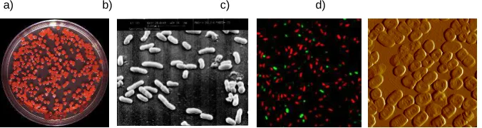
malonate. They have the capacity to reduce nitrates and ferment carbohydrates as maltose, D-mannose, salicin, sucrose and trehalose [8]. Figure 2 shows different images of S. marcescens from culture in Petri dish, scanning electron microscope, confocal microscope (dead-alive) and atomic force microscopy (AFM).
a) b) c) d)
Fig. 2. a) S. marcescens pigmented colonies growth in TSA. b) S. marcescens obtained by
Scanning Electron Microscope. c) S. marcescens from confocal microscope after staining
with LIFE/DEAD stain and d) S. marcescens obtained by AFM.
Serratia species occupy different habitats and are mainly isolated from
small mammals, water, plants and hospitalized patients.
Water is probably the principal habitat of S. plymuthica. However, many other species have been isolated from water. The 75% of 150 Serratia strains isolated from river water belonged to S. marcescens (F. Agbalika, F. Grimont, and P.D.A. Grimont, unpublished observations). In soil, S.
[image:49.499.85.423.362.452.2]mineralizing organic iron and dissolving gold and copper. Most of the strains isolated from soil and water are prodigiosin producers. This pigment is toxic to protozoa, so this may be an ecological advantage for them.
Plants are also a usual habitat for Serratia strains. S. ficaria is mainly find in figs, and S. rubidaea in coconuts. Serratia species were frequently found in plants (Grimont et al., 1981). Vegetables, mushrooms, mosses and wet plants are the most important habitats where Serratia is found. It is sometimes found in grasses and less in trees and shrubs. The most common Serratia strains isolated from plants other than figs and coconuts are S. liquefaciens and S. proteamaculans. S. marcescens is rarely isolated from plants.
Insects are mainly associated with S. marcescens, S. proteamaculans and S. liquefaciens, which are considered potential insect pathogens. The rarity of S. rubidaea in insects might be explained by its inability to produce chitinase, a virulence factor for insect-associated Serratia species.
Serratia strains can be isolated from many vertebrates as caws, turtles,
birds and wild rodents. Sometimes strains have been recovered from healthy vertebrates, but usually Serratia has been associated with chronic infections. For instance, red-pigmented strains are responsible for 0.2-1.5% of mastitis in cows. Serratia is not usually isolated from healthy people, whereas hospitalized patients are frequently colonized or infected. The opportunistic pathogen S. marcescens has been related to many infections. This fact will be widely described in subsequent sections [7].
1.2.3. TAXONOMY
The number of species that belong to the genus Serratia has increased from five species mentioned in the first edition of The prokaryotes to seven species described in Bergey´s Manual of Systematic Bacteriology until ten species known at present. The Serratia species are: S.
plymuthica, S. rubidaea, S. odorifera, S. ficaria, S. entomophila and S. fonticola.
1.2.4. PATHOGENICITY FACTORS
S. marcescens is an opportunistic pathogen that causes infections in
immunocompromised patients. The possible pathogenicity factors found in
Serratia are described below:
Fimbriae: are proteinaceous appendages that are thinner and shorter
than flagellum. Five types of fimbriae have been observed in Serratiae, and each strain can produce between one and three kinds of fimbrial hemaglutinin. The type 1 fimbriae produce mannose-sensitive hemagglutinin (MS-HA) and was found to be produced by all or almost all strains of S. marcescens whether environmental or clinical and also in some strains of other Serratia species. The production of MS-HA has been correlated with the ability of S. marcescens to attach to human bucal epithelial cells or to the human urinary bladder surface.
The type 3 fimbriae are associated with mannose-resistant hemagglutinin (MR/K-HA) and are produced by almost all Serratia strains. However, this type of fimbriae is more frequently produced by clinical strains than environmental strains. The type FGH MR/PHA fimbriae produced by strains of all species except S. plymuthyca and S. fonticola are associated with mannose-resistant hemaglutinin and these fimbriae are immunologically related in the different species [9].
The type F MR/P-HA fimbriae produced by some S. rubidaea are associated with mannose-resistant hemagglutinin and are immunologically unrelated with other hemagglutinins.
The type F MR/P-HA fimbriae produced by some S. fonticola strains are also associated with mannose-resistant hemagglutinin.
Siderophores: Siderophores are produced by clinical and environmental
S. marcescens strains. Serratia strains generally produce enterobactin but
O antigens: Although O-antigens cannot be considered truly
pathogenicity factors, they are located in LPS surface, and it is known that O6 and O14 serotypes of S. marcescens are predominant in infections.
Extracellular enzymes: Enzymes that play a prominent role in the
pathogenesis of experimental pneumonia and keratitis.
Plasmids: They probably play no rule in the virulence in experimental
models, but multiple drug resistance may affect the course and prognosis of infections. Non-pigmented strains of S. marcescens use to be more resistant because they often harbour resistance plasmids.
Relatively little is known about factors that contribute to S. marcescens pathogenesis within its host. However, some virulence factors of the human opportunistic S. marcescens have been recently identified by in
vivo screening [7, 10].
1.3. THE GENUS Pseudomonas
Pseudomonas spp. are aerobic, non-spore-forming Gram-negative bacilli,
which are usually motile by one or several polar flagella.
Pseudomonas is a genus including species that can use a wide range of
organic and inorganic compounds and can live under diverse environmental conditions. For that reason, they are ubiquitous in soil and water, plants, animals and humans (some isolates may infect immunocompromised patients).
This genus is well known for its metabolic and genetic plasticity. They generally grow rapidly and are able to metabolize a huge number of substrates including toxic organic chemicals, such as aliphatic and aromatic hydrocarbons [11].
They possess a strictly respiratory metabolism with oxygen as the terminal electron acceptor; but they are capable grow anaerobically due to its ability to use nitrate, an alternative electron acceptor [12].
One important consequence of its metabolic diversity is that
Pseudomonas spp., in particular P. aeruginosa, is common throughout the
This characteristic certainly contributes to the opportunistic nature of opportunistic infections [13].
Pseudomonas species, particularly the human pathogen P. aeruginosa,
not often colonize healthy humans. The throat, intact skin or stools of healthy individuals are densely colonized by normal flora, which do not include Pseudomonas species. Nevertheless, individuals that often received antibiotic therapies are at risk of gastrointestinal colonization with
P. aeruginosa and consequently, due to this reservoir the patient can
suffer septicemia caused by this microorganism. Patients undergoing anti-cancer chemotherapy or marrow ablation for bone marrow transplantation and patients with cystic fibrosis or receiving mechanical ventilation have greater risk to develop serious Pseudomonas infections [12].
1.3.1. TAXONOMY
Pseudomonas is a genus that was first proposed by Migula in 1894. It has
been subjected to many taxonomic revisions and identification methodologies. In the early 1970s this genus was classified into five unrelated groups according to RNA-DNA hibridization studies: I,
Proteobacteria; II, Burkholderia species; III, Comamonas, Acidovorax, and Hydrogenophaga genera; IV, Brevundimonas species; and V,
Stenotrophomonas and Xanthomonas genera. Currently, there are about
160 species within Pseudomonas genus, of which few species have clinical significance.
1.3.2. PATHOGENESIS
P. aeruginosa is the most important causative agent of opportunistic
infections among the genus Pseudomonas; this is the reason why we are basically going to focus on the pathogenesis of this particular microorganism [12,14]. It can cause both acute and chronic infections.
P. aeruginosa is now recognized as a common source of many
immunodeficiency, combined with a high incidence of antibiotic resistant strains, makes treatment of P. aeruginosa a serious medical challenge. There are many factors involved in the pathogenesis of P. aeruginosa. Thus, the virulence of this opportunistic pathogen cannot be attributed to a single factor; it has to be considered multifactorial. Some of the factors relevant in Pseudomonas pathogenesis are adhesins, endotoxins, proteases, hemolysins and a type III secretion system.
The virulence determinants with known roles in pathogenesis are summarized below:
Adherence: The two important types of adhesins used by P. aeruginosa
to colonize host tissues are fimbrial adhesins, such as type IV pili and type I fimbriae, and non-fimbrial adhesins, including lipopolysaccharide, flagella, outer membrane proteins and alginate.
Exotoxin A: It is the most potent exotoxin produced by almost all clinical
P. aeruginosa isolates.
Proteases and phospolipases: P. aeruginosa produces several
proteases including LasB elastase, LasA elastase, and alkaline protease. Las A, Las B and alkaline protease are associated with tissue damage in
P. aeruginosa infections. Besides, it produces two homologous
extracellular phospholipases, the hemolytic PlcH and the nonhemolytic PlcN, which has been demonstrated to be a virulence factor in a variety of
P. aeruginosa infections.
Type III secretion: This system is a major determinant of virulence in P.
aeruginosa. It is used to deliver four toxins that act in concert to inhibit
phagocytosis and promote tissue destruction. It is expressed in response to a variety of environmental signals (low Ca2+ concentrations, serum and contact with eukaryotic cell surfaces).
Quorum sensing: las and rhl are the two quorum sensing systems used
Biofilms: P. aeruginosa biofilms (surface-associated bacterial communities encased within a polymeric matrix) are mainly formed on the surfaces of medical devices, and can contribute to chronic infections. Moreover, inherent resistance of biofilms makes them difficult to treat and eliminate.
1.4. ANTIMICROBIAL AGENTS AND BACTERIAL RESISTANCE
1.4.1. ß-LACTAMS
ß-lactams are a group of antibiotics that contain a four-atom ring (ß-lactam) that determine their most relevant characteristics i.e. mechanism of action (inhibition and synthesis of the wall), main mechanisms of resistance (ß-lactamases) and low toxicity (strict inhibition of wall biosynthesis, since the structure do not exist in eukaryote cells). ß-lactam antibiotics include penicillins, cephalosporins, carbapenems and monobactams.
Since 1920, when penicillin was discovered, ß-lactams have been successfully used in the treatment of human infectious diseases. Due to their spectra of activity, effectiveness, low toxicity and wide therapeutic margin, they are the most used antimicrobial agents in the community, as well as in hospitals.
1.4.1.1. PENICILLINS
1.4.1.1.1. CLASSIFICATION AND STRUCTURE
Penicillins are natural or semisynthetic antibiotics containing the chemical nucleus 6-aminopenicillanic acid. The penicillins differ from one another in the substitution at position 6.
[image:55.499.178.322.538.610.2]1.4.1.1.2. TARGETS AND MECHANISM OF ACTION
Penicillins have the ability to inhibit a number of bacterial enzymes, specifically penicillin-binding proteins (PBPs) that are essential for peptidoglycan synthesis.
PBPs have enzymatic activity as carboxypeptidases and transpeptidases being directly responsible of the incorporation of new subunits into the growing peptidoglycan. Activity of penicillins is usually related to their ability to destroy bacterial wall by the triggering of membrane-associated autolytic enzymes. They can also inhibit endopeptidase and glycosidase enzymes, which are relevant in bacterial growth.
1.4.1.1.3. SPECTRUM OF ACTIVITY
Penicillins are active against most positive and many Gram-negative and on both aerobic and anaerobic organisms. Penicillin G is not active against enterococci, members of the family Enterobacteriaceae,
Pseudomonas spp. or members of the Bacteroides fragilis group.
Ampicillin and amoxicillin have similar spectra of activities than Penicillin G, and they are degraded by ß-lactamase and are inactive against many
Enterobacteriaceae and P. aeruginosa. Carboxypenicillins and ureidopenicillins are more stable against hydrolysis by the ß-lactamases of Enterobacteriaceae and P. aeruginosa.
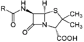
1.4.1.2. CEPHALOSPORINS
1.4.1.2.1. CLASSIFICATION AND STRUCTURE
Cephalosporins belong to ß-lactam antibiotics and originally derived from
Acremonium. They contain a 7-aminocephalosporanic acid nucleus, which
Cephalosporins are classified in generations. The evolution of the generations is related with an increment of their spectrum of activity and intrinsic activity against Gram-negative organisms.
Figure 4 shows the chemical structure of cephalosporins.
Fig.4. Cephalosporin core structure.
[image:57.499.80.420.330.521.2]Classification of cephalosporins is shown in table 1.
Table 1. Cephalosphorins classification
First generation (narrow-spectrum) Second generation (expanded spectrum) Third generation (broad-spetrum) Fourth generation (extended spectrum) Cefadroxil Cefazolin Cephalexin Cephaloridine Cephalotin Cephapirin Cephradine Cefaclor Cefamandole Cefonicid Ceforanide Cefuroxime Cefprozil Loracarbe Cefmetazole Cefotetan Cefoxitin Cefdinir Cefditoren Cefixime Cefoperazone Cefotaxime Cefpodoxime Ceftazidime Ceftibuten Ceftizoxime Ceftriaxone Cefepime Cefpirome
1.4.1.2.2. TARGETS AND MECHANISM OF ACTION
bactericidal effects by triggering autolytic enzymes in the envelope of bacteria.
1.4.1.2.3. SPECTRUM OF ACTIVITY
First generation cephalosporins (narrow spectrum) have good activity against positive organisms and modest activity against Gram-negative organisms.
Second generation cephalosporins (expanded spectrum) are more active against Gram-negative bacteria than first generation cephalosporins because of their stability to ß-lactamases, and less active against Gram-positive organisms. The activity also depends on the concrete antibiotic used.
Third generation cephalosporins (broad spectrum) are generally less active against Gram-positive cocci than the narrow spectrum agents, but they are more active against the Enterobacteriaceae and P. aeruginosa. Four generation cephalosporins (extended spectrum) are active against stably derepressed class I ß-lactamase mutants of Enterobacteriaceae and P. aeruginosa. Besides, these antibiotics penetrate well through the outer membrane of Gram-negative bacteria but they are inactive against enterococci or anaerobes.
1.4.1.3. MONOBACTAMS AND CARBAPENEMS
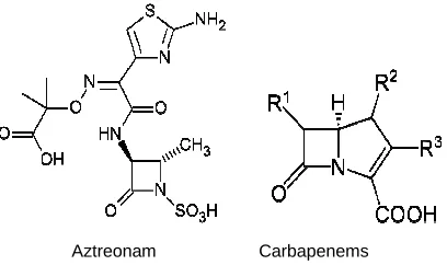
1.4.1.3.1. CLASSIFICATION AND STRUCTURE
Monobactams, such as aztreonam, are β-lactam compounds wherein the
β-lactam ring is alone and not fused to another ring.
[image:59.499.149.353.71.192.2]
Aztreonam Carbapenems
Fig. 5. Chemical structure of aztreonam and carbapenems.
1.4.1.3.2. TARGETS AND MECHANISM OF ACTION
Aztreozam binds to the PBP 3 of Gram-negative aerobes disrupting bacterial wall synthesis. It does not induce production of enzymes and it is not hydrolyzed by most common ß-lactamases.
Carbapenems bind to PBP 1 and PBP 2 of negative and Gram-positive bacteria, elongating the cell and causing lysis.
1.4.1.3.3. SPECTRUM OF ACTIVITY
Aztreonam has significant activity against Enterobacter spp. and S.
marcescens.
Carbapenems are active against aerobic Gram-positive species. More than 90% of Enterobacteriaceae are generally susceptible to carbapenems, including S. marcescens. Ertapenem is inactive against
Pseudomonas and Acinetobacter. Carbapenems are the most potent
ß-lactams against anaerobes. At present, there are some P. aeruginosa strains resistant to imipenem.
1.4.1.3.4. RESISTANCE TO ß-LACTAM ANTIBIOTICS
1. Penicillin-binding protein-mediated resistance
This kind of resistance takes several forms described below:
2. ß-lactamase-mediated resistance
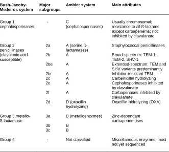
[image:60.499.86.417.157.454.2]There are two ß-lactamase classification systems currently used that are compared in table 2.
Table 2. ß-lactamase classification
Bush-Jacoby-Medeiros system
Major subgroups
Ambler system Main attributes
Group 1 cephalosporinases Group 2 penicillinases (clavulanic acid susceptible)
Group 3 metallo-ß-lactamase Group 4 - 2a 2b 2be 2br 2c 2e 2f 2d 3a 3b 3c - C (cephalosporinases)
A (serine ß-lactamases) A A A A A A D (oxacillin hydrolyzing) B (metalloenzymes) B B Not classified Usually chromosomal; resistance to all ß-lactams except carbapenems; not inhibited by clavulanate
Staphylococcal penicillinases
Broad-spectrum: TEM-1, TEM-2, SHV-1
Extended-spectrum: TEM and SHV variants predominantly Inhibitor-resistant TEM Carbenicillin hydrolyzing Cephalosporinases inhibited by clavulanate
Carbapenases inhibited by clavulanate
Oxacillin-hidrolyzing (OXA)
Zinc-dependant carbapenemases
Miscellaneous enzymes, most not yet sequenced
ß-lactamase mechanism: This mechanism has been studied in TEM-1 and SHV-2 Class A ß-lactamase. Disruption of the amide bond is done in a two-step reaction where the negatively charged carboxylate group of the ß-lactam antibiotic is attracted to the active site by the enzymes positively charged residues and then, ß-lactam becomes acylated.
Integrons are genetic elements of variable lengths that contain a 5’conserved integrase gene (int), mobile antibiotic resistance genes (cassettes), and an integration site for the gene cassette, attI (att, attachment). Class 1, 2 and 3 integrons are common in Gram-negative bacteria. Integrons capture antibiotic resistance gene cassettes by using a site-specific recombination mechanism. They act both as natural cloning systems and as expression vectors [15]. The capture and spread of antibiotic resistance determinants by integrons underlie the rapid evolution of multiple-antibiotic resistance in Gram-negative clinical isolates [16]. Integrons carrying ß-lactamases have been found in most Gram-negative bacteria.
ß-lactamases classes:
Class A ß-lactamases (Group 2b Penicillinases): The two commonly
class A ß-lactamases found in Enterobacteriaceae are TEM-1 and SHV-1, which are primarily penicillinases with diminished activity against cephalosporin substrates. They are the progenitors of the ESBLs and the inhibitor-resistant TEM ß-lactamases now common in many hospitals [12].
Bush group 2be (ESBLs): Extended-spectrum cephalosporins (a group
of potent ß-lactams) are poor substrates for hydrolysis by group 2b enzymes. Mutations at critical amino acids resulted in an expansion of the spectra of these enzymes enabling them to hydrolyze extended-spectrum cephalosporins. The number of enteric Gram-negative rods possessing ESBLs has increased recently and this has obliged to modify the choice of antimicrobial therapies.
Non-TEM, non-SVH ESBLs: CTX-M ß-lactamases commonly found in
Klebsiella pneumoniae, Escherichia coli, Salmonella spp., Shigella spp., Citrobacter freundii, Enterobacter spp., and S. marcescens are the most
Inhibitor-resistant class A ß-lactamases (Bush group 2br): This group
is formed by amino acid mutated TEM and SHV conferring resistance to inhibition by ß-lactamases inhibitors.
Class B lactamases (Bush group 3 enzymes): This class of
ß-lactamases is formed by metallo-enzymes that require zinc or another heavy metal for catalysis; their activities are inhibited by chelating agents such as EDTA. These ß-lactamases confer resistance to a wide variety of ß-lactam compounds, including carbapenems and cephamycins. They are resistant to inactivation by clavulanate, sulbactam and tazobactam. Some of them hydrolyse aztreonam. The majority of ß-lactamases are chromosomally encoded, and their expression may be constitutive or inducible. VIM and IMP class B lactamases are active against most ß-lactams and have been found in various Gram-negative clinical isolates. The majority of these enzymes are mobilized on integrons, transposons and mobile common regions.
Class C ß-lactamases (Bush-Jacoby-Medeiros group 1): ß-lactamases
chromosomally encoded by almost all Gram-negative bacteria except
Salmonella and Klebsiella. These enzymes are important in
Gram-negative clinical isolates, including S. marcescens. Class C ß-lactamases hydrolyze cephalosporins and penicillins. Most of them are resistant to inhibition by clavulanate, sulbactam and tazobactam. Some class C ß-lactamases of Gram-negative bacteria are plasmid mediated.
Class D lactamases (oxacillin-hydrolyzing): OXA enzymes
1.4.2. AMINOGLYCOSIDES
1.4.2.1. CLASSIFICATION AND STRUCTURE
Streptomycin was the first aminoglycoside introduced in 1944 for the treatment of serious Gram-negative infections. Structurally, each of this aminoglycosides contains two or more amino sugars linked by glycosidic bonds to an aminocyclitol ring nucleus.
There are two different groups of aminoglycosides. Streptomycin, neomycin, kanamycin, tobramycin and gentamicin belong to the group of natural aminoglycosides, and amikacin and netilmicin belong to the group of semisynthetic aminoglycosides (Figures 6 and 7).
Streptomycin Neomycin
Kanamycin
[image:63.499.126.366.281.560.2]Tobramycin Gentamicin
[image:64.499.138.358.78.200.2]
Amikacin Netilmicin Fig. 7. Chemical structures of semisynthetic aminoglycosides.
1.4.2.2. TARGETS AND MECHANISM OF ACTION
These bacterial agents inhibit bacterial protein synthesis by binding reversibly to the bacterial 30S ribosomal subunit preventing mRNA translation during protein synthesis and leading bacteria to death. Bacterial uptake of these agents is facilitated by ß-lactams and vancomycin, which inhibit bacterial wall biosynthesis.
1.4.2.3. SPECTRUM OF ACTIVITY
Aminoglycosides are generally active against aerobic Gram-negative rods (Enterobacteriaceae, P. aeruginosa and Acinetobacter spp.) and
Staphylococcus aureus (not recommended as single agents). They are
not active against anaerobes.
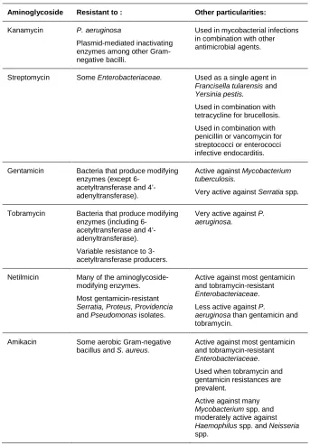
Table 3. Differences in antimicrobial spectra among aminoglycosides.
Aminoglycoside Resistant to : Other particularities:
Kanamycin P. aeruginosa
Plasmid-mediated inactivating enzymes among other Gram-negative bacilli.
Used in mycobacterial infections in combination with other antimicrobial agents.
Streptomycin Some Enterobacteriaceae. Used as a single agent in
Francisella tularensis and Yersinia pestis.
Used in combination with tetracycline for brucellosis.
Used in combination with penicillin or vancomycin for streptococci or enterococci infective endocarditis.
Gentamicin Bacteria that produce modifying
enzymes (except 6-acetyltransferase and 4’-adenyltransferase).
Active against Mycobacterium
tuberculosis.
Very active against Serratia spp.
Tobramycin Bacteria that produce modifying
enzymes (including 6-acetyltransferase and 4’-adenyltransferase).
Variable resistance to 3-acetyltransferase producers.
Very active against P.
aeruginosa.
Netilmicin Many of the
aminoglycoside-modifying enzymes.
Most gentamicin-resistant
Serratia, Proteus, Providencia
and Pseudomonas isolates.
Active against most gentamicin and tobramycin-resistant
Enterobacteriaceae.
Less active against P.
aeruginosa than gentamicin and
tobramycin.
Amikacin Some aerobic Gram-negative
bacillus and S. aureus.
Active against most gentamicin and tobramycin-resistant
Enterobacteriaceae.
Used when tobramycin and gentamicin resistances are prevalent.
Active against many
Mycobacterium spp. and
moderately active against
Haemophilus spp. and Neisseria
1.4.2.4. RESISTANCE TO AMINOGLYCOSIDES
There are three known mechanisms of resistance to aminoglycosides: 1. Reduction of intracellular aminoglycosides accumulation due to outer membrane permeability alteration, inner membrane transport decrease or active efflux increase.
2. Modification of the target site by mutation in the ribosomal proteins or 16S RNA.
3. Enzymatic modification of the drug.
All S. marcescens produce chromosomal AAC(6’)-Ic enzyme that may slightly affect the activity of all aminoglycosides except streptomycin and gentamicin.
Gentamicin is the most active aminoglycoside against Serratia. However, other aminoglycosides can be used when isolates do not produce aminoglycoside-modifying enzymes.
Resistance to aminoglycosides in P. aeruginosa usually results from drug inactivation by plasmid or chromosome-encoded enzymes harbored by resistant strains.
Enzyme-independent resistance can be due to defects in uptake/accumulation of the drug [17].
1.4.3. QUINOLONES
1.4.3.1. CLASSIFICATION AND STRUCTURE
The era of quinolones began in the early 1960s when Lesher and associates discovered nalidixic acid during the synthesis of the antimalarial agent chloroquine [18,19].
Table 4. Classification of quinolones by generations
First generation
Second generation
Third Generation
Four generation
Five generation
Nalidixic acid
Pipemidic acid
Cinoxacin
Flumequine
Ofloxacin
Enoxacin
Lomefloxacin
Ciprofloxacin
Pefloxacin
Levofloxacin
Sparfloxacin
Grepafloxacin
Gatifloxacin
Trovafloxacin
Moxifloxacin
Gemifloxacin
Sitafloxacin
Clinafloxacin
Garenofloxacin
DX-619, 771, WCK-1153,DW-286
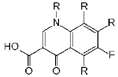
[image:67.499.78.422.94.224.2]The chemical structure of fluoroquinolones (Fig. 8) is based on the 1,4-dihydro-4-oxo-pyridine molecule, which has a carboxylic acid substituent at position 3. This substituent with the carbonyl group at position 4 appears to be essential for the activity of these antimicrobial agents. First generation quinolones (except flumequine) lack the fluorine atom at position 6. The second-generation quinolones have a cyclic diamine at the position 7 and fluorine atom at position 6 in the quinolone nucleus, and the third generation differs from the previous generation in the substituents located at positions 1, 7 and 8 of the quinolone nucleus [17, 20].
[image:67.499.188.310.426.506.2]1.4.3.2. TARGETS AND MECHANISM OF ACTION
DNA gyrase and topoisomerase IV are the protein targets of quinolones. On the one hand, DNA gyrase is a tetrameric enzyme with two A subunits and two B subunits that catalyzes the negative supercoiling of DNA. The energy required for this process is obtained from the hydrolysis of ATP. DNA gyrase is known to play a significant role in both transcription and replication of DNA. On the other hand, topoisomerase IV is a tetrameric enzyme with two A subunits and two B subunits encoded by the ParC and
ParE genes. It seems to be involved in decatenating daughter replicons
following DNA replication.
At present, the mechanism of action of quinolones is not fully understood. There are some controversies, despite the fact that the mechanism of action has been carefully studied. It is accepted that quinolones have two binding domains, one interacting with DNA and the other with DNA gyrase. Complex formation of the DNA gyrase-quinolone-DNA has been associated to inhibition of DNA replication.
1.4.3.3. SPECTRUM OF ACTIVITY
First generation quinolones are substantially less potent in vitro and have narrower antibacterial spectra than newer generations, having enhanced activities against Gram-negative bacilli, except P. aeruginosa. They lack activity against Gram-positive cocci and anaerobes. These quinolones reach high concentrations in the urinary tract and subsequently they are widely used to treat urinary tract infections [17, 21].
The third generation quinolones have improved activity against Gram-positive cocci and anaerobic bacteria. They are mostly used for the treatment of chronic bronchitis, acute sinusitis and community pneumonia. The fourth generation quinolones have similar spectrum of activity than third generation quinolones. They exhibit activity against anaerobes. They are used for the treatment of nosocomial pneumonia, intraabdominal and gynecologic infections and the same infections to which the previous generations are used but urinary tract infections.
Finally, some of the fifth generation quinolones have antimicrobial activity against the same microorganisms than those of fourth generation, whereas others are specifically designed to fight against infections caused by multidrug-resistant bacteria [20].
1.4.3.4. RESISTANCE TO QUINOLONES
Both chromosomal and plasmid-mediated quinolone resistance have been described. Chromosomal mutations comprise mutations in the topoisomerase genes (gyrA, gyrB, parC and ParE) and mutations that produce a reduction in drug accumulation (decrease uptake or increase efflux).
Table 5. Relevant factors favoring emergence of resistance to quinolones.
Factors dependent on the:
Drug Bacterium Host and others
Concentration of the drug achieved in the infection site or epithelium.
Time of exposure (concentration of the drug above the MIC).
Mutagenesis
Higher inoculum increase probability of spontaneous mutations.
Quorum sensing. (Control of virulence factors expression, the entry into stationary phase, the conjugal factor of transfer DNA, and spore formation and transformation competence).
Hypermutability.
Capability and capacity to form biofilm. (Bacterial density determines the gradients of nutrients and oxygen availability within the biofilm structure; bacteria located in deeper parts of biofilm have worst access to nutrients and oxygen; bacteria located in zones of the biofilm with high metabolic activity and oxygen concentration are better killed by antimicrobial agents).
Facility to acquire quinolone resistance. (Slow or no growth bacteria have increased resistance than exponential ones).
Pharmacokinetics of the drug at the infection or colonization sites.
Immune status.
Environment at the site infection such as pH, oxygen, nutrients, etc.
Many authors have shown that in S. marcescens, like in other
Enterobacteriaceae (but E.coli) a single substitution in the
quinolone-resistance determining region (QRDR) is enough to produce high level resistance. However, higher levels of fluoroquinolone resistance can be achieved when there is cooperation between QRDR alterations and low permeability (or active extrusion) limiting the intracellular accumulation of the drug [22].