Hyaluronic acid-coated gold nanoparticles as an anticancer drug delivery system - Biological characterization and efficacy
199
0
0
Texto completo
(2) UNIVERSITAT DE BARCELONA FACULTAT DE FARMÀCIA DEPARTAMENT DE FARMÀCIA I TECNOLOGIA FARMACÈUTICA PROGRAMA DE DOCTORAT: NANOCIÈNCIES. HYALURONIC ACID-COATED GOLD NANOPARTICLES AS AN ANTICANCER DRUG DELIVERY SYSTEM – BIOLOGICAL CHARACTERIZATION AND EFFICACY. Memòria presentada per Hanna Parkkola per optar al títol de doctor per la Universitat de Barcelona. Directora: Judith Sendra Cuadal. Doctoranda: Hanna Parkkola. Tutora: Ana Calpena Campmany. Hanna Parkkola Barcelona, 2014.
(3)
(4) UNIVERSITAT DE BARCELONA FACULTAT DE FARMÀCIA DEPARTAMENT DE FARMÀCIA I TECNOLOGIA FARMACÈUTICA. HYALURONIC ACID-COATED GOLD NANOPARTICLES AS AN ANTICANCER DRUG DELIVERY SYSTEM – BIOLOGICAL CHARACTERIZATION AND EFFICACY. Hanna Parkkola 2014.
(5)
(6) To my parents. To Tuomas. To Joan.
(7)
(8) ACKNOWLEDGEMENTS. I would like to thank the personnel of Endor Nanotechnologies for the opportunity to realize this doctoral thesis. Especially the CEO and founder of the company, Joaquin Querol, and the director of this thesis, Dr. Judith Sendra, from whom I have learned discipline, to pay plenty of attention on the smallest possible details and to think about EDS even in my sleep. I also want to thank Júlian Peña, for his professional advice and sharing the same interest in music, and Luciano Sobrevals and Rafa Miñana for their friendship, for teaching me so much about the flora and the fauna and for sharing the cell culture hood during the long hours of time consuming experiments. Very special thanks to Laura Vivero, for sitting next to me every day during almost five years, I appreciate your help, patience and friendship deeply, thank you for sharing this part of my life with me.. During this project we have collaborated with various entities, including Leitat Technological Centre, PCB Toxicology Unit and CIBBIM Nanomedicine, which deserve my gratitude. Thank you for your professionalism, advice and sharing your knowledge.. Thank you also all my “new” friends that I have met in Barcelona for sharing amazing experiences with me, for showing me new places and for making me feel like home. You are too numerous to mention one by one, and I don’t want to leave anyone out, but you know who you are!. Suvi, Katrin, Natalia, Michela, Dr. Katrin and Antonija, I want to express my appreciation for the coffees, lunches, pints of wine and all the precious moments that our group of “guiris” has shared. Thank you also Adam and Erik.. Thank you my friends back in Finland: my “business partners” Marja, Heli and Mari, and my loyal companions Veera, Reeta, Iida and Janika, I miss you very dearly and value you for receiving me with opens arms even after long silences..
(9) I want to thank Montse and Jaume for welcoming me warmly into their family and further making me feel like home in Barcelona.. Finally, a big thank you to my family, to my brother Tuomas for putting up with me and my sunny personality all these years, for our long talks and for listening to me when I’ve been having difficult times, and to my parents Riitta and Kai for providing me such a wonderful start for life, for believing in me and supporting me in every single step of my life. You made me the person I am. Thank you Joan for sharing my ups and downs, for teaching me to appreciate new places and experiences, for everything we have shared and for showing me what happiness is.. Kiitos.
(10) The experimental work of this thesis has been executed in the laboratory of Endor Nanotechnologies, S.L. in Parc Científic de Barcelona (C/ Baldiri Reixac 15; 08028 Barcelona).. The work has been financed by Ajuts Talent Empresa (TEM DGR: 2009 TEM 00023), Agència de Gestió d’Ajuts Universitaris i de Recerca (AGAUR: Generalitat de Catalunya)..
(11)
(12) PUBLICATIONS. Parkkola H., Vivero L., Ramis M., Querol J., Sendra J. 2012. Proceed with caution? Concept and application of the precautionary principle in nanobiotechnology: Gold Nanoparticles and Cell Viability Evaluation. Editors: Ach J.S., Lüttenberg B., Paslack R., Weltring K-M. LIT Verlag Vivero L., Parkkola H., Sendra J., Querol J., Cambero J.P.G., Ramis M. 2012. Proceed with caution? Concept and application of the precautionary principle in nanobiotechnology: Gold Nanoparticle Stability Studies in Physiological Media. Editors: Ach J.S., Lüttenberg B., Paslack R., Weltring K-M. LIT Verlag Sobrevals L., Parkkola H., Vivero L., Miñana R., Sendra J. 2014. Nanotechnology: Recent Trends, Emerging Issues and Future Directions. Chapter: Nanomedicine: The New Age of Therapeutics. Editor: Islam N. Nova Science Publishers, Inc. Posters:. Parkkola H., Vivero L., Vega M., Ramis M., Querol J., Sendra J. 2010. Metallic Nanoparticles and Cytotoxicity Assessment. International Congress of Toxicology. Patents:. Ramis Castelltort M., Querol Sastre J., Vivero Sanchez L., Sendra Cuadal J., Parkkola H. System for the release of a therapeutic agent, pharmaceutical composition containing it, the preparation and medical use thereof. International Publication number WO 2012/089768A1. Applicant: Endor Nanotechnologies, S.L..
(13)
(14) Table of contents. ABBREVIATIONS ...................................................................................................... 7 ABSTRACT ............................................................................................................... 11 RESUM ...................................................................................................................... 12 1.. INTRODUCTION............................................................................................... 15 1.1. Cancer ........................................................................................................... 15. 1.1.1. Cancer – an introduction ...................................................................... 15. 1.1.2. Treatment ............................................................................................. 17 1.1.2.1 Cancer chemotherapy.......................................................... 17. 1.1.2.1.1. Platinum-based drugs.............................................................. 19. 1.1.2.2 Personalized medicine ........................................................ 20 1.1.3. Cancer biomarkers ............................................................................... 21 1.1.3.1 CD44 .................................................................................. 22. 1.1.3.1.1. CD44 structure ....................................................................... 22. 1.1.3.1.2. CD44 functions....................................................................... 24. 1.1.3.1.2.1. CD44 in cancer ................................................................... 26. 1.1.3.2 Hyaluronic acid .................................................................. 26 1.1.3.2.1. Structure and functions of HA ................................................ 27. 1.1.3.2.2. HA interactions with cell surface receptors ............................. 28. 1.1.3.2.3. HA degradation ...................................................................... 29. 1.1.3.2.4. HA in cancer........................................................................... 30. 1.1.3.2.5. HA signaling .......................................................................... 31. 1.1.3.3 HA-CD44-targeted therapies ............................................... 34 1.2. Nanomaterials in biomedicine ....................................................................... 36. 1.2.1. Cancer nanomedicine ........................................................................... 38 1.2.1.1 Imaging and drug delivery .................................................. 39 1.2.1.2 In vitro diagnostics ............................................................. 40 1.
(15) 1.2.1.3 Drugs and therapy ............................................................... 41 1.3. Gold nanoparticles......................................................................................... 42. 1.3.1. History of medicinal gold ..................................................................... 42. 1.3.2. AuNP synthesis .................................................................................... 43. 1.3.3. Properties of AuNPs ............................................................................. 44. 1.3.4. Biomedical AuNP-applications in cancer.............................................. 45 1.3.4.1 In vitro assays ..................................................................... 46 1.3.4.2 Imaging .............................................................................. 46 1.3.4.3 Therapy .............................................................................. 47 1.3.4.4 Drug delivery ...................................................................... 48. OBJECTIVES ............................................................................................................. 53 2.. MATERIALS AND METHODS ......................................................................... 57 2.1. Nanosystem synthesis .................................................................................... 58. 2.1.1. AuNP synthesis .................................................................................... 58. 2.1.2. AuNP conjugation ................................................................................ 59 2.1.2.1 HA thiolation ...................................................................... 59 2.1.2.2 AuNP-HA conjugation........................................................ 59 2.1.2.3 EDSfl synthesis ................................................................... 59 2.1.2.4 PEGNP synthesis ................................................................ 60. 2.1.3. EDS-CIS conjugation ........................................................................... 61 2.1.3.1 L1 ....................................................................................... 61 2.1.3.2 Mua .................................................................................... 62 2.1.3.3 Encapsulation ..................................................................... 62. 2.1.4 2.2. Purification and concentration of NM ................................................... 62. Chemical characterization of the nanosystems ............................................... 64. 2.2.1. TEM .................................................................................................... 64. 2.2.2. DLS ..................................................................................................... 64. 2.2.3. Z-potential............................................................................................ 65 2.
(16) 2.2.4. ICP-MS for Au & Pt quantification ...................................................... 65. 2.2.5. EA ....................................................................................................... 66. 2.2.6. Stability studies .................................................................................... 67 2.2.6.1 Visual examination ............................................................. 67 2.2.6.2 UV-VIS .............................................................................. 68. 2.3. In vitro-characterization of the nanosystems .................................................. 69. 2.3.1. Cell line characterization ...................................................................... 69 2.3.1.1 Cell culture ......................................................................... 69 2.3.1.2 Cell line characterization by qPCR...................................... 70 2.3.1.3 Cell line characterization by Western blot ........................... 71 2.3.1.4 Cell line characterization by flow cytometry ....................... 72. 2.3.2. Internalization studies........................................................................... 72 2.3.2.1 TEM for cells ...................................................................... 72 2.3.2.2 Immunofluorescence ........................................................... 74 2.3.2.3 Internalization by ICP-MS .................................................. 75 2.3.2.4 siRNA transfection and ICP-MS ......................................... 75. 2.3.3. Toxicity studies .................................................................................... 76 2.3.3.1 Cell viability assays & IC50................................................ 76 2.3.3.2 Ames test ............................................................................ 77 2.3.3.3 Comet assay ........................................................................ 80 2.3.3.4 ROS production .................................................................. 81. 2.3.4. Drug release ......................................................................................... 82. 2.3.5. Efficacy studies .................................................................................... 82 2.3.5.1 Colony formation assay ...................................................... 82. 2.4. In vivo-characterization of the nanosystems ................................................... 84. 2.4.1. Animals ................................................................................................ 84 2.4.1.1 Subcutaneous xenograft models .......................................... 85 2.4.1.2 EDS studies ........................................................................ 85. 3.
(17) 2.4.1.2.1. EDS blood circulation............................................................. 85. 2.4.1.2.2. EDS biodistribution ................................................................ 85. 2.4.1.2.3. Blood biochemical analysis after EDS administration ............. 86. 2.4.1.3 EDS-Drug studies ............................................................... 86 2.4.1.3.1. In vivo stability of double conjugated candidates .................... 86. 2.4.1.3.2. EDS-CIS biodistribution ......................................................... 87. 2.4.1.3.3. In vivo tumor growth .............................................................. 87. 2.4.2. Histology ............................................................................................. 88 2.4.2.1 Immunohistochemistry ....................................................... 88 2.4.2.2 H&E ................................................................................... 89 2.4.2.3 Silver staining ..................................................................... 89. 2.4.3 2.5. Animal weight ...................................................................................... 89. Statistical analysis ......................................................................................... 90. ANNEX: solutions ...................................................................................................... 91 3.. RESULTS ........................................................................................................... 99 3.1. Characterization of EDS .............................................................................. 100. 3.1.1. Chemical characterization .................................................................. 100 3.1.1.1 TEM ................................................................................. 100 3.1.1.2 DLS .................................................................................. 101 3.1.1.3 Z-potential ........................................................................ 102 3.1.1.4 ICP-MS ............................................................................ 102 3.1.1.5 EA .................................................................................... 103 3.1.1.6 Stability ............................................................................ 103. 3.1.1.6.1. Visual analysis ...................................................................... 103. 3.1.1.6.2. UV-VIS ................................................................................ 104. 3.1.2. Internalization of EDS ........................................................................ 106 3.1.2.1 CD44 expression in different cell lines .............................. 106 3.1.2.2 Internalization ................................................................... 108 4.
(18) 3.1.2.2.1. TEM ..................................................................................... 108. 3.1.2.2.2. Immunofluorescence............................................................. 109. 3.1.2.2.3. ICP-MS ................................................................................ 110. 3.1.3. Toxicity of EDS ................................................................................. 112 3.1.3.1 In vitro toxicity ................................................................. 113. 3.1.3.1.1. Cell viability ......................................................................... 113. 3.1.3.1.2. IC50 ..................................................................................... 114. 3.1.3.1.3. Ames test .............................................................................. 114. 3.1.3.1.4. Comet assay ......................................................................... 116. 3.1.3.1.5. ROS production ................................................................... 117. 3.1.3.2 In vivo toxicity .................................................................. 119. 3.2. 3.1.3.2.1. CD44 expression in Panc-1 xenografts.................................. 120. 3.1.3.2.2. Blood circulation .................................................................. 122. 3.1.3.2.3. Biodistribution ...................................................................... 122. 3.1.3.2.4. Animal weight ...................................................................... 124. 3.1.3.2.5. Histology: tissue morphology (H&E and silver staining) ...... 124. 3.1.3.2.6. Blood biochemical analysis .................................................. 126. Characterization of EDS-Drug ..................................................................... 128. 3.2.1. Drug conjugation strategies ................................................................ 128 3.2.1.1 ICP-MS ............................................................................ 128 3.2.1.2 Biological characterization of double conjugates ............... 129. 3.2.1.2.1. In vitro characterization ........................................................ 129. 3.2.1.2.2. In vivo characterization ......................................................... 130. 3.2.2. Chemical characterization of EDS-CIS ............................................... 132 3.2.2.1 TEM ................................................................................. 132 3.2.2.2 UV-VIS ............................................................................ 132 3.2.2.3 DLS .................................................................................. 133 3.2.2.4 Z-potential ........................................................................ 134 5.
(19) 3.2.2.5 ICP-MS & EA .................................................................. 134 3.2.3. Stability of EDS-CIS .......................................................................... 135 3.2.3.1 In vitro drug release .......................................................... 135 3.2.3.2 In vivo stability ................................................................. 136. 3.2.4. Internalization of EDS-CIS................................................................. 137. 3.2.5. Efficacy & toxicity of EDS-CIS ......................................................... 139 3.2.5.1 Colony formation assay .................................................... 139 3.2.5.2 Cell viability & IC50 ........................................................ 140 3.2.5.3 EDS-CIS biodistribution ................................................... 141 3.2.5.4 In vivo antitumoral efficacy............................................... 142 3.2.5.5 Drug accumulation in tumor ............................................. 144 3.2.5.6 Au & Pt accumulation in tumor and liver .......................... 145 3.2.5.7 Liver histology.................................................................. 146. 4.. 5.. DISCUSSION ................................................................................................... 151 4.1. Chemical characterization ........................................................................... 153. 4.2. Stability....................................................................................................... 156. 4.3. Internalization ............................................................................................. 158. 4.4. Efficacy ....................................................................................................... 160. 4.5. Toxicity....................................................................................................... 162. 4.6. Future prospects .......................................................................................... 165. CONCLUSIONS ............................................................................................... 169. REFERENCES ......................................................................................................... 173. 6.
(20) ABBREVIATIONS. AuNP. Gold nanoparticle. CD44. Principal receptor for hyaluronic acid. CFA. Colony formation assay. CIS. Cisplatin. CSC. Cancer stem cell. DLS. Dynamic light scattering. EA. Elemental analysis. ECM. Extracellular matrix. EDS. Endor Delivery System. EDS-drug. Endor Delivery System conjugated with an anticancer drug. EGFR. Epidermal growth factor receptor. EMT. Epithelial to mesenchymal transition. EPR. Enhanced permeability and retention. ErbB2. Human epidermal growth factor receptor 2. ERK. Extracellular signal-regulated kinase. ERM. ERM protein family: ezrin, radixin, moesin. ESF. European Science Foundation. FAK. Focal adhesion kinase. FDA. US Food and Drug Administration. HA. Hyaluronic acid/Hyaluronan. HARE. Hyaluronan receptor for endocytosis. HAS. Hyaluronan synthase. H&E. Hematoxylin & eosin. HER2. (see ErbB2). HEX. Hexosaminidase. HMW. High molecular weight. HYAL. Hyaluronidase. ICP-MS. Inductively coupled plasma mass spectrometry. ID. Initial dose. IF. Immunofluorescence. JNK. c-Jun N-terminal kinase 7.
(21) LMW. Low molecular weight. LYVE-1. Lymphatic vessel endothelial hyaluronan receptor 1. MAPK. Mitogen-activated protein kinase. MPS. Mononuclear phagocyte system. mTOR. Mechanistic target of rapamycin (serine/threonine kinase). NCL. Nanotechnology Characterization Laboratory. oHA. HA oligosaccharide. PDGF/PDGFR. Platelet-derived growth factor/receptor. PDT. Photodynamic therapy. PEG. Polyethylene glycol. PI3K. Phosphoinositide 3-kinase. PKC. Protein kinase C. Pt. Platinum. RHAMM. Receptor for hyaluronan-mediated motility. ROS. Reactive oxygen species. RTK. Receptor tyrosine kinase. SPR. Surface plasmon resonance. TEM. Transmission electron microscopy. TLR. Toll-like receptor. UV-VIS. Ultraviolet-visible spectroscopy. VEGF/VEGFR. Vascular endothelial growth factor/receptor. 8.
(22) ABSTRACT/RESUM. 9.
(23) 10.
(24) ABSTRACT. Novel stategies are needed to improve the limited efficacy of current cancer therapies. High expectations are directed towards nanotechnology-based applications in cancer medicine. We have developed a drug delivery system based on hyaluronic acid (HA) coated gold nanoparticles (AuNPs) that targets CD44, a cell surface glycoprotein overexpressed by various cancer cells. This nanocarrier (EDS: Endor Drug Delivery System) provides a platform for the conjugation of anticancer agents for a more efficient cancer treatment.. We show that the unique characteristics of AuNPs remain unchanged when the targeting ligand and a chemotherapeutic agent are conjugated. HA-coating stabilizes AuNPs and improves their biocompatibility. EDS is shown to target CD44 and is internalized through this receptor. The nanocarrier does not cause toxic effects in vitro or in vivo. After conjugating an anticancer agent (CIS: cisplatin) to the nanocarrier, EDS shows gradual drug release in physiological media due to changes in pH and hyaluronidase activity, and the drug reaches its target cells in its active form. In vivo efficacy of EDSCIS conjugate is higher than that of free cisplatin. However, more safety and toxicological studies must be conducted. New drug candidates are being tested in order to validate EDS as a platform for antitumoral drugs.. 11.
(25) RESUM. Els actuals tractaments oncòlogics presentan una eficàcia limitada que forcen la recerca de noves teràpies mes eficients. En aquest sentit, la nanotecnologia ha generat moltes expectatives. Durant aquesta tesi, hem desenvolupat un sistema d’alliberació de fàrmacs formats per nanopartícules d’or (AuNP) recobertes amb àcid hialurònic (HA), dirigit a CD44, una glicoproteïna que es sobreexpresa a la superfície cel.lular de diferents tipus de tumor. Aquest sistema (EDS: Endor Delivery System) proporciona una plataforma per a la conjugació de diferents fàrmacs antitumorals per tal de tractar el càncer d’una manera més eficient.. Aquesta tesi demostra que les propietats úniques que aporten les AuNP es mantenen quan la molècula que dirigeix al tumor (HA) i l’agent antitumoral són conjugats. El recobriment d’HA estabilitza les AuNP, millorant la seva biocompatibilitat. A més, també es demostra que l’EDS va dirigit a CD44, al qual s’uneix i és internalitzat a les cèl.lules. El nanotransportador no presenta efectes tòxics ni in vitro ni in vivo. Quan el nanotransportador s’uneix a un agent antitumoral (Cisplatí, CIS), es va alliberant de manera gradual en medis fisiològics, a través de l’acció de les hialuronidases i del canvi de pH, alliberant el fàrmac amb la seva forma activa. L’eficàcia in vivo del CIS conjugat a l’EDS ha mostrat ser superior a la del fàrmac lliure. No obstant això, són necessaris més estudis de seguretat y toxicitat. Nous fàrmacs antitumorals estan sent estudiats per tal de validar l’EDS com una plataforma per a l’alliberació de fàrmacs antitumorals.. 12.
(26) INTRODUCTION. 13.
(27) 14.
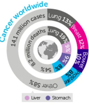
(28) 1.. INTRODUCTION. 1.1 Cancer. 1.1.1 Cancer – an introduction. According to the World Health Organization, approximately 14,1 million new cancer cases were diagnosed in 2012, and by the year 2030 the cancer burden is expected to increase to 23,6 million new cases per year. Cancer is also a leading cause of death and led to 8,2 million mortalities in 2012 (International Agency for Research on Cancer and Cancer Research UK, 2014).. Figure 1. Worldwide cancer statistics in the year 2012. Source: Cancer Research UK.. The creation of cancer, i.e. tumorigenesis, is a complex multistep process, with multiple genetic alterations that leads to progressive transformation of healthy cells into malignant derivatives. The dynamic changes in the cancer cell genome can occur through gain of function mutations in oncogenes and loss of function mutations in tumor suppressor genes that result in growth advantage. In 2000, Hanahan & Weinberg described six essential alterations in cancer cell phenotype compared to normal cells, acquired during tumor development. These alterations are: self-sufficiency in growth signals, evasion of apoptosis, insensitivity to anti-growth signals, limitless replicative 15.
(29) potential, sustained angiogenesis, and tissue invasion and metastasis. The alterations occur when genetic reprogramming in various extensively interconnected signaling cascades (such as PI3K, different MAPKs and SMAD among others) takes place, leading to cancer progression (Hanahan & Weinberg, 2000).. In 2011, Hanahan & Weinberg broadened the cancer hallmark concept by introducing two new emerging hallmarks: reprogrammation of energy metabolism and evasion of immune destruction; and two enabling characteristics of neoplasia: genome instability and tumor-promoting inflammation (Figure 2). They also highlighted the importance of the tumor microenvironment with its normal tissue derived cells – endothelial cells, immune inflammatory cells and cancer-associated fibroblasts – and the dynamic extracellular matrix (ECM) that they collectively remodel in cancer progression (Hanahan & Weinberg, 2011).. Figure 2. Hallmarks and enabling characteristics of cancer. Modified from Hanahan & Weinberg, 2011.. To tackle the global cancer burden, treatment strategies including medical, surgical, radiation, physical therapies and pain and palliative care should be designed. Thorough screening and preventive protocols should be developed and awareness increased, especially in the less developed world, where the cancer occurrence rate is growing and where there are limited resources and lack of access to quality health care. The state-of16.
(30) the-art of technology should be employed to create new innovative techniques and costefficient approaches tailored to the variable biology of the disease (Are et al., 2013).. 1.1.2 Treatment. Cancer can be locally treated by radiation, hyperthermia, laser or removing the tumor surgically. Radiation therapy uses high energy radiation to kill cancer cells by damaging their DNA, while hyperthermia exposes tissues to high temperatures to kill or damage cancer cells. Systemic cancer treatments include chemotherapy, hormone therapy that uses drugs to inhibit the growth of hormone-dependent tumors (breast, ovarian and prostate cancer among others), and biological therapies which include gene therapy, antibodies and cytokines (American Cancer Society, National Cancer Institute, Cancer Research UK).. 1.1.2.1 Cancer chemotherapy. Cancer chemotherapeutics are divided into alkylating agents (including platinum compunds), antibiotics, antimetabolites, topoisomerase inhibitors, mitotic inhibitors and others, depending on their chemical structure and mechanism of action (American Cancer Society, Table 1).. Most classical chemotherapeutic agents mediate their antitumoral effects through interactions with DNA. They act on DNA by breaking the helix itself or interfering with DNA-related proteins. Alkylating agents and some antibiotics (bleomycin and mitomycin C) form DNA adducts through strand crosslinking. Topoisomerase inhibitors, anthracycline antiobiotics and antimetabolites interfere with DNA synthesis by inhibiting different enzymes involved in DNA replication. Mitotic inhibitors act on tubulin in different ways: vinca alkaloids prevent polymerization of tubulin dimers while taxane binding stabilizes microtubules inhibiting the normal reorganization of the tubule network (Espinosa et al., 2003).. 17.
(31) Table 1. Chemoterapeutic agent families. Source: American Cancer Society.. 18.
(32) 1.1.2.1.1 Platinum-based drugs. The accidental discovery of the biological properties of cisplatin in 1965 initiated the development of platinum drugs for anticancer therapy. Cisplatin rapidly proceeded into clinical trials and was approved by FDA in 1978 for the treatment of bladder and testicular cancer. After that, platinum-based agents have been used to treat ovarian, colorectal, prostate and non-small cell lung cancer among others (Kelland, 2007).. Cisplatin is a neutral complex that in order to execute its cytostatic effects, needs to be activated by replacement of the cis-chloro ligands with water molecules. The monoaquated form of cisplatin is a highly reactive species that interacts with DNA primarily purine bases (adenosine and guanine) - and proteins and forms DNA adducts (Figure 3) (Kelland, 2000). This intra- and interstrand crosslinking leads to physical contortions in the DNA structure that is recognized by various damage recognition proteins which transduce DNA damage signals to their downstream effectors. The activated signaling pathways include p53 and MAPK (ERK, JNK & p38) among others, and through crosstalk between the different pathways involved, cisplatin-caused DNA damage leads to apoptosis (Siddik, 2003).. Figure 3. The mechanism of action of cisplatin.. 19.
(33) Besides cisplatin, the other two world-wide approved platinum-based anticancer drugs are carboplatin and oxaliplatin. Since toxicity of platinum-based drugs is directly related to the facility of the replacement of the ligands, and chloride as a leaving group is highly labile, these compounds with less reactive groups were developed (Figure 4).. Figure 4. The structure of world-wide approved Pt-based anticancer drugs.. Nevertheless, all Pt-based drugs though express severe dose-limiting side-effects, such as neurotoxicity and myelosuppression, due to uptake into all rapidly dividing cells, not just tumors. Besides, Pt-based drugs are secreted through the kidneys, which causes rigorous nephrotoxicity. For this toxicity, big effort has been directed to development of less toxic Pt-derivatives, but many of them have been discontinued in clinical trials. In order to reduce this toxicity, during the past decade the focus has shifted towards design of delivery vehicles for the already approved Pt drugs (Wheate et al., 2010).. 1.1.2.2 Personalized medicine. The aim of personalized medicine is the treatment of a disease based on the genetic characteristics of a population. This means individualized treatments of patients based on their gene and proteome expression analysis. Patients with cancers that show expression of validated cancer biomarkers can receive a treatment tailored to target their oncologic signaling profile instead of receiving classical chemotherapy that does not distinct cancer cells from normal cells.. 20.
(34) According to Cancer Research UK, there are over 200 different types of cancer. The clinical outcome depends on traditional clinico-pathological variables (patient’s age, tumor stage, grade and e.g. in the case of breast cancer, estrogen-receptor status), but also on the genetic heterogeneity of the tumors. Besides, cancer is a dynamic disease and tumor cells go through clonal evolution and changes in mutation signatures over time, which further complicates the discovery of the proper treatment. During the past 30 years, gene expression profiling has enabled definition of molecular subgroups of cancer that respond to treatment differently, which has contributed to the emergence of personalized medicine (Baird & Caldas, 2013).. In order to optimize the efficacy of personalized cancer therapy, new diagnostic and predictive biomarkers and therapeutic targets need to be discovered and validated. Personalized cancer medicine may be the answer for treating cancers with limited chemotherapeutic efficacy and decreasing chemoresistance of certain tumors. Finding the correct treatment for each patient could improve clinical outcomes greatly.. 1.1.3 Cancer biomarkers. As the molecular understanding of cancer increases – owing largely to genome sequencing – the rational development of targeted cancer drugs is gaining ground. An example of biomarker-targeted cancer treatment are small molecule kinase inhibitors, with a total of 17 approved inhibitors used as anticancer therapeutics in 2013 and an estimate of nearly 400 in clinical development. The most common kinases targeted in oncology include the growth factors and/or their receptors: VEGF/VEGFR, HER2 (ErbB2), EGFR and PDGF/PDGFR; and regulators of altered metabolism: PI3K and mTOR. Probably the most known example of biomarker-targeted cancer therapy is trastuzumab, a HER2-targeting antibody, the pioneer of targeted therapy that is now widely used in the treatment of breast cancer (Gonzalez de Castro et al., 2013).. Besides kinases, other potential cancer cell biomarkers include epithelial-mesenchymal transition-associated (EMT) molecules: vimentin, N-cadherin, snail and twist; multidrug resistance proteins; pluripotency-associated transcription factors and cancer stem cell (CSC) markers CD133, ALDH, CD21 and CD44 (Mimeault & Batra, 2014). 21.
(35) 1.1.3.1 CD44. CD44 is a transmembrane glycoprotein that mediates the response of cells to the microenvironment that surrounds them. CD44 is the main receptor of hyaluronic acid (HA) but can interact also with other lingands such as osteopontin, collagens, fibronectin and laminin. The interactions between CD44 and its ligands mediate intercellular and cell-matrix interactions, cell migration – by modulating secretion and activation of proteolytic enzymes – and migration of lymphocytes to inflammatory sites (Isacke & Yarwood, 2002).. Furthermore CD44 has a broad function in cellular signaling cascades. Through ligand binding and interactions with other cell surface receptors, it can regulate cellular growth, survival, and differentiation.. 1.1.3.1.1 CD44 structure. CD44 interacts with various components of the extracellular matrix, including glycosaminoglycans (GAGs), collagen, laminin and fibronectin. CD44 consists of an amino-terminal domain that binds its ligands, a stem structure, a transmembrane region and a cytoplasmic tail region (Figure 5).. 22.
(36) Figure 5. The structure of CD44 receptor. Modified from Ponta et al., 2003.. The amino-terminal globular domain contains motifs in its link domain (amino acids 32-123) that bind HA and other glycosaminoglycans. The link domain contains four highly conserved cysteine residues that form interchain disulphide links that account for stability of the module. Outside the link motif there are two additional cysteine residues thought necessary for correct folding and GAG binding. The stem structure separates the amino-terminal domain from the transmembrane region and is consisted of 46 amino acids. It contains alleged proteolytic cleavage sites. The transmembrane region of CD44 consists of 23 hydrophobic amino acids and is thought to have a role in the grouping of CD44 in lipid rafts. The carboxy-terminal cytoplasmic domain binds proteins associated to signaling and cytoskeletal organization, such as ankyrin and ERM family proteins (ERM: ezrin, radixin and moesin). The cytoplasmic tail contains two serine residues the phosphorylation of which, and therefore binding of the proteins, is controlled by various kinases, including protein kinase C (PKC) and Rho kinase (Ponta et al., 2003).. The human CD44 gene consists of 20 exons. The standard isoform (CD44s) expresses 10 exons being the smallest of the isoforms. The remaining 10 exons give rise to multiple CD44 variants (CD44v) formed through alternative splicing of the pre-mRNA (Figure 6).. 23.
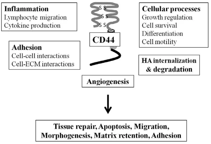
(37) Figure 6. CD44 pre-mRNA structure (above) and CD44 standard and variant structures (below). Modified from Goodison et al., 1999.. Alternative splicing of CD44 pre-mRNA and addition of different combinations of variant exons in the extracellular structure of the receptor can alter the binding of its ligands. Besides alternative splicing, post translational modifications, such as glycosylation, addition of heparan and keratin sulphate and sialic acid residues on the standard region and variant sites further modifies ligand binding characteristics and affects the function of the receptor (Goodison et al., 1999).. 1.1.3.1.2 CD44 functions. CD44 has various important roles in physiological and pathophysiological conditions (Figure 7). The functions of CD44 include cell aggregation and migration, proliferation, repair and regeneration, uptake and degradation of HA, lymphocyte activation and lymph node homing, myelopoiesis, lymphopoiesis, angiogenesis and cytokine release. CD44 shows high expression levels especially in proliferating cells, such as cells involved in tissue repair, morphogenesis, organogenesis and angiogenesis. The expression patterns and distributions of CD44s and CD44v in different tissues suggest differing functions for the standard and variant isoforms (Sneath & Mangham, 1998; Underhill, 1992). 24.
(38) CD44 mediates cell aggregation through anchoring HA/proteoglycan rich pericellular matrices. Being a transmembrane receptor, CD44 can communicate cell-matrix interactions into the cell, but may also conversely alter the associated matrix in response to intracellular signals. Thus CD44 provides means for interaction and communication between cells and the surrounding matrix. Besides, CD44 can serve as a co-receptor with other classical signaling receptors, physically linking to receptors such as c-Met and ErbB family of receptor tyrosine kinases, hence mediating the activation of various signaling pathways (Knudson & Knudson, 1999; Knudson & Knudson, 2004).. CD44 participates in cytoskeletal organization, binding directly to ankyrin, an adaptor protein that is attached to cytoskeletal actin. Binding of ligands promotes CD44 interactions with ankyrin as well as small GTP-binding proteins – such as RhoA that mediates phosphorylation of various cytoskeletal proteins – leading to their activation and producing changes in actin assembly and reorganization of the cytoskeleton (Bourguignon, 2008).. Figure 7. Some of the various processes in which CD44 participates.. CD44 has also been shown to be expressed in high levels in embryonic, hematopoietic, mesenchymal and epithelial stem cells as well as cancer stem cells (CSC) (Williams et al., 2013). 25.
(39) 1.1.3.1.2.1 CD44 in cancer. CD44 has been shown to contribute to neoplasia. High CD44 levels are associated with various types of malignant tumors and the invasiveness of certain tumors. Due to its functions in normal tissues, CD44 can contribute to different stages of cancer. The functions involved in tumorigenesis, invasion and metastasis are:. Cell growth promoting activity Production of autocrine factors caused by CD44-ligand interactions Locomotory signaling Mitogenic signaling Contributing to invasion via adherence to ECM through its ligands Invasion to HA-rich tissues by degradation of HA Tumor angiogenesis (Sneath & Mangham, 1998). CD44 has also been established a CSC marker and used to isolate and enrich CSCs from different types of tumors (Al-Hajj et al., 2003; Chu et al., 2009). Cancer stem cells are characterized as stem cell-like cells within a tumor, capable of self-renewal, differentiation and able to form tumors when transplanted into a host. They are defined by asymmetric cell division, in which one progeny maintains the stem cell properties and the other one undergoes further cell division and differentiation. CSCs are believed to be more resistant to chemo- and radiation therapy than other cancer cells and are associated to metastasis (Yu et al., 2012).. 1.1.3.2 Hyaluronic acid. Hyaluronic acid is the main ligand for the cell surface receptor CD44. HA is a sugarchain macromolecule that displays different properties depending on its size; high molecular weight (HMW) HA of the ECM links its binding molecules into aggregates and regulates cell adhesion, motility, and growth while HA oligosaccharides (low molecular weight: LMW) influence the same cell behaviors by interacting differently 26.
(40) with cell surface receptors. Like its main receptor, HA levels are increased in many cancers (Itano & Kimata, 2008).. 1.1.3.2.1 Structure and functions of HA. HA is a polysaccharide composed of repeating disaccharide units of glucuronic acid and N-acetylglucosamine (Figure 8). The size of a HA polysaccharide under normal physiological conditions is 1000 – 10 000 kDa and it consists of up to 25 000 disaccharides. HA is a negatively charged polymer and synthesized by three related hyaluronan synthases: HAS1, HAS2 and HAS3, multipass transmembrane enzymes that extrude HA into the cell surface or extracellular matrix while synthesizing it (Toole, 2004).. Figure 8. Structure of a HA disaccharide unit (left) and interchain hydrogen bonds within polymer consisting of 4 units (right), source: Jackson, 2009.. HA takes part in a variety of cell functions as well as in the maintenance of tissue structure. It is an important part of the ECM present in most vertebrate tissues and binds multiple proteoglycans – such as aggrecan, versican, and neurocan – that contribute to its assembly and regulate its functions. HA solutions are viscoelastic, and because of this property HA is prominent in synovial fluid and soft connective tissues. The physiological roles of HA in the ECM include water homeostasis, prevention of fluid flux through tissues and regulation of substance transport in the intercellular space, as well as lubrication and protection (Laurent et al., 1996).. Through cell surface receptors, HA also operates as a signal transducer regulating adhesion, cell motility, growth and differentiation activating various intracellular signaling cascades, such as c-Src, Ras and MAPKs. The actions of HA on the signaling 27.
(41) cascades are dictated by tissue concentration and size of HA molecules that interact with the receptors on cell surface. These properties are regulated by cooperating biosynthesis by HAS enzymes and the degradation of HA by hyaluronidases (HYALs) (Itano, 2008).. 1.1.3.2.2 HA interactions with cell surface receptors. HA is involved in tissue homeostasis and integrity, and acts as a template for macromolecular interactions in the assembly of the extracellular matrix. For these reasons HA has been widely used in the fields of dermatology (Gold, 2009) and regenerative medicine (Diamond et al., 2012; Prestwich, 2011).. Besides CD44, HA interacts with other cell surface HA-binding proteins, hyaladherins, including:. RHAMM/CD168 (receptor for HA-mediated motility) LYVE-1 (lymphatic vessel endothelial receptor 1) HARE/stabilin 2 (HA receptor for endocytosis) TLR2 & TLR4 (toll-like recpetors 2 and 4). RHAMM is a soluble cytoplasmic HA receptor that does not contain a transmembrane domain, but still performs extracellular functions, such as forming signaling complexes with CD44 and ERK1,2 thus activating the ERK1,2 kinase pathway (Hamilton et al., 2007). RHAMM is known to bind HA, mediate HA-signaling and cell motility, but its involvement in HA-uptake has not been shown (Hall et al., 1994). This receptor promotes cell motility and invasion as a result of interaction with HA. RHAMM expression, as that of CD44, is increased in tumorigenesis. Intracellular RHAMM associates with centrosomes and mitotic spindles and through these associations has the potential to promote genomic instability and neoplastic progression. (Maxwell et al., 2008) RHAMM has also been shown to be able to compensate for CD44 in binding HA, supporting cell migration and up-regulating inflammation-related genes (Nedvetzki et al., 2004).. 28.
(42) Systemic HA turnover is executed by the scavenger receptors LYVE-1 and HARE. LYVE-1 is expressed in lymphatic vessels and endothelia of lymph node sinuses and it binds HA, but not other glycosaminoglycans. LYVE-1 plays a role in HA homeostasis by uptaking HA to the lymph for partial degradation. HARE can be found with LYVE-1 in endothelial cells in the medullary sinuses of the lymph nodes and also in the sinusoidal epithelium of spleen and liver where HA fragments that exit lymph nodes are finally terminally degraded. Besides HA, HARE scavenges other glycosaminoglycans and takes part in their systemic clearance from the circulatory and lymphatic systems through clathrin-mediated endocytosis (Jackson, 2004, 2009).. HA also interacts with Toll-like receptors that play a role in pathogen recognition and inititation of immune responses. HA oligosaccharides have been shown to induce dendritic cell maturation and MAPK activation leading to TNF-α production. HA fragments stimulate metalloproteinase and inflammatory cytokine expression and NFкB activation. HA-TLR interaction has as well been shown to have a protective effect against tissue injury (Black et al., 2013; Jiang et al., 2011).. 1.1.3.2.3 HA degradation. HA is degraded in humans by five hyaluronidases: HYAL1, HYAL2, HYAL3, HYAL4 and PH-20/Spam1. The main hyaluronidases in charge of HA degradation in somatic tissues are HYAL1 and HYAL2. Degradation of HA is an endolytic, non-processive event where the HA fragment formed from glycosidic cleavage turns into a substrate for additional degradation. The final degradation products are tetrasaccharides, which can be further degraded intracellularly by lysosomal enzymes (Stern & Jedrzejas, 2008).. HA is degraded locally in tissues by the same cells that synthesize it. HA is internalized in a CD44-dependent mechanism that is associated with neither clathrin-coated vesicles, caveolae nor pinocytosis. The uptake of HA has been shown to depend on CD44 expression and partially degraded HA-oligosaccharides have been shown to be internalized faster than HMW HA. CD44-bound HA is degraded by HYAL2 to LMW fragments and endocytosed. After internalization, HA is delivered to lysosomes for further degradation by lysosomal HYAL1. The mechanism for CD44-mediated HA 29.
(43) uptake is possibly due to CD44 interactions with the actin cytoskeleton or regulated by partial ECM degradation and CD44 turnover (Knudson et al., 2002; Sironen et al., 2011).. 1.1.3.2.4 HA in cancer. HA is associated with carcinogenesis and it has been shown to be produced in excess by tumor cells as well as the surrounding stromal cells. The levels of HA are elevated in many different types of tumors and high HA levels have been linked to malignant progression. Through binding its primary receptor, CD44, HA has been shown to induce various intracellular signal transduction pathways, generally associated with tumorigenesis and the disruption of the CD44-HA connection has been found to alter the activities of these pathways. The various different tumor types in which high HA concentrations have been related to malignancy include:. Breast carcinoma Ovarian carcinoma Lung adenocarcinoma Prostate cancer Gastric and colorectal cancers Bladder cancer Head and neck cancer (Toole, 2004). HA has various roles in the initiation and progression of cancer. HA forms part of the tumor extracellular matrix linking together macromolecules and creating a meshwork favorable for tumorigenesis, allowing crosstalk between tumor cells and the stroma. This tumor microenvironment rich with HA can also act in recruiting inflammatory cells and in activation of cancer-related signaling pathways, accelerating through these mechanisms of action angiogenesis, lymphangiogenesis and inflammation. Aberrant HA production has been shown to increase cell growth and migration by decreasing contact inhibition, and over expression of HA synthase genes enhances tumorigenic abilities of some cancer cells (Itano et al., 2008).. 30.
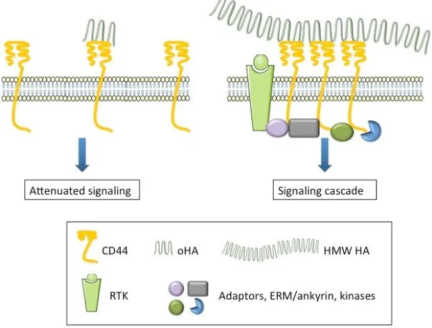
(44) 1.1.3.2.5 HA signaling. CD44-meadiated HA signaling differs between malignant cells and normal tissue cells. In normal tissues, where HA can be found in abundance, endogenous HMW HA binds CD44 receptors multivalently causing a quiescent state. When this binding is disrupted, signaling cascades are activated (Figure 9, A & B). In migrating cells – such as embryonic, endothelial and malignant cells – signaling is induced by the binding of the endogenous HA and inactivated by disruption of this binding (Figure 9, C & D) (Knudson & Knudson, 2004).. Figure 9. Normal tissue HA signaling through CD44 is induced by disruption of HA binding (A & B) while in malignant cells the situation is the opposite (C & D). Modified from Knudson & Knudson, 2004.. Besides CD44, HA has been shown to interact with other tumorigenesis-related signaltransducing receptors, including RHAMM. The binding of HA causes the cytoplasmic tail of CD44 to interact with various molecules – such as SRC kinases, RHO GTPases, VAV2, GAB1, ankyrin and ezrin – having regulatory roles in various intracellular signaling pathways. RHAMM, as well as CD44, binds HA on the cell surface and transduces signals related to malignant behavior. Interactions of HA with CD44 and RHAMM lead to alterations in the activities of tyrosine kinases, protein kinase C, focal adhesion kinase (FAK), phosphatidylinositol 3-kinase (PI3K) and various components. 31.
(45) of the cytoskeleton (Figure 10). The effects of HA involved in tumorigenesis through CD44 regulate especially cell survival through ErbB-signaling (Toole, 2004).. Figure 10. Cellular responses resulting from HA-CD44/RHAMM interactions and the intracellular components involved .. When native, HMW HA starts oncogenic signaling through CD44, it is thought to happen through multivalent binding of an individual HA polysaccharide to multiple receptors. Small oligosaccharides of HA (oHA) on the other hand can bind CD44 monovalently and they have been shown to have the opposite effects on CD44-signaling and tumor progression compared to the endogenous HA. oHA has been described to act as an antagonist of the native HA competing for CD44 and replacing the multivalent, cooperative binding thus interfering with tumorigenesis (Figure 11) (Toole et al., 2008).. 32.
(46) Figure 11. oHA binding compared to HMW HA binding to CD44 and the cellular response. Modified from Toole et al., 2008.. Besides exhibiting antitumoral effects, HA oligosaccharides have been shown to produce opposite effects as well (Dang et al., 2013; Sugahara et al., 2006). There is a considerable amount of literature regarding cellular responses to HA and plenty of controversy in results of studies. In some cases the size of the HA or the signaling molecules involved have not been monitored. Different sizes of HA fragments can cause diverse and often contrary effects.. In general elevated HA production by cells can promote oncogenic characteristics leading to increased tumor growth and metastasis, while reduction in HA production inhibit these processes. Variance can also occur due to deviations in HA turnover that produces low molecular weight fragments that can induce angiogenesis, as studied extensively by Professor Mark Slevin (Gaffney et al., 2009; Slevin, Kumar, & Gaffney, 2002), but detailed understanding of HA turnover and its effects on cancer cell behavior remain unclear (Toole et al., 2008).. 33.
(47) 1.1.3.3 HA-CD44-targeted therapies. HA and the entire complex system associated to its synthesis, binding and degradation offers a variety of targets for cancer therapy. Both CD44 and HA are expressed in normal tissues, not only in the tumor microenvironment, but carrier molecules extravasate and localize primarily into tumor tissue.. Strategies for HA-CD44-targeted cancer therapy include:. Targeting drugs to CD44 Targeting drugs to ECM HA Interfering with matrix HA-CD44 interactions Interfering with HA synthesizing or degrading enzymes. Antibody-based anticancer therapies rely on overexpression of the epitope on the target cells and the access of the carrier into the tumor tissue is facilitated by the enhanced permeability and retention (EPR) effect that results from the abnormal, leaky vasculature of the tumor compared to normal tissue. The lack of lymphatic clearance further favors localization of carriers in the tumor. Various CD44-antibodies conjugated to cytotoxic agents (for cancer therapy) and radioisotopes (for cancer detection) have reached clinical trials. Some of these anti-CD44 conjugates have shown promising results, but some have resulted in dose-limiting toxicity (Colnot et al., 2002, 2003; Rupp et al., 2007).. HA is a water soluble polysaccharide with various functional groups for conjugation, features required from a successful drug carrier. Anticancer agent-loaded HA-carriers have been widely studied in vitro and they show both CD44-specific uptake by cancer cells and anticancer effect (Platt & Szoka, 2008).. Professor Bryan P. Toole, one of the most respected authorities on HA, has conducted extensive research on oHA, showing that these oligosaccharides can interfere with matrix HA-CD44 interactions, inhibiting tumorigenic behavior of cancer cells. His studies have also shown that disrupting the CD44-HA interaction by oHA can even 34.
(48) suppress drug resistance and that oHA is a noteworthy candidate in fighting cancer (Slomiany et al., 2009a, 2009b).. Yet another target for HA-focused therapy are the HA synthases, overexpressed in many highly malignant cells. Inhibition of HAS1, HAS2 and HAS3 has been shown to decrease tumor cell growth and affect motility in vitro and in vivo (Teng et al., 2011; Wang et al., 2013). These studies prove that HASs may be valuable potential targets for anticancer therapy.. There is also evidence that HA degrading enzymes, HYALs, can act as tumor suppressors; their overexpression leads to reduction of tumorigenicity and HYAL administration in vivo has resulted in decreased tumor volumes (Shuster et al., 2002). On the other hand, data contrary to HYALs exhibiting tumor suppressing effects exists (Bouga et al., 2010; Franzmann et al., 2003; Tan et al., 2011). HYALs have been found overexpressed in many cancers. Their knockdown can result in decreased cell malignancy while forced overexpression can promote tumor growth. These opposite results suggest HYALs to have roles as both tumor suppressors and promoters. This dual role of HYALs seems to be a concentration-dependent phenomenon, naturally occurring levels in tumor environment acting as a promoter and exceeding levels causing tumor suppression (Karbownik & Nowak, 2013).. Studies have shown that HA can be used to target tissue microenvironments, such as those of a tumor, where an increase in HA synthesis, cellular uptake and metabolism occur. HA coupled to a variety of imaging contrast agents targeted sites of elevated HA metabolism, which shows that HA-based imaging probes could be used as diagnostic as well as therapeutic tools (Veiseh et al., 2012).. Conventional cancer therapy is starting to develop towards personalized medicine that exploits various cancer biomarkers. Special interest is directed to targeted therapies. This approach still requires validation of therapy targets, but there are already various that have been extensively studied, such as HA and the complex system associated to its synthesis and catabolism.. 35.
(49) 1.2 Nanomaterials in biomedicine. Nanomaterial (NM) – as defined by the European Comission Recommendation in 2011 – is a natural, incidental or manufactured material containing particles of which one or more dimensions is in the size range of 1 nm - 100 nm. Various national and international standardization bodies, organizations and authorities have pursued on developing a definition for nanomaterials, but many of the released non-normative definitions can be conflicting and we still lack a comprehensive global or an agreed EU definition.. Despite the controversy of the correct nanomaterial definition, during the recent years there has been a rapid growth in the number of products containing nanomaterials, introduced to the market in different sectors, such as the pharmaceutical, biotechnology and energy among others (Lövestam, 2010).. Engineered nanomaterials consist of various different materials, shapes and sizes. At nanoscale, their physical, chemical and biological properties can differ greatly from the corresponding bulk material. These unique size-dependent characteristics make NM very attractive for biomedical applications. Besides size, properties that contribute to NM activity include shape, hydrophobicity and electronic configuration. NM can also be easily derivatized through surface modifications to further adjust them to serve their purpose in pharmaceutical applications (Liang et al., 2008).. Nanomedicine uses nanosized tools for the prevention, diagnosis and treatment of disease. Nanomedicine aims to achieve medical benefit and to improve quality-of-life through monitoring, control, construction, repair, defense and improvement of human biological systems, as defined by the European Science Foundation’s (ESF) Forward Look in Nanomedicine in 2005. The opportunities that nanomedicine offers are improved diagnostics, biosensors and imaging agents on molecular as well as in vivo level, and new advanced therapeutics and technologies for disease and tissue regeneration and repair (ESF Forward Look on Nanomedicine 2005).. 36.
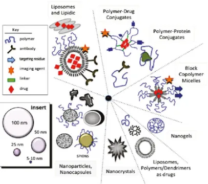
(50) Nanomedicine is a relatively young concept, even though medical applications of colloids have a long history. The birth of the nanomedicines on the second half of the 20th century was enabled by the discovery of synthetic polymer chemistry a few decades earlier. The pioneers of nanomaterials include Faraday and his studies with colloidal gold in the 19th century and Mechnikov and Ehrlich who received the Nobel Prize in 1908 for their work on phagocytosis and cell-targeted therapies. The main classes of nanomedicines today in the market or clinical development include liposomes, polymer conjugates, block copolymer micelles, dendrimers, nanocrystals, nanoparticles (NPs) and nanocapsules, that can act as drug carriers, imaging agents or have therapeutic activity on their own (Figure 12). Emerging nanomedicines encompass fullerenes, carbon nanotubes, metallic NPs, metal oxides, quantum dots and silicone-based NPs. New innovative nanomedicines arise from rational design and require the coordination of various different disciplines, such as chemists, biologists, pharmacologists, toxicologists and clinicians (Duncan & Gaspar, 2011).. 37.
(51) Figure 12. Main classes of nanomedicines in clinical trials or clinical use. Source: Duncan & Gaspar, 2011.. 1.2.1 Cancer nanomedicine. Nanomaterials offer new strategies to diagnoe and treate cancer owing to their unique properties. Their multiple promising characteristics make them a desirable target for investigation in cancer biomedicine. NMs can be localized and detected in specific sites of diseases. Due to their small size they can evade clearance through the kidneys and exploit the EPR effect. The blood circulation time of NMs can be increased by modifying their size, composition and surface coating. Besides, the high surface-areato-volume ratio of NMs results in a high loading capacity of ligands such as imaging and therapeutic agents (Wang et al., 2012).. 38.
(52) Cancer nanomedicine includes applications in imaging, diagnostics and drug delivery and NMs may be used as drugs on their own or in enhancement of the efficacy of existing drugs.. 1.2.1.1 Imaging and drug delivery. The unique features of NMs make them an intriguing platform for optimized therapy. NMs possess a large surface area that permits loading substantial amounts of agents or drugs. They can be targeted to disease sites by functionalizing their surface with targeting molecules. In addition, multiple different agents can be integrated on the NM surface creating multifunctional platforms. Besides, NM blood circulation time can be enhanced and escape from the mononuclear phagocyte system (MPS) ensured by modifying their size and surface. All these advantageous properties make NMs attractive for drug delivery as well as biomedical imaging (Lee et al., 2012).. NMs can reach tumors in two ways; through passive or active targeting, that can be improved by optimizing size and surface characteristics. Passive targeting exploits the EPR effect to achieve tumor accumulation while active targeting reaches the tumor owing to NM surface-integrated ligands for cell surface receptors. Probably the most utilized surface coating for passive targeting is polyethyleneglycol (PEG), which decreases blood protein interactions and increases stability. The coating for actively targeting NMs can vary from antibodies to nucleic acid ligands (Sultana et al., 2013). Specific tumor characteristics, such as genetic expression patterns, altered protein expression, uncontrolled growth, invasion and tumor microenvironment can be targeted and exploited (Shin et al., 2013).. The most innovative NMs do not function only as imaging or therapeutic agents, but are combinations of these two, acting simultaneously as therapeutic and diagnostic tools, known as theranostics. These theranostic applications can exploit among others gold NPs or quantum dots, that can be optically imaged without any modifications, and allow a high drug load. Besides, NMs that do not possess the optical features for imaging can be functionalized using fluorescent ligands among others (Doane & Burda, 2012).. 39.
(53) Theranostic nanoplatforms hold great promise of developing cancer therapy towards personalized medicine. Imaging probes combined with therapeutic NMs or NM-based drug delivery systems are expected to simultaneously diagnose, deliver drugs and monitor therapeutic effect (Kelkar & Reineke, 2011). However, theranostic NMs have not yet reached clinical settings: safety and efficacy still need great attention.. 1.2.1.2 In vitro diagnostics. The unique electrical, magnetic, luminescent and catalytic properties of various different NMs can be exploited in diagnostic devices. NMs can improve sensitivity and enable measurement of previously undetectable analytes. They can be used to analyze complex media samples (e.g. blood and urine) in user friendly assays reducing sample preparation and long read-out times. NM use in diagnostic assays can lead to device miniaturization and thus reduction of production costs. NM-based diagnostic devices can also allow multiple signal detection simultaneously, which can improve detection of complex diseases, such as cancer (Kurkina & Balasubramanian, 2012; Minelli et al., 2010; Shinde et al., 2012).. Inorganic metallic NPs are a class of nanomaterials that has been widely employed by in vitro cancer diagnostics due to their plasmon surface resonance and magnetic, photothermal and optical properties. Metallic NP-based strategies in cancer diagnostics include detection of enzyme activity, SNPs (single nucleotide polymorphisms) and tumor-related antigens. An example of a successful clinical application of NP-based diagnostic tool is an FDA-approved technology (Mirkin group, commercialized with the Verigene system from Nanosphere, Inc.) that utilizes magnetic and gold nanoparticles in a sandwich assay for multiplex detection of cancer markers. Briefly, antibodyfunctionalized magnetic microparticles (MMPs) are incubated with sample. Antibodyand ssDNA-functionalized gold NP-probes are added to MMPs and MMP-target-gold sandwiches are formed and separated from unbound gold with a magnetic field. ssDNA is then released from the gold NPs and this barcode DNA that corresponds to specific antibodies on the NP surfaces is analyzed by DNA array (Minelli et al., 2010).. 40.
(54) 1.2.1.3 Drugs and therapy. NMs used in medicine most often have an enabling function, i.e. they are used to enhance the characteristics of a traditional drug. However, there are NMs that convey the therapeutic effect themselves. These NMs have unique medical effects compared to conventional small molecule drugs due to their structure. An example of a therapeutic nanomedicine is NP-based magnetic hyperthermia developed by Magforce, to treat cancer. This treatment uses magnetic nanoparticles that are injected into the tumor and subjected to a magnetic field that causes the NPs to oscillate developing heat that destroys the tumor cells that have internalized the NPs (Wagner et al., 2006).. In addition to hyperthermia, metallic NPs have been reported in anti-tumorigenic applications using photodynamic therapy (PDT). An existing cancer treatment that could exploit the features of metallic NPs is ionizing radiation: for example X-ray irradiated gold NPs have been shown to induce apoptosis. Because the NPs are targeted to the tumor, damage of healthy tissue by radiation and elevated temperatures can be minimized. These techniques provide noninvasive treatment possibilities against cancer (Arvizo et al., 2013).. Nanotechnology applied to medicine can enhance greatly existing cancer drugs or create new ones with superior efficacy. It can also facilitate diagnosis due to its versatility and multifunctionality. Even though nanomedicines currently constitute only a fraction of the pharmaceutical market, they have the potential to revolutionize the industry.. 41.
(55) 1.3 Gold nanoparticles. 1.3.1 History of medicinal gold. Gold has a long history of therapeutic use. The late medieval alchemists introduced the concept of the “Elixir of life”, a solution containing gold that had been considered a magico-religious substance since the era of classical antiquity. Variations of this potable gold however often contained no gold, since aqua regia, needed to dissolve gold, was unknown to some of the supporters of medicinal gold. Nevertheless, potable gold was claimed to have salubrious effects on the heart and one of its most famous advocates was Paracelsus. In the 17th century potable gold entered pharmacopeias and the most common method of preparation was dissolving elemental gold in aqua regia, followed by heating to remove the solvent. The 19th century saw the development of double chloride of gold: a mixture of sodium chloride and gold chloride that was used to treat syphilis in a pill form. Double chloride injections were also used to treat alcoholism and experimented in the treatment of various incurable diseases, such as tuberculosis, diabetes and epilepsy until the 20th century. Then colloidal gold found its use in the treatment of rheumatoid arthritis and radioisotypes of gold in cancer treatment (Higby, 1982).. In eastern cultures, including China and India, the therapeutic benefits of gold preparations have been reported since 2500 BC. Today, colloidal gold is widely used in Ayurvedic medicine in India for rejuvenation and revitalization. In western medicine, intramuscularly administered gold compound, sodium aurothiomalate and orally administered auranofin are still used for treatment of rheumatoid arthritis. They act as immunomodulators, but their precise mechanisms of action remain unknown. Besides,198Au, a radioisotype of gold has been used during the 20th century as intratumoral injections to deliver large amounts of ionizing radiation into a tumor without damaging neighboring tissues due to the low penetrating ability of beta rays, the predominant radiation from 198Au (Bhattacharya & Mukherjee, 2008).. 42.
(56) 1.3.2 AuNP synthesis. AuNPs can be synthesized by various methods. They can be produced by so-called “top-down” methods, such as mechanical grinding of bulk metal or using metal vapor techniques. The “bottom-up” synthesis builds NPs from smaller entities. These chemical approaches use chemical reduction of gold salts, electrochemical pathways or controlled decomposition of metastable organometallic compounds. To prevent agglomeration, a variety of stabilizing agents are used. Protection can be achieved through electrostatic stabilization, based on repulsion between particles, or steric stabilization, that uses bulky organic molecules to protect the surface of the NPs. The main classes of protective groups are:. Polymers P, N and S donors (e.g. phosphanes, amines, thioethers) Solvents (e.g. THF, THF/MetOH) Long chain alcohols Surfactants Organometals (Zhou et al., 2009). Two of the most popular methods for spherical AuNP synthesis were introduced by Turkevich in 1951 and by Brust and Schiffrin in 1994. The Turkevich method uses citrate reduction of HAuCl4 in water. The size of the resulting AuNPs can be controlled by varying the proportion of the reducing agent compared to the gold salt. Besides reducing agent, citrate acts as a capping agent as well, stabilizing the NPs after synthesis. The Brust-Schiffrin method produces AuNPs of low size distribution. This method allows synthesis of AuNPs in organic solutions. It involves a reaction of HAuCl4 with toluene in the presence of tetraoctylammonium bromide as the phasetransfer reagent and sodium borohydride as the reducing agent. Dodecanethiol is used as stabilizer in this reaction (Daniel & Astruc, 2004).. Besides chemical syntheses, greener biosynthetic approaches have been described. These approaches rely on bioreduction of gold salts using either cell-free extracts from. 43.
Figure


+7


Documento similar
Importantly, the person is able to perform the task correctly (not worse). The effects of training are not transferable; even though the untrained condition was
The analysis of the biological effects of gold nanoparticles generated by biological reduction of HAuCl 4 gold precursor in HeLa cells and the comparison with
Simultaneous expression of PI3K and any of the three Aβ peptides also produced an increase in the number of active zones, boutons and branches when compared to Aβ express- ing NMJs
Tumor cells (and other cells in the tumor) deplete nutrient levels (glucose, glutamine, amino acids, O 2 , etc.) in the TME, increase the levels of some metabolites, such as lactic
We describe the exfoliation of these materials and the formation of stable suspensions in water, suitable to interact and detect a variety of potential water contaminants such as
No obstante, como esta enfermedad afecta a cada persona de manera diferente, no todas las opciones de cuidado y tratamiento pueden ser apropiadas para cada individuo.. La forma
Mesoporous Silica Nanoparticles present well-defined and tunable physicochemical properties, including particle size, pore size, pore volume, surface area, volume area, pore
Administration of darolutamide (600 mg twice daily for 5 days) prior to co-administration of a single dose of rosuvastatin (5 mg) together with food resulted in approximately
