oa
México
Publicación Digital de la Facultad de Medicina Veterinaria y Zootecnia
OA
http://veterinariamexico.unam.mx/Accepted: 2018-08-03 Published: 2019-02-05 Additional information and declarations
can be found on page 12
Copyright 2018 José Luis Martínez-Ibarra et al.
open access
Distributed under Creative Commons CC-BY 4.0
Abstract
The effect of resveratrol on the in vitro maturation (IVM) of ovine (Ovis aries) oocytes and the development of handmade cloned embryos was eval-uated. The nuclear maturation and reactive oxygen species (ROS) levels in the oocytes, as well as the early development and morphological cloned embryo quality, were evaluated under different resveratrol concentrations (0, 0.5, 2 and 5 μM). After IVM, no significant difference was observed in the maturation rate of oocytes treated with 0.5 μM (81.3 %) and 2 μM (72 %) resveratrol compared to that of the control group (0 μM) (74.2 %), but the rate significantly decreased at 5 μM (56 %) (p < 0.05). When the oocyte ROS levels were determined, no significant differences among the groups were observed (p > 0.05). For cloned embryo development, the embryos obtained from the oocytes treated with 0.5 μM resveratrol showed higher (p < 0.05) compacted morula rates (10.7 %) compared to the embryos obtained from the oocytes treated with 0, 2 and 5 μM (6.2, 0 and 0 %, re-spectively). Regarding embryo morphological quality, the embryos from the oocytes treated with 0.5 μM resveratrol showed a lower rate of poor quality morulae (4.7 %) in comparison to those treated with 0, 2 and 5 μM (23.8, 23.3 and 33.3 %, respectively) (p < 0.05). In conclusion, resveratrol showed no significant improvement on the IVM or ROS levels in domestic ovine oocytes. However, treatment with 0.5 μM resveratrol during IVM improved embryo quality and promoted morulae compaction of Ovis aries handmade cloned embryos.
Keywords: Resveratrol, Ovis aries, In vitro maturation, Reactive oxygen species, Embryo development, Handmade cloning.
Effect of resveratrol on the in vitro
maturation of ovine (Ovis aries)
oocytes and the subsequent
development of handmade cloned
embryos
José Luis Martínez-Ibarra1* Eugenia Adriana Espinoza-Mendoza1 Raymundo Rangel-Santos2 Demetrio Alonso Ambriz-Garcia1 María del Carmen Navarro-Maldonado1
1 Departamento de Biología de la Reproducción. División de Ciencias Biológicas y de la Salud. Universidad Autónoma Metropolitana. Unidad Iztapalapa. Av. San Rafael Atlixco 186, Col. Vicentina, Iztapalapa, 09340, Ciudad de México, México. 2 Posgrado en Producción Animal,
Departamento de Zootecnia. Universidad Autónoma Chapingo. Km 38.5 Carretera México-Texcoco, Chapingo, 56230, Estado de México, México.
*Corresponding author:
Email address: [email protected]
Cite this as:
Introduction
In vitroembryo production by somatic cell nuclear transfer (SCNT) is a reproductive biotechnology with great potential for the propagation of genetically valuable indi-viduals, farm animal production and the preservation of endangered wild species.1 This technology can also be a useful tool for basic research into knowledge on embryo development and nuclear reprogramming processes. SCNT can be defined as the process that makes it possible to obtain one or more genetically identical individuals from a single nucleus or somatic cell.2 Diverse domestic animals, such as sheep, monkeys, cattle, mice, goats, pigs, cats, rabbits, rats, mules, horses, dogs and ferrets,3 and wild species, such as gaurs,4 European mouflon,5 buffalo,6 gray wolves,7 and camels, have been cloned.8
Traditionally, in SCNT, micromanipulators are used to enucleate oocytes and introduce genetic material from the individuals to be cloned. A variant of the technique is handmade cloning (HMC), which does not use micromanipulators, making it an accessible and less complex technique.9 Using this technique, por-cine,10 bovine11 and ovine12 clones have been obtained. For both cloning techniques, efficiency is still low; depending on the species, only 1–5 % of the transferred embryos develop into viable offspring, and cloned animals have shown developmental problems in other cases.13 These problems could be due to the complexity of the technique, which involves several factors that can affect embryo development.14 In this regard, Camargo et al.15 stated that oocyte in vitro matura-tion is the main factor for obtaining optimal in vitro embryo development.
An important factor that can affect in vitro maturation (IVM) is oxidative stress, which causes an imbalance between oxidant and antioxidant levels inside the cell.16 It has been reported that oocyte maturation is strongly influenced by gluta-thione and reactive oxygen species (ROS) levels.17 ROS are oxygen metabolism de-rivatives formed by the breakdown, excitation or reduction of molecular oxygen.18 ROS overproduction during IVM and early embryo development can produce lipid peroxidation in the cell membrane, DNA fragmentation, and alterations in RNA transcription and protein synthesis.16 These events can cause oocyte and embryo damage and may even cause cell death. Therefore, it is necessary to establish an IVM system that is capable of reducing intracellular ROS levels as well as oxidative damage that reduce oocyte quality, contributing to improved embryo development. The use of several antioxidants, such as β-mercaptoethanol, cysteine, cystine and L-carnitine, has been proposed,17,19 as well as ascorbic acid and α-tocopherol (vita-min C and vita(vita-min E, respectively).20 Recently, it has been reported that resveratrol (trans-resveratrol) used during IVM increases embryo development in porcine, bo-vine and caprine species.21-23
Resveratrol (3,4’,5-trihydroxystilbene) is a natural polyphenol present in plants such as polygonum (Polygonum cuspidatum) and the common grape vine (Vitis vinifera), and it has biological activity to protect plants from the attack of pathogens, fungi and bacteria. Resveratrol has antioxidant, anti-inflammatory, cardioprotective and anti-cancer effects,24 and it regulates gene expression related to apoptosis by activating sirtuin 1 (SIRT1) and increasing mitochondrial function and the ATP content in oocytes.25
Material and methods
Unless otherwise specified, all chemicals and reagents used in this study were purchased from Sigma-Aldrich Chemical Company (St. Louis, MO, USA). All tech-niques were performed under sterile conditions in a laminar flow hood. Incubation was always at 38 °C and 5 % CO2 in a 95 % humidified atmosphere. The present experiments complied with relevant institutional animal welfare guidelines, such as the Guidelines for the Ethical Conduct of Research, Teaching and Diffusion of the Biological and Health Sciences Division from the Universidad Autónoma Metropoli-tana and were approved by the Divisional Council of Biological and Health Sciences Division in Session 8.10 on May 18th, 2010 in accordance with the national animal welfare guidelines (NOM-062-ZOO-1999; NOM-033-ZOO-1995) and the policies and ethics committee approval (Academic Commission of Ethics of the Biological and Health Sciences Division at Universidad Autónoma Metropolitana).
Oocyte collection and in vitro maturation
The ovaries were obtained from adult Ovis aries Criollo sheep from a slaughter-house in Nezahualcoyotl, Estado de Mexico and transported to the laboratory at 28-35 °C in saline solution [0.9 % NaCl and 1 % antibiotics (10, 000 UI/mL pen-icillin, 10 mg/mL streptomycin sulfate and 25 μg/mL amphotericin) (Antibac-Anti-fun 100x, In Vitro, SA)] within two hours. The ovaries were washed three times in isothermal saline solution. The cumulus oocyte complexes (COCs) were punctured within a 2–6 mm diameter of the ovary follicles and aspirated using a 20 gauge hypodermic needle attached to a 10 mL disposable syringe containing 1 mL of HEPES (4-(2-hydroxyethyl)-1-piperazineethanesulfonic acid) buffered TCM-199 (In Vitro, SA) and 100 UI/mL heparin.26 Once the follicular fluid was obtained, the COCs with a homogeneous cytoplasm and at least three layers of cumulus cells were selected for IVM, based on the Kakkassery et al.27 criteria. The COCs were washed three times in maturation medium [TCM-199 containing 10 % fetal bovine serum (FBS) (Microlab), 1 % epidermal growth factor (EGF), 5 μg/mL follicle stim-ulating hormone (FSH) (Folltropin-V, Bioniche), 5 UI/mL chorionic gonadotrophin (CG) (Loeffler), and 0.6 % antibiotics]. Groups of 25 to 30 COCs were cultured in four-well dishes with 500 μL of maturation medium under 300 μL of mineral oil and incubated for 24 hours.28 During IVM, the COCs were treated with different concentrations of resveratrol (0, 0.5, 2 and 5 μM). The resveratrol was dissolved in dimethyl sulfoxide (DMSO) as a 500 μM stock solution and stored at −20 °C before being added to the maturation medium.23
Assessment of nuclear maturation
Measurement of intracellular ROS levels
The matured oocytes were sampled to measure intracellular ROS levels by a 2’,7’-dichlorodihydrofluorescein diacetate (H2DCFDA) fluorescence assay. An av-erage of six oocytes from each treatment group were incubated in darkness in TCM-199 containing 10 μM of H2DCFDA for 30 minutes. Following incubation, the oocytes were washed three times in 100 μL of HEPES-buffered TCM-199 and fixed onto slides. The fluorescence was observed under an epifluorescence microscope equipped with a 460 nm filter. The fluorescent images were saved as JPG files, and the fluorescent intensities of the oocytes were analyzed by ImageJ software (Version 1.41; National Institute of Health, Bethesda, MD, USA).
Handmade cloning
Preparation of the recipient cytoplast
The cytoplasts were prepared as described by Vajta et al.30 Mature oocytes with expanded cumulus cells were transferred into a microcentrifuge tube containing 0.5 mg/mL hyaluronidase in HEPES-buffered TCM-199, incubated for a minute and vortexed at maximal speed for three minutes. Then, the oocytes were transferred to a culture dish containing T2 (T denotes HEPES-buffered TCM-199, and the number denotes the percentage of FBS), incubated with 10 μg/mL demecolcine in TCM-199 for 40 min and washed twice in T2. The denuded oocytes were placed into 30 μL pronase (2 mg/mL in T10) for 5–10 min to digest the zona pellucida and then transferred into 20 μL T20 to stop the activity of the pronase enzyme. The zona-free oocytes were examined by use of a stereoscopic microscope, and those with a homogeneous cytoplasm and first polar body extrusion were selected. The selected zona-free oocytes were transferred into 20 μL T10 and enucleated by manual bisection using an ultrasharp microblade (Ultra-Sharp Splitting Blade, Bion-iche) to remove the polar body and nuclear material. The enucleated oocytes were transferred to T20 and incubated until the enucleation of all oocytes was finished.
Preparation of the donor cells (karyoplast)
were incubated with 700 μL of trypsin-versene (0.05/0.05 %) (In Vitro, SA) for five minutes. Then, 700 μL of supplemented DMEM was added, and the cells were centrifuged at 150/g for four minutes to produce a cell pellet. The pellet was recovered and divided into two halves. One half was resuspended in three millili-ters of supplemented DMEM and incubated for seven days until the cells reached confluence, and then the cells were passaged. The other half was resuspended in 500 μL of DMEM and kept at room temperature until cell fusion.28 The cells were then ready for use as nucleus donors or karyoplasts.
Pairing and electrofusion of karyoplasts and cytoplasts
Two cytoplasts were immersed in 15 μL phytohemagglutinin (5 mg/mL in HEPES-buffered TCM-199) for 5–10 seconds and then transferred to 15 μL T2 containing approximately 30 karyoplasts previously placed into the solution. The two cytoplasts were attached to a karyoplast forming a triplet, which was transferred to 15 μL fusion medium (5.46 g D-mannitol and 0.1 g polyvinyl alcohol in 98 mL H2O) and placed between the two electrodes in the fusion chamber (BTX Micros-lide. Model 450; BTX, San Diego, CA) covered with 200 μL fusion medium. Then, the triplet was aligned between both electrodes of the chamber with an AC pulse (4 V) using the BLS electrofusion instrument (CF-150/B. Budapest, Hungary), and a single DC pulse (105 V) was applied for 9 μs for fusion. The fused triplets that acquired a spherical form were considered cloned embryos and were transferred to SOF medium (In Vitro, SA) containing 5 % FBS and incubated for two hours for reprogramming.30
Activation and in vitro culture of cloned embryos
The chemical activation of the cloned embryos was achieved by incubation in 5 μM Ca2+ ionophore in T2 for five minutes, followed by washing three times in T20. Then, the embryos were individually incubated in 2 μL droplets of SOF containing 5 % FBS and 2 mM 6-dimethylaminopurine (6-DMAP) covered with mineral oil for four hours and washed three times with SOF containing 5 % FBS. Finally, the activated embryos were cultured using the well of the wells (WOW) system, as described by Vajta et al.30 The microwells were prepared with a v-shaped needle in the bottom of each well of a four-well dish and contained 500 μL SOF with 15 % FBS and covered with 300 μL mineral oil. The embryos were carefully loaded indi-vidually into the microwells and incubated for seven days.
Assessment of embryo development
and morphological embryo quality
Experimental design
In experiment 1, the effect of different concentrations (0, 0.5, 2 and 5 μM) of res-veratrol on the IVM of ovine oocytes was determined. In experiment 2, the effect of different resveratrol concentrations during IVM on intracellular ROS levels was evaluated. In experiments 3 and 4, the effect of resveratrol treatment during IVM on the subsequent early embryonic development and morphological embryo quality of handmade cloned embryos was examined.
Statistical analysis
All statistical analyses were performed using NCSS 2007 (NCSS, LLC. Kaysville, Utah, USA). The percentage data for maturation rate, cleavage and embryonic yield were compared by one-way ANOVA followed by Duncan’s multiple range test. The data are presented as the mean ± standard deviation. Differences were considered significant at p < 0.05. The experiments were replicated at least five times.
Results and discussion
Effect of different concentrations of resveratrol
on ovine oocyte IVM
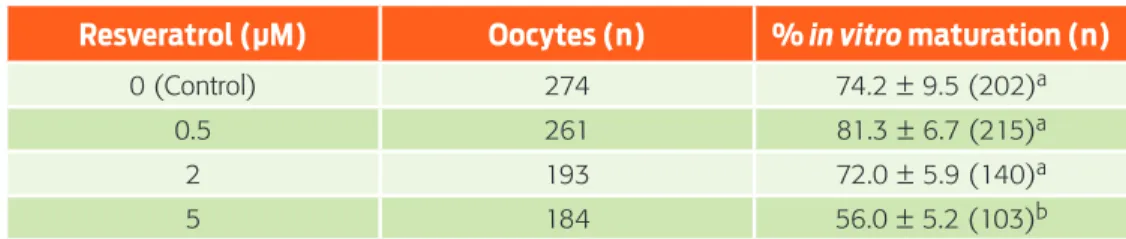
The effect of different concentrations (0, 0.5, 2 and 5 μM) of resveratrol on the maturation rate of ovine oocytes was assessed; the results are presented in Table 1. No significant difference was observed between the 0.5 and 2 μM resveratrol groups (81.3 and 72 %, respectively) compared with the control (74.2 %). Howev-er, the maturation rate of the oocytes treated with 5 μM resveratrol was decreased (56 %) compared to that of the control (F(3,18) = 13.939, p = 6.085).
Our results revealed that resveratrol treatment during IVM resulted in concen-tration-dependent effects. Although the results were not significant, the treatment with 0.5 μM resveratrol was considered optimal because it tended to increase the IVM rate. In contrast, the 5 μM resveratrol treatment decreased the IVM rate signifi-cantly compared to other concentrations. Similar to these results, Mukherjee et al.23 found a beneficial effect with 0.25 and 0.5 μM resveratrol and a detrimental effect with 5 μM on goat oocyte IVM. However, Kwak et al.21 when working with porcine oocytes, reported that resveratrol treatment during IVM had a concentration-depen-dent effect; 2 μM resveratrol increased the percentage of mature oocytes; however, with the highest concentration (10 μM), the percentage significantly decreased. We suggest that 5 μM resveratrol during IVM decreases the nuclear maturation rate due to an imbalance between the levels of oxidants and intracellular antioxidants. Table 1. Effect of different resveratrol concentrations during IVM
Resveratrol (µM) Oocytes (n) % in vitro maturation (n)
0 (Control) 274 74.2 ± 9.5 (202)a
0.5 261 81.3 ± 6.7 (215)a
2 193 72.0 ± 5.9 (140)a
5 184 56.0 ± 5.2 (103)b
Furthermore, the decrease in oocyte nuclear maturation may be related to the abil-ity of resveratrol to inhibit phosphodiesterase activabil-ity, which results in an increase in the concentration of cAMP,32 and high levels of cAMP inhibit the progression of meiosis, while low levels activate it.33 Considering that, resveratrol affects other functions such as cytoplasmic maturation and the regulation of apoptotic gene expression (Bax, Bak, and Caspase-3) through an optimal oxidation-reduction bal-ance, which could contribute to optimal oocyte maturation and better embryonic and fetal development.21,23
Effect of resveratrol on intracellular ROS levels during IVM
The intracellular ROS level in in vitro matured oocytes treated with different resvera-trol concentrations is shown in Figure 1. The highest ROS levels were found at 5 μM (95.5 %), while they were lower at 0.5 and 2 μM (88.3 and 84.4, respectively) compared with the levels found in the control (0 μM). However, these differences were not significant (F(3,20) = 0.53, p = 0.6625) (Figure 2).
These results differ from those reported by Mukherjee et al.,23 who evaluated ROS levels in matured goat oocytes; these authors found the highest levels with 0 and 5 μM and the lowest with 0.25 and 0.5 μM resveratrol, with a significant differ-ence of 30 % between them. Similarly, in porcine oocytes, Kwak et al.21 obtained the highest levels of ROS with 0 and 10 μM resveratrol and the lowest levels of ROS with 0.2 and 2 μM resveratrol, with decreases of 30 % and 60 %, respectively. These authors concluded that resveratrol decreases ROS levels in matured oocytes because, at optimum concentrations, it increases intracellular GSH levels, which are strongly associated with cytoplasmic maturation and may play roles in nuclear maturation. The results obtained in this study show that, although resveratrol also decreased ROS levels, these levels were reduced by only 11.7 % to 15.6 %, which differs from the previous results of 30 %21 and 30 % to 60 %.23 These results show that resveratrol application during IVM has different effects among species, which is most likely caused by the different sensitivity of each species to this an-tioxidant. It is known that resveratrol enhances SIRT1 expression in oocytes and increases the expression level of phospho-5’-adenosine monophosphate-activated protein kinase in oocytes.25 However, in our study, resveratrol showed no benefits on IVM in oocytes; however, its beneficial effect was observed at the morula stage.
Effect of resveratrol treatment during IVM on the
in vitro
development of cloned embryos
Figure 1. Epifluorescence microscope photographs of ovine oocytes treated with different resveratrol concentrations and stained with 2’,7’-dichlorodihydrofluorescein (H2DCFDA) (10x). In bright field (a-d) and with 460 nm filter (e-h).
80,000
70,000
60,000
10,000 20,000 30,000 40,000 50,000
0.0
0 (control) 0.5 2 5
a
a
a
a
Fl
uor
es
cenc
e unit
s
(pix
el/
o
vocit
o
)
Resveratrol (µM)
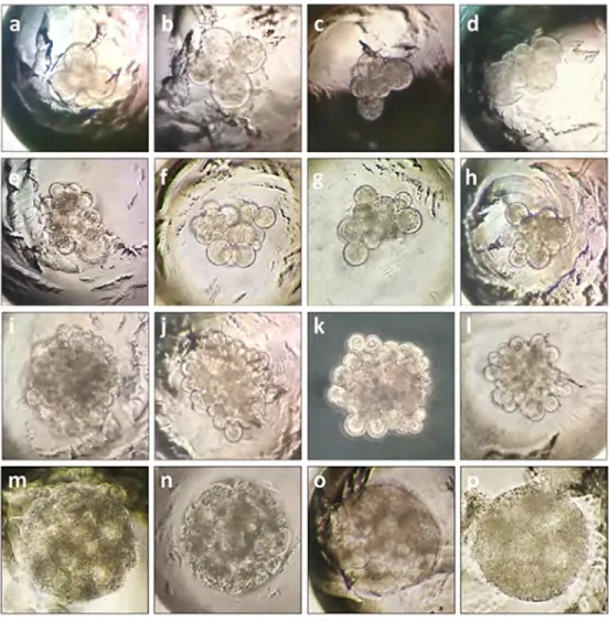
Figure 3. Different developmental stages of the HMC sheep embryos cultured in the WOW system. Cleaved embryos (a-d), 8–16 blastomere embryos (e-h), morulae (i-l) and compact morulae (m-p) (200X).
Table 2. Effect of resveratrol treatment during IVM on handmade cloned embryo development
Resveratrol
(µM) cultured (n)Embryos embryos (n)% Cleaved % 8–16 blastomere embryos (n) % Morulae (n) morulae (n)% Compact
0 (Control) 64 81.2 ± 5.1 (52)a 65.6 ± 3.6 (42)a 32.8 ± 7.8 (21)a 6.2 ± 0 (4)a
0.5 57 84.4 ± 9.9 (48)a 70.2 ± 6.2 (40)a 35.1 ± 5.9 (20)a 10.7 ± 4.1 (6)b
2 45 80.1 ± 8.1 (36)a 64.3 ± 7.5 (29)a 28.7 ± 3 (13)a 0 ± 0 (0)c
5 44 77.2 ± 5.2 (34)a 63.6 ± 7.4 (28)a 6.8 ± 4.5 (3)b 0 ± 0 (0)c
treated with 0.5 μM resveratrol (10.7 %) was higher (F(3,12) = 10.14, p = 0.001) than the control group (6.2 %), while, for the groups treated with 2 and 5 μM res-veratrol, there were no compact morulae.
During the IVC of the cloned embryos, there were no significant differences in the cleaved embryo rate among resveratrol concentrations; however, 5 μM resver-atrol tended to reduce the embryonic yield at the morula stage, and this reduction was more notable at the compact morulae stage. This result shows that treatment with 5 μM resveratrol during the IVM of ovine oocytes has a deleterious long-term impact that manifests during early embryonic development. In contrast, the posi-tive effect of the 0.5 μM resveratrol treatment during IVM was maintained during embryonic development, generating greater morulae compaction. This finding may be related to the properties of E-cadherin, which is a principal calcium-dependent molecular component of adherent junctions that is associated with the compaction of morulae and blastocyst formation.34 E-cadherin plays a key role in the differenti-ation of the trophectoderm and supports further embryonic development,35 and it is known that resveratrol increases E-cadherin expression in the cell by promoting its transcriptional activity.36 Similar to the results of this study, Mukherjee et al.23 reported that a 0.5 μM resveratrol concentration favors embryonic development, whereas 5 μM reduces it. In contrast, Wang et al.22 reported that a 1 μM resveratrol treatment favors bovine embryonic development, which differs from the results obtained in this work in sheep and those reported by Mukherjee et al.23 in goats. It is clear that the optimal concentration of resveratrol for each species is different, which is possibly due to the sensitivity of each species to this antioxidant. Howev-er, it is evident that there is greater similarity between the results obtained in the present work in sheep and those obtained in goats. These similarities are probably because these two species (Ovis aries and Capra hircus) are phylogenetically close since they both belong to the subfamily Caprinae and since there are many ana-tomical and morphological similarities between them.37
Effect of resveratrol treatment during IVM
on the morphological quality of cloned embryos
After 120 h of IVC, the morphological quality of the cloned embryos showed no differences among groups. However, after 168 h of IVC, as shown in Table 3, the morulae treated with 0.5 μM resveratrol during IVM showed an increase in the percentage (30.1 %) of excellent quality embryos and a significant decrease in the percentage of poor quality embryos (4.7 %) compared to the control group (23.8 % and 23.8 %, respectively) (F(3,7) = 24.59, p < 0.0001). In contrast, with 5 μM resveratrol, the percentage of excellent quality embryos decreased (F(3,11) = 25.88, p < 0.0001).
increased, reaching a maximum at the compact morulae stage, possibly because the antioxidant effect of resveratrol improves oocyte nuclear and cytoplasmic mat-uration at low concentrations, maintaining the oxidative balance within the cell.23 The positive long-term impact of resveratrol may be because, at low concentrations of resveratrol during IVM, the expression of pro-apoptotic genes in the oocytes de-creases.21 Resveratrol probably promotes cytoplasmic maturation, which is critical for achieving optimal embryonic development.21 Other authors reported that the beneficial effect of resveratrol is due not only to its antioxidant activity but also to its ability to regulate SIRT1 expression.22 SIRT1 is involved in the regulation of mito-chondrial biogenesis, including the generation of ATP and the regulation of AMPK, which increases β-oxidation and, consequently, the consumption of fatty acids, thus improving embryo development.25 However, further studies could confirm these observations.
Conclusions
The effects of resveratrol on oocyte IVM and ROS concentrations are controversial. In this work, during sheep oocyte IVM, resveratrol did not significantly improve the nuclear maturation rate or intracellular ROS levels. However, the results sug-gest that 0.5 μM resveratrol during IVM improves oocyte quality and promotes morulae compaction of Ovis aries handmade cloned embryos during early devel-opment. This finding may be because resveratrol affects the expression of calci-um-dependent cell adhesion molecules, such as E-cadherin, that are required for morulae compaction.
Table 3. Effect of resveratrol treatment during IVM on the morphological quality of cloned morulae
Resveratrol (µM) Evaluated embryos
% Embryo quality (n) 1
(Excellent) (Good)2 (Regular)3 (Bad)4
0 (Control) 21 23.8 (5)a 28.5 (6)a 23.8 (5)a 23.8 (5)a
0.5 20 30.1 (6)a 39.6 (8)a 19.8 (4)a 4.7 (2)b
2 13 23.3 (3)a 30.0 (4)a 19.8 (3)a 23.3 (3)a
5 3 0.0 (0)b 33.3 (1)a 33.3 (1)a 33.3 (1)a
Acknowledgements
We thank Dr. Enrique Canchola Martínez for his theoretical contribution on the usefulness of resveratrol and Dr. Edith Arenas Ríos for her theoretical contribution to the determination of ROS levels in oocytes. Both researchers belong to the De-partment of Reproductive Biology, UAM-I.
We thank CONACYT for the scholarship number 302861 granted to the main author during his studies at the Biology of Animal Reproduction Master.
Conflicts of interest
None of the authors have any conflicts of interest to declare.
Author contributions
JLMI conceived the study, performed the experiments, interpreted the results and performed the statistical analysis. EAEM performed the experiments and interpret-ed the results. RRS contributinterpret-ed to the manuscript preparation. DAAG collectinterpret-ed the samples, interpreted the results and performed the statistical analysis. MDCNM directed the research, conceived the study and interpreted the results. All authors read, revised and approved the final manuscript.
References
1. Folch J, Cocero MJ, Chesné P, Alabart JL, Domínguez V, Cognié Y, et al. First birth of an animal from an extinct subspecies (Capra pyrenaica pyre-naica) by cloning. Theriogenology. 2009;71:1026–34. doi: 10.1016/j. theriogenology.2008.11.005.
2. Bren L. Cloning: revolution or evolution in animal production. FDA Consum. 2003;37:28–33.
3. Meissner A, Jaenisch R. Mammalian nuclear transfer. Dev Dyn. 2006;235:2460– 9. doi: 10.1002/dvdy.20915.
4. Lanza RP, Cibelli JB, Diaz F, Moraes CT, Farin PW, Farin CE, et al. Cloning of an endangered species (Bos gaurus) using interspecies nuclear transfer. Cloning. 2000;2:79–90. doi: 10.1089/152045500436104.
5. Loi P, Ptak G, Barboni B, Fulka J, Cappai P, Clinton M. Genetic rescue of an en-dangered mammal by cross-species nuclear transfer using post-mortem somat-ic cells. Nat Biotechnol. 2001;19:962–4. doi: 10.1038/nbt1001-962.
6. Shi D, Lu F, Wei Y, Cui K, Yang S, Wei J, et al. Buffalos (Bubalus bubalis) cloned by nuclear transfer of somatic cells. Biol Reprod. 2007;77:285–91. doi: 10.1095/ biolreprod.107.060210.
7. Kim MK, Jang G, Oh HJ, Yuda F, Kim HJ, Hwang WS, et al. Endangered wolves cloned from adult somatic cells. Cloning Stem Cells. 2007;9:130–7. doi: 10.1089/clo.2006.0034.
8. Wani NA, Wernery U, Hassan FA, Wernery R, Skidmore JA. Production of the first cloned camel by somatic cell nuclear transfer. Biol Reprod. 2010;82:373–9. doi: 10.1095/biolreprod.109.081083.
10. Du Y, Kragh PM, Zhang Y, Li J, Schmidt M, Bogh IB. Piglets born from handmade cloning, an innovative cloning method without micromanipulation. Theriogenol-ogy. 2007;68:1104–10. doi: 10.1016/j.theriogenolTheriogenol-ogy.2007.07.021.
11. Taylor-Robinson AW, Walton S, Vajta G. Production of a healthy farm-born calf by modified somatic cell nuclear transfer. Int J Livest Res. 2014;4:99–104. doi: 10.5455/ijlr.20140301122524.
12. Zhang P, Liu P, Dou H, Chen L, Chen L, Lin L, et al. Handmade cloned transgenic sheep rich in Omega-3 fatty acids. PloS ONE. 2013;8:e55941. doi: 10.1371/ journal.pone.0055941.
13. Oback B. Climbing mount efficiency–small steps, not giant leaps towards high-er cloning success in farm animals. Reprod Domest Anim. 2008;43(Suppl 2):407–16. doi: 10.1111/j.1439-0531.2008.01192.x.
14. Han YM, Kang YK, Koo DB, Lee KK. Nuclear reprogramming of cloned em-bryos produced in vitro. Theriogenology. 2003;59:33–44. doi: 10.1016/ S0093-691X(02)01271-2.
15. Camargo LS, Viana JH, Sa WF, Ferreira AM, Ramos AA. Factors influencing in vitro embryo production. Anim Reprod. 2006;3:19–28.
16. Combelles CM, Gupta S, Agarwal A. Could oxidative stress influence the in-vi-tro maturation of oocytes? Reprod Biomed Online. 2009;18:864–80. doi: 10.1016/S1472-6483(10)60038-7.
17. You J, Kim J, Lim J, Lee E. Anthocyanin stimulates in vitro development of cloned pig embryos by increasing the intracellular glutathione level and inhibit-ing reactive oxygen species. Theriogenology. 2010;74:777–85. doi: 10.1016/j. theriogenology.2010.04.002.
18. Turrens JF. Mitochondrial formation of reactive oxygen species. J Physiol. 2003;552:335–44. doi: 10.1113/jphysiol.2003.049478.
19. De Matos DG, Furnus CC. The importance of having high glutathione (GSH) level after bovine in vitro maturation on embryo development: effect of β -mer-captoethanol, cysteine and cystine. Theriogenology. 2000;53:761–71. doi: 10.1016/S0093-691X(99)00278-2.
20. Tao Y, Chen H, Tian NN, Huo DT, Li G, Zhang YH, et al. Effects of l–Ascor-bic acid, α–Tocopherol and co–culture on in vitro developmental potential of porcine cumulus cells free oocytes. Reprod Dom Anim. 2010;45:19–25. doi: 10.1111/j.1439-0531.2008.01129.x.
21. Kwak SS, Cheong SA, Jeon Y, Lee E, Choi KC, Jeung EB, et al. The effects of resveratrol on porcine oocyte in vitro maturation and subsequent embryonic development after parthenogenetic activation and in vitro fertilization. Theriog-enology. 2012;78:86–101. doi: 10.1016/j.theriogTheriog-enology.2012.01.024. 22. Wang F, Tian X, Zhang L, He C, Ji P, Li Y, et al. Beneficial effect of
resvera-trol on bovine oocyte maturation and subsequent embryonic development after in vitro fertilization. Fertil Steril. 2014;101:577–86. doi: 10.1016/j. fertnstert.2013.10.041.
24. Gambini J, López-Grueso R, Olaso-González G, Inglés M, Abdelazid K, El Alami M, et al. Resveratrol: distribución, propiedades y perspectivas. Rev Esp Geriatr Gerontol. 2013;48:79–88. doi: 10.1016/j.regg.2012.04.007.
25. Itami N, Shirasuna K, Kuwayama T, Iwata H. Resveratrol improves the quality of pig oocytes derived from early antral follicles through sirtuin 1 activation. Theriogenology. 2015;83:1360–7. doi: 10.1016/j.theriogenology.2015.01.029. 26. Robledo-Verduzco JM, Herrera-Camacho J, Cajero-Juárez M, Navarro-Maldonado
MC, García-Valladares A. Evaluación de dos medios de maduración in vitro para la producción de embriones ovinos. Trop Subtrop Agroecosyt. 2009;10:95–9. 27. Kakkassery MP, Vijayakumaran V, Sreekumaran T. Effect of cumulus oocyte
complex morphology on in vitro maturation of bovine oocytes. J Vet Anim Sci. 2010;41:12–7.
28. Navarro-Maldonado MDC, Hernández-Martínez S, Martínez-Ibarra J, Vázquez-Ave-daño R, Ambríz-García DA, Rangel-Santos R, et al. Clonación de embriones de Ovis aries utilizando fibroblastos criopreservados durante 14 meses. R Iberoam Cienc. 2016;3:45–53.
29. Rienzi L, Balaban B, Ebner T, Mandelbaum J. The oocyte. Hum Reprod. 2012;27(Suppl 1):i2–i21. doi: 10.1093/humrep/des200.
30. Vajta G, Bartels P, Joubert J, De la Rey M, Treadwell R, Callesen H. Production of a healthy calf by somatic cell nuclear transfer without micromanipulators and carbon dioxide incubators using the Handmade Cloning (HMC) and the Submarine Incubation System (SIS). Theriogenology. 2004;62:1465–72. doi: 10.1016/j.theriogenology.2004.02.010.
31. Stringfellow DA, Seidel SM. Manual of the International Embryo Transfer Society: a procedural guide and general information for the use of embryo transfer tech-nology, emphasizing sanitary precautions. 2a ed. Savoy, IL: International Embryo Transfer Society; 1990.
32. Tennen RI, Michishita-Kioi E, Chua KF. Finding a target for resveratrol. Cell. 2012;148:387–9. doi: 10.1016/j.cell.2012.01.032.
33. Tarazona A, López A, Olivera-Ángel M. La competencia del ovocito: ¿qué, cómo y cuándo? Acta Biol Colomb. 2010;15:3–18.
34. Nagafuchi A, Shirayoshi Y, Okazaki K, Yasuda K, Takeichi M. Transformation of cell adhesión properties by exogenously introduced E-cadherin cDNA. Nature. 1987;329:341–3. doi: 10.1038/32934la0.
35. Watson AJ, Westhusin ME, DeSousa PA, Betts DH, Barcroft LC. Gene expression regulating blastocyst formation. Theriogenology. 1999;51:117–33.
36. Qing J, Xuan L, Zhifen H, Lihong Z, Hua S, Linlin Y, et al. Resveratrol suppress-es epithelial-to-msuppress-esenchymal transition in colorectal cancer through TGF-ß1/ Smads signaling pathway mediated Snail/E-cadherin expression. BMC Cancer. 2015;15:97. doi 10.1186/s12885-015-1119-y.
37. Halstead P, Collins P, Isaakidou V. Sorting the sheep from the goats: morpholog-ical distinctions between the mandibles and mandibular teeth of adult Ovis and Capra. J Archaeol Sci. 2002;29:545–53. doi: 10.1006/jasc.2001.0777.