Otras secciones de este sitio:
☞ ☞ ☞ ☞
☞ Índice de este número ☞
☞ ☞ ☞
☞ Más revistas ☞
☞ ☞ ☞
☞ Búsqueda
Others sections in this web site:
☞ ☞ ☞ ☞
☞ Contents of this number
☞ ☞ ☞ ☞
☞ More journals
☞ ☞ ☞ ☞ ☞ Search
Artículo:
Evaluation of the DIRAMIC system for detection of urinary tract infections and for Escherichia coli identification
Derechos reservados, Copyright © 2003: Asociación Mexicana de Microbiología, AC
Revista Latinoamericana de Microbiología
Número
Number3 - 4
Julio-Diciembre
July-December 2 0 0 4
Volumen
Volume 4 6
edigraphic.com
Evaluation of the DIRAMIC system for detection of
urinary tract infections and for
Escherichia coli
identification
Fernando Travieso Ruiz,* Gloria Roura Carmona,* Cheyla Romay Penabad,* Rolando Contreras Alarcón*
Rev
ista
Lat
inoamer
MG Vol. 46, Nums. 3-4 July - September. 2004 October - December. 2004 pp. 67 - 71
* Diagnostic Division, National Center for Scientific Research, Havana, Cuba.
Received October 27, 2003; received in revised form April 14, 2004; accepted May 17, 2004.
ABSTRACT. The use of the DIRAMIC system for the detection of
urinary tract infections (UTI) and the possibility to identify
Escheri-chia coli in the same culture media was evaluated. The results from
DIRAMIC detection system were compared to counts of colony forming units per milliliter (CFU/ml) of urine inoculated in CLED
Medium; 884 urine specimens were processed taking ≥104 CFU/ml
as criteria of positive urine culture counts. For E. coli identification,
substrates for the determination of β-glucuronidase and
tryptopha-nase were incorporated to the culture medium and named DETID-Ec. Outputs were compared to those from API RAPIDEC-ur strips. The DIRAMIC system can detect UTI, with a sensitivity and speci-ficity of 82.25 and 94.49%, respectively. It was possible to identify
E. coli during detection with 87.50% of sensitivity and 95.96% of
specificity. The small volumes of culture medium used in the DIRAMIC system as well as the short times for the detection make the system a rapid and economical method for screening UTI. Fur-thermore, by using DETID-Ec culture medium the time and the number of biochemical tests necessary for the E. coli identification are lowered.
Key words: Urine, urinary tract infection, Escherichia coli,
detec-tion, identification.
RESUMEN. Se evaluó el uso de sistema DIRAMIC en la detección
de las infecciones del tracto urinario (ITU) y la posibilidad de iden-tificar Escherichia coli en el mismo medio de cultivo. Los resultados de la detección por el DIRAMIC fueron comparados con el recuen-to de unidades formadoras de colonias por mililitro de orina inocu-lado (UFC/ml) en Medio CLED; se procesaron 884 muestras
toman-do como criterio de urocultivo positivo los recuentos ≥104 UFC/ml.
Para la identificación de E. coli, se incorporaron al medio los
sustra-tos para la determinación de la β-glucuronidasa y la triptofanasa y
se denominó DETID-Ec, los resultados se compararon con los de ti-ras API RAPIDEC-ur. El sistema DIRAMIC pudo detectar las ITU con una sensibilidad y una especificidad de 82.25 y 94.49%, respec-tivamente. Fue posible identificar E. coli durante la detección con un 87.50% de sensibilidad y un 95.96% de especificidad. Los resul-tados de la evaluación del sistema DIRAMIC unido a los pequeños volúmenes de medio de cultivo utilizado y al corto tiempo requerido en la detección, lo convierten en una alternativa rápida y económica para el monitoreo de las ITU. Con el uso del medio de cultivo DE-TID-Ec, disminuye el tiempo y el número de pruebas bioquímicas necesarias para la identificación de E. coli.
Palabras clave: Orina, infección del tracto urinario, Escherichia
coli, detección, identificación.
INTRODUCTION
UTI are the most common bacterial infections that af-fect humans and the most frequent urological diseases, in both inpatients and outpatients. For this reason urine is the specimen more frequently processed in clinical microbiol-ogy laboratories.2,4,25 Nevertheless, approximately 20.00%
of specimens in urine cultures are positive6,20 being rather
necessary economic and rapid methods for performing this analysis. DIRAMIC is a turbidimetric semiautomatic sys-tem developed in the National Center for Scientific Re-search (DIRAMIC, Cuba) for the detection of UTI just in four hours. This system is being also used in antibiotic
sus-ceptibility studies and evaluating microbial quality of raw and pasteurized milk.8,16
The microbial enzyme detection indicating the presence of pathogens in clinical specimens has been largely used in microbiological diagnosis, since it allows to save resources and avoid wasting time for results. Escherichia coli is the most common microorganism associated with UTI (70.00 – 80.00% of cases).6,23,25 A wide amount of diagnostic kits
use the detection of enzymes β-glucuronidase and tryp-tophanase as indicators of E. coli. On the other hand, dem-onstration of both enzymes makes possible identification of almost all E. coli isolates.1,3,12,22 The culture medium
used for the detection of UTI through the DIRAMIC sys-tem was supplemented with the substrates of these en-zymes and was named DETID-Ec Medium.
Travieso et al Evaluation of the DIRAMIC system for detection of urinary tract infections and for Escherichia coli identification
Rev Latinoam Microbiol 2004; 46 (3-4): 67-71 MG
68
edigraphic.com
sustraídode-m.e.d.i.g.r.a.p.h.i.c cihpargidemedodabor
MATERIAL AND METHODS
Clinical specimens
A total of 884 urine specimens from outpatients (557) and inpatients (327) from five hospitals in Havana with symptoms of UTI were processed, 603 specimens from women and 281 from men. These urine samples were ob-tained mainly by midstream specimens (811), so that the correct method of collection was indicated to the patients; the rest were obtained either by bags applied around the perineum or indwelling catheters (73) using syringes for collection. Specimens were appropriately transported and processed before two hours after collecting.
Evaluated methods
The DIRAMIC system (DIRAMIC, Cuba) harbour as measuring unit a turbidimetric reader calibrated with the Mc Farland turbidity standards. It allows to detect as opti-cal transmittance, the turbidimetric changes produced by the microbial growth in culture media.
The substrates of β-glucuronidase and tryptophanase (4-methylumbelliferyl-β-D-glucuronide and L-tryptophan (OXOID, UK), respectively) were added to the culture medium used for the diagnostic kit of DIRAMIC system. The resulting medium, DETID-Ec, makes possible E. coli identification.
Before using DIRAMIC, the system was calibrated with fresh culture media. For urine analysis, 500 µl of urine were inoculated in vials with 4.5 ml of culture medium, a turbidimetric reading was carried out at zero hour and vials were incubated at 37 °C during four hours. Then, a second reading was carried out in order to detect positive speci-mens by increasing turbidity of cultures; for doubtful re-sults the incubation was lengthened one hour further.
After the infection was detected in a urine specimen, in-cubation continues up to six hours (that is, one or two
hours more). Thus, vials were exposed to ultraviolet light (365 nm) and observed for fluorescence due to 4-methy-lumbelliferone. Finally, 200 µl of indole detecting reagent were added to detect tryptophanase activity, using the re-agent containing p-dimethylaminocinnamaldehyde, which is the most sensitive.7,13
Reference methods
The count of CFU/ml in CLED Medium (OXOID, UK) was used as a reference method for the determination of UTI; five µl of urine was delivered to the medium. After 18 h of incubation at 37 oC, those cultures presenting amounts
of ≥104 CFU/ml were considered infected specimens.
The identification of all urine isolates was made using API RAPIDEC-ur strips (bioMériux, France). For inoculat-ing strips, homogenized suspensions from several colonies (two-five colonies) showing the same morphology were used. The results are evaluated after two hours of incuba-tion at 37 oC.
Sensitivity and specificity of the methods
The sensitivity and specificity of both methods were calculated as follows:
% Sensitivity = True positives
True positives + False negatives × 100
% Specificity = True negatives
True negatives + False positives × 100
RESULTS
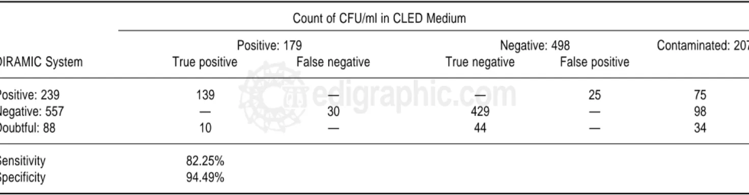
The results of urine cultures from the 884 urine speci-mens are shown in Table 1. The evaluation of DIRAMIC system in the detection of UTI generated 139 true positive
Table 1. Comparison of both urinary tract infections detection methods.
Count of CFU/ml in CLED Medium
Positive: 179 Negative: 498 Contaminated: 207
DIRAMIC System True positive False negative True negative False positive
Positive: 239 139 — — 25 75
Negative: 557 — 30 429 — 98
Doubtful: 88 10 — 44 — 34
Sensitivity 82.25%
edigraphic.com
results, 25 false positive, 429 true negative and 30 false negative for a sensitivity of 82.25% and specificity of 94.49%. Contaminated samples were not taken into ac-count.
Through the classical method, 179 urine cultures were positive. From them, 190 isolates were totally or partially identified (Table 2). In 11 samples, two different microor-ganisms were isolated from agar plates; six cases of them, two bacteria produced mixed infection of samples and in the other five, a second bacterium was identified as lacto-bacillus from normal vagina microbiota.
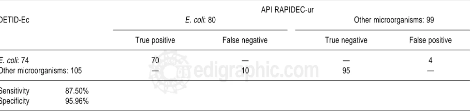
The results obtained in E. coli identification were 74 us-ing DETID-Ec and 80 by API RAPIDEC-ur (Table 3). Other microorganisms, including E. coli glucuronidase and/or indole negative, were 105 with DETID-Ec and 99 by API RAPIDEC-ur. The evaluation of DETID-Ec pro-duced 70 true positive results, 4 false positive, 95 true neg-ative and 10 false negneg-ative; the sensitivity and specificity were 87.50 and 95.96%, respectively.
DISCUSSION
The 20.25% of positive specimens obtained using the reference method are in agreement with reports of some other authors,6,14,20 also the 3.35% of multiple isolates.5,6,11
In this survey, 23.42% of the specimens were contaminat-ed, being their quality especially important in the reliabili-ty of results by rapid diagnostic methods.
Contaminated specimens must be discarded in the refer-ence method, because in solid culture media it is possible to notice colony morphological differences, so these sam-ples should be reanalyzed. Since in DIRAMIC system reading is made in liquid media, contaminated specimens became distributed among positive, negative or doubtful results. Therefore, contaminated samples taken as doubtful or negative can be discarded, being unnecessary further analysis. For instance, it was observed that the 88.64% of all doubtful results were negative (50.00%), nevertheless they include some colonies, or contaminated specimens (38.64%). In the remaining 11.36% growth indexes reached after four hours hampered positive results.
False positive results are in correspondence with con-taminated specimens or with low colony numbers in CLED Agar plates (<104 CFU/ml). On the other hand, false
nega-tive results were those where growth indexes did not reach required values, possibly due to low multiplication veloci-ties of microorganism, as well as intrinsic characteristics or presence of any inhibitory growth substance in urine.
The high specificity achieved (94.49%), together with small volumes of culture medium used and short times in analysis make this system a valuable alternative for screen-ing a higher amount of specimens in any clinical microbi-ology laboratory. Despite of a sensitivity not so high (82.25%), the use of the system as screening allows to dis-card just in few hours (four – five hours) negative speci-mens and therefore focusing work and resources in posi-tive cases.
Table 3. Comparison of both identification methods.
API RAPIDEC-ur
DETID-Ec E. coli: 80 Other microorganisms: 99
True positive False negative True negative False positive
E. coli: 74 70 — — 4
Other microorganisms: 105 — 10 95 —
Sensitivity 87.50%
Specificity 95.96%
Results refers to the 179 positive urine cultures in Petri dishes, not for the 190 isolates from these specimens. The four specimens with E. coli isolated in mixed infections were considered as “E. coli”.
Table 2. Identification of bacteria isolated from the 179 positive urine cul-tures.
Microorganism Number of isolates (%)
Escherichia coli 80 (42.10)
Escherichia coli glucuronidase negative 9 (4.74) Escherichia coli indole negative 1 (0.53)
Enterococcus 18 (9.47)
Klebsiella pneumoniae 14 (7.37)
Pseudomonas 11 (5.79)
Staphylococcus aureus 8 (4.21)
Enterobacter-Citrobacter 8 (4.21)
Proteus mirabilis 7 (3.68)
Other microorganisms 34 (17.89)
Travieso et al Evaluation of the DIRAMIC system for detection of urinary tract infections and for Escherichia coli identification
Rev Latinoam Microbiol 2004; 46 (3-4): 67-71 MG
70
edigraphic.com
:rop odarobale FDP VC ed AS, cidemihparG
arap
acidémoiB arutaretiL : cihpargideM sustraídode-m.e.d.i.g.r.a.p.h.i.c
As other commercial systems, sensitivity was higher when the threshold was ≥105 CFU/ml (86.67%), but the
necessary specificity of screening detection systems fell down to 92.81%. So, the use of one or the other threshold must be choice according to specific laboratory policies.
Regarding identification, the combinations for the six cases of mixed infection were as follows: two E. coli - En-terococcus, two Staphylococcus aureus - EnEn-terococcus, one E. coli - Staph. aureus and one E. coli - Pseudomonas. Although E. coli was the most frequent microorganism isolated (47.37%), its incidence as the main pathogen in urinary sepsis was rather low, compared to those reports from various authors indicating this bacterium as respon-sible for more than 70.00% of the cases. It should be con-sidered that a great number of specimens in this survey belongs to patients with anatomical or functional abnor-malities in their urinary tracts or with underlying illness-es, where some other microorganisms play a more impor-tant role.6,19,25
The identification method used as control detected 9 E.
coli glucuronidase negative strains. Since isolation cultures
medium CLED Agar contains lactose, β-glucuronidase ac-tivity is lowered or even inhibited in microorganisms growing there because of the acid produced during fermen-tation.2,3 This fact may be the cause of the high percentage
of glucuronidase negative strains (10.00%). This kind of strains is reported as “other microorganisms” by DETID-Ec diagnostic kit. They must be subsequently identified as
E. coli by conventional biochemical tests. So,
glucu-ronidase negative and false negative isolates can be cor-rectly identified but to a larger extent.
Moreover, the use of the DETID-Ec diagnostic kit makes possible to discard with 95.96% of specificity the microorganisms different from E. coli in infected urine specimens. The three false positive results observed were, according to RAPIDEC-ur strips, one Staph. aureus - En-terococcus, one Klebsiella oxytoca and one Proteus
vulgar-is - Morganella-Providencia. Previous surveys have
report-ed the presence of β-glucuronidase activity in some strains of Enterococcus and some other members of
Enterobacte-riaceae different from E. coli that are also urinary
patho-gens.3,17,21 The ability to produce indole from tryptophan is
also present in Kl. oxytoca and Pr. vulgaris.9,18
The occurrence of 10 false negative results obviously affected sensitivity. The results may be explained as insuf-ficient incubation times for a right expression of the en-zyme β-glucuronidase in assays and consequently a lack of release detectable amounts of 4- methylumbelliferone. Some other authors have observed the need of incubating some isolates overnight for fluorescence expression.15,17,24
Furthermore, rests of antibiotics in specimens from previ-ous therapies possibly delay the activity of β
-glucu-ronidase.10 Detection of trytophanase activity was
unaf-fected.
The 87.50% of the E. coli isolated were correctly identi-fied in six hours using the DETID-Ec diagnostic kit, with a great specificity, being valuable for this purpose. In addi-tion identificaaddi-tion of E. coli by means of β-glucuronidase and tryptophanase activities detection represents a promis-ing and economical option. Since rapid results in E. coli identification depend on microbial concentration, positive specimens having contaminants at low concentration can be detected through DETID-Ec.
In conclusion, rapid detection of UTI and identification of E. coli in direct urine specimens were possible with the use of DIRAMIC system and the DETID-Ec diagnostic kit. These functions are fulfilled with an acceptable sensitivity and high specificity. The reduction of workload, saving re-sources and time are benefits of the use of DIRAMIC sys-tem and DETID-Ec diagnostic kit.
ACKNOWLEDGEMENTS
We thank the staff of the Microbiology Laboratory of DIRAMIC Group for the specimens processed. We are also grateful to Celso Pérez and Jesús Núñez for their help in the manuscript’s translation and Iliana Vinardell for the analysis of data.
REFERENCES
1. Batchelor, B I F. 1995. Identification of urinary pathogens by the
RAPIDEC ur system. J. Med. Microbiol. 43:72-74.
2. Blondeau, J M., Y. Yaschuk, D. Galenzoski, D. Hrabok, M.
Isac-son, L. Lee, H. Link & L. Walshaw. 1995. Evaluation of the Cult-Dip Plus dip slide method for urinary tract infection. J. Clin. Pathol. 48:710-713.
3. Brenner, K P., C C. Rankin, Y R. Roybal, G N. Stelma, P V.
Scarpi-no & A P. Dufour. 1993. New medium for the simultaneous detec-tion of total coliforms and Escherichia coli in water. Appl. Environ. Microbiol. 59:3534-3544.
4. Bukharie, H A. & I M. Saeed. 2001. Antimicrobial Resistance
Among Pathogens Causing Acute Uncomplicated UTIs. Infect. Med. 18:358-362.
5. Doern, G V., R. Vautour, M. Gaudet & B. Levy. 1994. Clinical
im-pact of rapid in vitro susceptibility testing and clinical identifica-tion. J. Clin. Microbiol. 32:1757-1762.
6. Eisenstadt, J. & J A. Washington. 1996. Diagnostic microbiology
for bacteria and yeasts causing urinary tract infections, pp.29-66. In H L T. Mobley & J W. Warren (Eds). Urinary tract infections. Mo-lecular pathogenesis and clinical management. ASM Press. Wash-ington D.C.
7. Hawkey, P M. & D A. Lewis. 1989. Appendix III: Principles of
bio-chemical test for the identification of bacteria, pp.299-306. In Med-ical bacteriology, a practMed-ical approach. IRL Press. Oxford, New York and Tokyo.
8. Hernández, J E., O R. Contreras, P. Ponce, M. Armenteros & F.
edigraphic.com
9. Holt, J G., N R. Krieg, P H A. Sneath, J T. Staley & S T. Williams.
1994. Bergey’s Manual of Determinative Bacteriology, 9th ed. The Williams & Wilkins Co. Baltimore, Philadelphia, Hong Kong, Lon-don, Munich, Sydney and Tokyo.
10. Huang, S W., J J. Wu & T C. Chang. 1994. Enzyme capture assay for rapid identification of Escherichia coli in blood cultures. J. Clin. Microbiol. 32:1444-1448.
11. Khalifa, M A., A A. Abdoh, F G. Silva & D J. Flournoy. 1995. In-terpretation of multiple isolate urine cultures in adult male patients. J. Nat. Med. Assoc. 87:141-147.
12. Kodaka, H., M. Ishikawa, M. Iwata, F. Kashitani, S. Mizuochi & K. Yamaguchi. 1995. Evaluation of new medium with chromogenic substrates for members of the family Enterobacteriaceae in urine samples. J. Cli. Microbiol. 33:199-201.
13. Lowrance, B L., P. Reich & W H. Traub. 1969. Evaluation of two spot-indole reagents. Appl. Microbiol. 17:923-924.
14. Moya, G. & N. Armaignag. 1985. Evaluación de los resultados de urocultivos realizados a 1758 pacientes de consulta externa. Rev. Cub. Med. 24:463-467.
15. Palmer, C J., Y L. Tsai, L A. Lee & L R. Sangermano. 1993. Evalu-ation of Colilert-Marine Water for detection of total coliforms and
Escherichia coli in the marine environment. Appl. Environ.
Micro-biol. 59:786-790.
16. Serufo, J C., A. Barbosa, S. Goncalves, O. Guimaraes, I. Uehara, L R. Valle, H G. De Campos, V J. Diorio, O R. Contreras & A. Pas-cual. 1995. Diagnóstico Rápido da Infecção do Trato Urinário. Estudo Comparativo com o Método Convencional. J. Bras. de Mi-crobiol. 69:155-164.
17. Shadix, L C., M E. Dunnigan & E W. Rice. 1993. Detection of
Es-cherichia coli by the nutrient agar plus 4-methylumbelliferyl β -glu-curonide (MUG) membrane filter method. Can. J. Microbiol. 39: 1066-1070.
18. Sneath, P H A., N S. Mair, M E. Sharpe & J G. Holt. 1986. Bergey’s Manual of Systematic Bacteriology, Vol. 2, Section 12. The Will-iams & Wilkins Co. Baltimore, Hong Kong, London and Sydney.
19. Sobel, J D. & D. Kate. 1985. Urinary tract infections, pp.426-452. In G L. Mandell, R G. Douglas & J E. Bennett (Eds). Principles and practice of infectious diseases, 2nd ed. John Wiley & Sons. New York, Chichester, Brisbane, Toronto and Singapore. 20. Soro, O., M. Raggi & G C. Schito. 1996. Performance of
Uro-Quick, a new automated method for determination of bacteriuria. Galeno 4:31-40.
21. Tryland, I. & L. Fiksdal. 1998. Enzyme characteristics of β
-glucu-ronidase-positive bacteria and their interference in rapid methods for detection of waterborne coliforms and Escherichia coli. Appl. Environ. Microbiol. 64:1018-1023.
22. Turner, K M., L. Restaino & E W. Framptom. 2000. Efficacy of chromocult coliform agar for coliform and Escherichia coli detec-tion in foods. J. Food Prot. 63:539-541.
23. Vázquez, A. 1995. Infección urinaria en el adulto. Rev. Cub. Med. 34:106-117.
24. Villari, P., M. Iannuzzo & I. Torre. 1997. An evaluation of the use
of 4-methylumbelliferyl-β-D-glucuronide (MUG) in different solid
media for the detection and enumeration of Escherichia coli. J. Appl. Microbiol. 24:286-290.
25. Warren, J W. 1996. Clinical presentations and epidemiology of uri-nary tract infection, pp.3-27. In H L T. Mobley & J W. Warren (Eds). Urinary tract infections. Molecular pathogenesis and clinical management. ASM Press. Washington, D.C.
Correspondence to:
Dr. Rolando Contreras Alarcón.
Diagnostic Division, National Center for Scientific Research,
25 Ave and 158, Cubanacan, Playa, Havana, Cuba. P.O. Box 6990.
Phone: (537) 208-0959. Fax: (537) 208-7358.