PERMANYER www.permanyer.com
ORIGINAL ARTICLE Rev Inves Clin. 2015;67:250-7
Parecoxib Increases Blood Pressure
Through Inhibition of Cyclooxygenase-2
Messenger RNA in an Experimental Model
Ángel Antonio Vértiz-Hernández
1*, Flavio Martínez-Morales
2, Roberto Valle-Aguilera
2,
Pedro López-Sánchez
3, Rafael Villalobos-Molina
4and José Pérez-Urizar
51Coordinación Académica Región Altiplano, Universidad Autónoma de San Luis Potosí, Matehuala, S.L.P., México; 2Faculty of Medicine, Universidad Autónoma de San Luis Potosí, S.L.P., México; 3School of Medicine, Instituto
Politécnico Nacional, México, D.F., México; 4Faculty of Chemical Sciences, Universidad Autónoma de San Luis Potosí,
S.L.P., México; 5Faculty of Advanced Studies-Iztacala, Universidad Nacional Autónoma de México, Tlalnepantla,
Edo. de México, México
Corresponding author:
*Ángel Antonio Vértiz Hernández
Universidad Autónoma de San Luis Potosí Coordinación Académica Región Altiplano Carretera Cedral, Km. 5.600
Ejido San José de las Trojes
C.P. 78700, Matehuala, S.L.P., México
E-mail: [email protected] and [email protected] Received for publication: 03-06-2015 Accepted for publication: 11-08-2015
ABSTRACT
Background: Cyclooxygenase-2 selective inhibitors have been developed to alleviate pain and inflammation; however, the use of a selective cyclooxygenase-2 inhibitor is associated with mild edema, hypertension, and cardiovascular risk. Aim: To evaluate, in an experimental model in normotensive rats, the effect of treatment with parecoxib in comparison with diclofenac and aspirin and L-NAME, a non-selective nitric oxide synthetase, on mean arterial blood pressure, and cyclooxygenase-1 and -2 messenger RNA and protein expression in aortic tissue. Methods: Rats were treated for seven days with parecoxib (10 mg/kg/day), diclofenac (3.2 mg/kg/day), aspirin (10 mg/kg/day), or L-NAME (10 mg/kg/day). Mean arterial blood pressure was evaluated in rat tail; cyclooxygenase-1 and -2 were evaluated by reverse transcription-polymerase chain reaction and Western blot analysis in aortic tissue. Results: Parecoxib and L-NAME, but not aspirin and diclofenac, increased mean arterial blood pressure by about 50% (p < 0.05) without changes in cardiac frequency. Messenger RNA cyclooxygenase-1 expression in aortic tissue was not modified with any drug (p < 0.05). L-NAME and parecoxib treatment decreased messenger RNA cyclooxygenase-2 and cyclooxygenase-2 (p < 0.05). While cyclooxygenase-1 protein decreased with the three drugs tested but not with L-NAME (p < 0.05), the cyclooxygenase-2 protein decreased only with aspirin and parecoxib (p < 0.05). Conclusion: Parecoxib increases the blood pressure of normotensive rats by the suppression of COX-2 gene expression, which apparently induced cardiovascular control. (REV INVES CLIN. 2015;67:250-7)
Key words: Parecoxib. Hypertension. Cyclooxygenase-2. Prostaglandin I2. Thromboxane A2. mRNA inhibition.
.r
e
hsi
l
b
u
p
e
ht
f
o
n
ois
si
mr
e
p
ne
tti
r
w r
oir
p
e
ht
t
u
o
hti
w
g
ni
y
p
oc
ot
o
h
p r
o
de
c
u
d
or
pe
r
e
b
ya
m
n
oit
aci
l
b
u
p s
i
ht
f
o t
ra
p
o
N
INTRODUCTION
Cyclooxygenase-2 (COX-2) selective inhibitors have been developed to alleviate pain and inflammation, based on the finding that cyclooxygenase-1 (COX-1) is involved in the physiology of the gastrointestinal mucosa1,2 and is different from COX-2, which is
in-duced by inflammation3. Therefore, selective
inhibi-tion of COX-2 could dissociate anti-inflammatory activity from the gastrointestinal side effects of the steroidal anti-inflammatory drugs (NSAID, non-selective COX)4,5.
The selective inhibition induced by COX-2 may cause gastrointestinal side effects that are of lesser impor-tance compared with the cardiovascular side effects in relation to the physiological balance between the prothrombotic and vasoconstrictor actions of COX-1 and COX-2. The COX-1 is derived from thromboxane A2 (TXA2) in platelets, and the anti-aggregation and vasodilation actions of COX-2 derive from pros-taglandin I2 (prostacyclin, PGI2). Inhibition of COX-2 in endothelium in favor of platelet aggregation may implicate cardiovascular risk6,7. The original
hypoth-esis was that COX-2 inhibition only affects proin-flammatory prostaglandins; however, this was soon questioned and the discovery that COX-2 inhibition in humans suppressed the systemic biosynthesis of prostacyclin represented a breakthrough in the
risk-benefit assessment of coxibs8. Prostaglandin I2 is
a potent vasodilator and platelet inhibitor produced in blood vessels by the enzymatic activity of COX-1 and COX-2 and prostacyclin synthase. Thrombox-ane A2 has been shown in vitro and in vivo to modulate vasoconstrictor and platelet aggregator activities, and this COX-derived prostanoid is
pro-duced mainly by platelets activated via COX-1 dur-ing hemostasis9.
The use of a selective COX-2 in healthy individuals is associated with mild edema and hypertension due to modest sodium retention in the first days of therapy10.
Thus, studies have been conducted to analyze the risks arising from these drugs, including Vioxx Gastro-intestinal Outcomes Research (VIGOR; rofecoxib vs. naproxen)11, the Celecoxib Long-term Arthritis Safety
Study (CLASS; celecoxib vs. diclofenac and ibupro-fen)12, the Multinational Etoricoxib and Diclofenac
Ar-thritis Long-term (MEDAL; etoricoxib vs. diclofenac in a pooled analysis), and the Therapeutic Arthritis
Research and Gastrointestinal Event Trial (TARGET; lumiracoxib vs. ibuprofen and naproxen)4. The studies
showed that COX-2 inhibitors celecoxib and rofecoxib increase cardiovascular risks compared with placebo, although the studies do not clearly define the mecha-nisms for cardiovascular risk development. Parecoxib is one of the drugs in this group and is the first inject-able COX-2 selective inhibitor indicated for the treat-ment of acute postoperative pain; it is an inactive pro-drug that undergoes rapid amide hydrolysis in vivo into the pharmacologically metabolite-active valdecoxib13.
This drug has had widespread use because it offers an advantage over the other coxibs and has good anal-gesic effect in postoperative pain. As such, it com-prises a good therapeutic option in acute dental pain and in orthopedic and gynecological pain; however, many authors suggest that valdecoxib and parecoxib are both efficacious and well tolerated14. At present,
three coxibs (celecoxib, etoricoxib, and parecoxib) are authorized and marketed in several countries, with parecoxib preferably for hospital use15.
The adverse cardiovascular events observed in experi-mental models and in patients under selective COX-2 inhibition therapy could be explained by either a TXA2/ PGI2 imbalance or changes in the expression and/or activity of the COX isoforms. Therefore, in this study we developed an experimental model to evaluate the effect of the selective COX-2 inhibitor parecoxib in comparison with the non-selective COX inhibitors di-clofenac and aspirin, as well as water (negative con-trol) and L-NAME, a non-selective nitric oxide synthe-tase (as positive control of hypertension) on mean arterial blood pressure (MABP), and 1 and COX-2 messenger RNA (mRNA) and protein expression in the aortae of normotensive rats.
MATERIALS AND METHODS
Animals
Male Wistar Kyoto rats weighing 200-250 g each were housed in an environmentally controlled room with a 12-hour/12-hour light/dark cycle; they were given standard rodent chow and tap water ad libitum. All experimental procedures described here were approved by the local Animal Care Committee of the Autono-mous University of San Luis Potosi and followed the Declaration of Helsinki principles.
.r
e
hsi
l
b
u
p
e
ht
f
o
n
ois
si
mr
e
p
ne
tti
r
w r
oir
p
e
ht
t
u
o
hti
w
g
ni
y
p
oc
ot
o
h
p r
o
de
c
u
d
or
pe
r
e
b
ya
m
n
oit
aci
l
b
u
p s
i
ht
f
o t
ra
p
o
N
Treatments
The rats were acclimated to handling by humans pri-or to randomization, and then were divided into the following four groups of six rats each: (i) the control group received untreated drinking water; (ii) parecoxib group (Dynastat®, Pfizer Co., México), 10 mg/kg/day;
(iii) diclofenac group (Artrenac, Merck Co., México), 3.2 mg/kg/day; (iv) aspirin group (Pisa Laboratorios, Méxi-co), 10 mg/kg day; and (v) L-NAME group (L-NAME, Sigma-Aldrich Co.), 10 mg/kg day. All experimental drugs were administered in the animals’ drinking water, and the treatments were given for up to seven days.
Mean arterial blood pressure
measurements
Mean arterial blood pressure was measured daily by using the tail-cuff method with an LE 5002 Storage Pressure Meter (Letica Scientific Instruments, USA). Previous to treatment, the rats underwent a one-week adaptation period to avoid stress during measure-ments. Rats were placed in a temperature-controlled restriction chamber at 28°C during 10 minutes. The cuff was inflated automatically by means of a ret-roserver, and the pressures measured fell within the range of 30-300 mmHg. From each rat, heart rate, systolic blood pressure (SBP), and diastolic blood pres-sure (DBP) values were obtained. The MABP was cal-culated by the instrument that integrates the differ-ence between SBP and DBP in terms of time, which is calculated by means of the following equation: MABP = (DBP + SBP)/3. This procedure was repeated five times, every two minutes, during the 10-minute re-striction time. Results are expressed in mean mmHg ± standard error of the mean (SEM) (n = 6).
Total RNA isolation reverse transcription
At the end of the treatment period, the animals were euthanized by overexposure to the anesthetic (ether vapor). The thoracic aorta was immediately excised and placed on a phosphate-buffered saline (PBS)-pre-cooled plate. The aortic tissue was dissected from the adherent fat and connective tissues and was maintained on ice, then frozen in liquid nitrogen, and maintained at –80°C for subsequent analysis. Fifty milligrams of tho-racic aorta was dissected and immediately homoge-nized in 1 ml of Trizol™. Total RNA was isolated using 200 µl of chloroform, and the samples were centrifuged
at 12,000 rpm for 15 minutes at 4ºC. The superna-tant was collected, the RNA was precipitated with 500 µl of isopropanol for 10 minutes at –20ºC, and the integrity of the nucleic acid was evaluated by denaturing electrophoresis.
Reverse transcription of 5 µg of total RNA was per-formed using Oligo (dT) 12-18 primer (Invitrogen™) and the Moloney murine leukemia virus (M-MLV) re-verse transcriptase enzyme (Invitrogen™) during one hour at 37ºC. Subsequently, the complementary DNA (cDNA) of COX-1 and COX-2 was amplified by a spe-cific reverse transcription-polymerase chain reaction (RT-PCR) using the following primers (Life Technolo-gies), and glyceraldehyde 3-phosphate dehydrogenase (GAPDH) was used as the in-house control gene: 1 forward, 5’TAA-GTA-CCA-GGT-GCT-GGA-TGG; COX-1 reverse, 5’GGT-TTC-CCC-TCT-AAG-GAT-GAG-G; COX-2 forward, 5’TAC-AAG-CAG-TGG-CAA-AGG-C; COX-2 reverse, 5’CAG-TAT-TGA-GGA-GAA-CAG-ATG-GG; GAPDH forward, 5’AAC-ACA-GTC-CAT-GCC-ATC-AC, and GAPDH reverse, 5’TTC-ACC-ACC-CTG-TTG-CTG-TA. Thirty cycles of amplification were performed, consisting of denaturing at 94ºC for 60 seconds, anneal-ing at 64ºC for 60 seconds, and extension at 72 ºC for 60 seconds. The PCR amplification products (264 pb, 303 pb, and 360 pb for COX-1, COX-2, and GAPDH, respectively) were analyzed by the optical density of the bands. Results were expressed in arbitrary den-sity units (mean ± SEM) vs. the denden-sity of GAPDH.
Protein extraction and Western blot
Homogenates were prepared from 100 mg of tissue as follows: frozen rat thoracic aortae were suspended in 1 ml of cold Tris-hydrochloride buffer (100 mM Tris, pH 7.4) containing a protease inhibitor cocktail (9.9 × 10⁻³ mM PMSF, 0.09 mM TLCK, 2.07 × 10⁻³ mM,
and 1.25 × 10⁻³ mM IAA). The tissues were
homog-enized at 10,000 rpm for two minutes and immedi-ately centrifuged at 1,000 rpm for 10 minutes at 4 ºC in a refrigerated centrifuge (Sorvall Biofuge, Fresco). The pellet was discarded and the supernatant was stored in aliquots at –80 ºC. Total protein concentration was determined by the Bradford micromethod assay (Biorad). Immunoblotting was performed according to a standard protocol. Twenty micrograms per sample of total protein was subjected to sodium dodecyl sul-fate-polyacrylamide gel electrophoresis (SDS-PAGE), and the resolved proteins were electrotransferred onto
.r
e
hsi
l
b
u
p
e
ht
f
o
n
ois
si
mr
e
p
ne
tti
r
w r
oir
p
e
ht
t
u
o
hti
w
g
ni
y
p
oc
ot
o
h
p r
o
de
c
u
d
or
pe
r
e
b
ya
m
n
oit
aci
l
b
u
p s
i
ht
f
o t
ra
p
o
N
0 25 50
MABP (mmHg)
75 100 125 150 175
* *
Contr ol
Aspirin Diclof
enac Parec
oxib
L-NAME polyvinylidene difluoride (PVDF) membranes in a
Trans-Blot Cell (BioRad Labs, Hercules, CA, USA). The mem-branes were blocked with fresh TBS-T buffer (20 mM Tris-HCl, 150 mM NaCl, pH 7.5, 0.5% Tween 20) containing 5% fat-free milk for one hour at room temperature. The membranes were washed in TBS-T buffer and incubated overnight at 4ºC with the primary antibody against COX-1 (1:200), COX-2 (1:200), and
β-actin (1:1,000) (Santa Cruz Laboratories). After three
washes, the membranes were incubated for two hours with the horseradish peroxidase (HRP)-secondary anti-body diluted at 1:1,000, and a chemiluminescent sub-strate was added (Luminol, Santa Cruz Laboratories). The bands were quantified by densitometry, and the amount of each product was normalized with respect to the amount of β-actin (load control in Western blot).
Statistical analysis
Data are expressed as mean ± SEM and were ana-lyzed with the Dunnett test for analysis of variance (ANOVA) for multiple comparisons. A value of p < 0.05 was considered statistically significant.
RESULTS
Daily oral intake of parecoxib and L-NAME, but not of aspirin and diclofenac, resulted in a progressive in-crease in BP in normal rats from day 3 of treatment. By day 7 of treatment, MABP was 96 ± 14 mmHg in the control group, 102 ± 7 mmHg in the aspirin group, 97 ± 5 mmHg in the diclofenac group, 150 ± 17 mmHg in the parecoxib group (p < 0.05 vs. control), and 155 ± 13 mmHg in the L-NAME group (p < 0.05 vs. control). Parecoxib and L-NAME treatments increased MABP by 50% compared with the control group (Fig. 1). The heart rate was not modified throughout the observa-tion period: 409 ± 8 beats/minute in control vs. 380 ± 24 beats/minute in the L-NAME group and 439 ± 16 beats/minute in the parecoxib group, while there were no differences in the aspirin and diclofenac groups vs. the control group (data not shown).
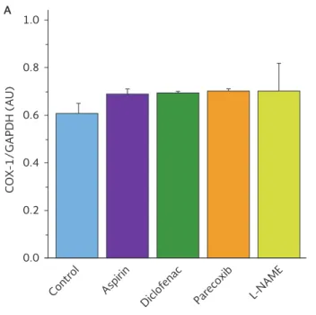
Expression of COX-1 mRNA in aortic tissue was un-changed by any of the treatments (Fig. 2 A). However, COX-2 gene expression was completely suppressed by parecoxib and L-NAME decreased COX-2 mRNA levels by about 50%. The non-selective COX inhibitors aspirin and diclofenac did not modify COX-2 mRNA expres-sion (Fig. 2 B).
In contrast to the mRNA results, COX-1 protein in aortic tissue was significantly reduced by all COX in-hibitors, that is, by parecoxib 68%, diclofenac 40%, and aspirin 72% (p < 0.05 vs. control), but no effect was observed with L-NAME (Fig. 3 A). With regard to COX-2, protein expression was reduced by 50% with aspirin and 22% with parecoxib (p < 0.05 vs. control), although no changes were observed with the diclof-enac and L-NAME treatments (Fig. 3 B). Protein ex-pression of COX-1 was significantly reduced (50%) with aspirin, diclofenac, and parecoxib compared with the control group (p < 0.05) (Fig. 3 A).
DISCUSSION
This experimental model was designed to compare the effect of mid-term exposure to selective and non-selective COX inhibitors on BP, and gene/protein ex-pression of COX-1 and COX-2 mRNA in aortic tissue of normotensive rats. Parecoxib increased MABP by sup-pressing COX-2 at the protein and mRNA levels (Fig. 1). Our study shows that parecoxib is associated with an important increase of MABP, contrary to the study by Chan, et al. in 200916, in which the authors reported Figure 1. Mean arterial blood pressure (mmHg) of rats (n = 6 per group) after seven days of treatment with the selective cyclooxygenase-2 inhibitor parecoxib compared with the non-selective cyclooxygenase inhibitors diclofenac and aspirin or the non-selective nitric oxide synthetase L-NAME (positive control for hypertension) or water (negative control for hyper-tension). Bars represent mean ± standard error of the mean (SEM). *Significant differences vs. control group (p <0.05). MABP: mean arterial blood pressure.
.r
e
hsi
l
b
u
p
e
ht
f
o
n
ois
si
mr
e
p
ne
tti
r
w r
oir
p
e
ht
t
u
o
hti
w
g
ni
y
p
oc
ot
o
h
p r
o
de
c
u
d
or
pe
r
e
b
ya
m
n
oit
aci
l
b
u
p s
i
ht
f
o t
ra
p
o
N
Contr ol
Aspirin Diclof
enac Parec
oxib
L-NAME
1.0
A B
0.8
0.6
0.4
C
O
X-1/GAPDH (A
U)
0.2
0.0
Contr ol
Aspirin Diclof
enac Parec
oxib
L-NAME
C
O
X-2/GAPDH (A
U)
*
*
0.0 0.2 0.4 0.6 0.8 1.0
Figure 2. A: messenger RNA (mRNA) expression of cyclooxygenase-1 determined by reverse transcriptase polymerase chain reac-tion in rat aortic tissue on day 7 of treatments. B: messenger RNA (mRNA) expression of cyclooxygenase-2 determined by RT-PCR in rat aortic tissue on day 7 of treatments. Bars represent the mean ± standard error of the mean (SEM) (n = 6). *Significant differences vs. control (p < 0.05).
COX: cyclooxygenase; GAPDH: glyceraldehyde 3-phosphate dehydrogenase; AU: arbitrary optical density units.
that rofecoxib and etoricoxib were related with hyper-tension but coxibs had little effect on blood pressure.
Thus, the final effect in MABP appears to be related with PGI2 and TXA2. Rudic, et al. in 200517
docu-mented that suppression of COX-2-derived PGI2 or deletion of the PGI2 receptor (IP) profoundly influ-ences the morphological response of the vasculature to hemodynamic stress. Mechanism-based vascular remodeling may interact with a predisposition to hy-pertension and atherosclerosis, contributing to the gradual transformation of cardiovascular risk during extended periods of treatment with selective COX-2 inhibitors. The COX-2 inhibitors suppress PGI2 biosyn-thesis without concomitant inhibition of TXA2, which derives predominantly from platelet COX-1.
Indeed, the seven-day treatment with the non-selec-tive COX inhibitors diclofenac or aspirin did not modify MABP. The effect observed may be explained by the selective inhibition of these drugs. Parecoxib produced an increase of nearly 50% in MABP, similar to that ob-served with the non-selective nitric oxide (NO) synthe-tase inhibitor L-NAME. It is known that the endothelium regulates vascular tone through the production of NO,
and also by prostacyclin and hyperpolarizing factors, thus strengthening the conclusions of studies in hu-mans or in obese Zucker rats, which have reported endothelium dysfunction in several vascular beds as-sociated with reduced NO bioavailability and hyper-tension18. The absence of NO is also associated with
reduced cardiac output, cardiac hypertrophy, large ar-eas of fibrosis, and myocardial necrosis, changes in myocardial contractility, and cardiomyocyte and vas-cular smooth-muscle remodeling19.
It appears that parecoxib may only be related with inhibition of COX-2, but not of COX-1 (Fig. 2 B and 2 A). COX-2 is an enzyme that may cause metabolic imbal-ance, resulting in an overproduction of harmful by-products that may damage the arterial wall and in-duce arterial blood clotting, increasing the risk for thromboembolic events4. When COX-2 is inhibited,
less PGI2 is synthesized from arachidonic acid and more leukotriene B4 and TXA2 are produced. The PGI2 is both vasodilator and anti-aggregator, while TXA2 is vasoconstrictor and pro-aggregator, and this tip of the balance allows TXA2 to function unopposed, leading to an increased risk for adverse cardiovascular events20. These changes may explain the increase in
.r
e
hsi
l
b
u
p
e
ht
f
o
n
ois
si
mr
e
p
ne
tti
r
w r
oir
p
e
ht
t
u
o
hti
w
g
ni
y
p
oc
ot
o
h
p r
o
de
c
u
d
or
pe
r
e
b
ya
m
n
oit
aci
l
b
u
p s
i
ht
f
o t
ra
p
o
N
0.0
80 KD –
80 KD – 41 KD –
41 KD –
80 KD –
80 KD – 41 KD –
41 KD – 0.2
0.4
C
O
X-1/
β
-a
ctin (A
U)
0.6 0.8 1.0 1.2 1.4
A B
0.0 0.2 0.4 0.6 0.8 1.0 1.2 1.4
* *
*
Contr ol
Aspirin Diclof
enac Parec
oxib L-NAME
C
O
X-2/
β
-a
ctin (A
U)
*
*
Contr ol
Aspirin Diclof
enac Parec
oxib L-NAME
COX-2 69.09 KD
β-actin 42 KD
COX-1 69.05 KD
β-actin 42 KD
Figure 3. A: expression of cyclooxygenase-1 in rat aortic cells. Cyclooxygenase-1 expression was determined by Western blot of protein amounts and semi-quantitated by densitometry. Results are the average of six separate experiments expressed as the cyclooxygenase-1/beta actin ratio. B: expression of cyclooxygenase-2 in rat aortic cells. Cyclooxygenase-2 expression was deter-mined by Western blot of protein amounts and semi-quantitated by densitometry. Results are the average of six separate experi-ments expressed as the cyclooxygenase-2/beta actin ratio. Bars represent the mean ± standard error of the mean (SEM) (n = 6). *Significant differences vs. control (p < 0.05).
COX: cyclooxygenase; AU: arbitrary optical density units.
heart rate, MABP, and COX-2 mRNA in the parecoxib and L-NAME groups in our study. The cardiovascular safety of coxibs is an important public health issue, considering the large number of predominantly elderly patients with osteoarthritis who present with a rela-tively high incidence of cardiovascular comorbidity, particularly hypertension, as has been shown21.
We also found in this study that parecoxib produced total suppression of COX-2 mRNA (Fig. 2 B), and that protein expression is decreased by 30% in the aorta of normotensive rats (Fig. 3 B); this same drug did not induce changes in COX-1 mRNA levels (Fig. 2 A). These
results were similar to those obtained with L-NAME, in-dicating that chronic suppression of NO synthase (NOS) may result in greater dependence on COX-2-derived PGI2 synthesis in terms of maintenance of vascular tone. Aspirin decreases the expression of the COX-1 protein, as well as COX-2, by 50 and 22%, respec-tively (Fig. 3 A and 3 B), the latter probably due to a 60% homology between the amino acids structure of COX-1 and COX-2. Aspirin binds to the residues of Ser 516 at the active site of COX-2, as well as to the residues of Ser 530 at the active site of COX-1, main-taining in apparent balance the products of the COX isoforms and their cardiovascular effects22. This may
.r
e
hsi
l
b
u
p
e
ht
f
o
n
ois
si
mr
e
p
ne
tti
r
w r
oir
p
e
ht
t
u
o
hti
w
g
ni
y
p
oc
ot
o
h
p r
o
de
c
u
d
or
pe
r
e
b
ya
m
n
oit
aci
l
b
u
p s
i
ht
f
o t
ra
p
o
N
explain the slight increase of MABP in our study (Fig. 1). In addition, a dynamic balance between the prosta-glandins PGI2 and TXA2 (and many other mediators) is crucial in maintaining cardiovascular homeostasis and has critical pathophysiological and therapeutic impli-cations23. Thus, some authors have found significant
individual variations in the response to coxibs due to a number of candidate genes, including, in many indi-viduals, CYP2C9, which is associated with a marked variability in the response to coxibs, although the im-portance of genetic variations with respect to cardio-vascular risk remains unknown24,25.
Some evidence from COX-2 inhibition trials has been published, suggesting that reduction in PGI2 may be associated with systemic hypertension in human sub-jects26, exacerbating the TXA2 function27. At any rate,
the results of our study showed that a decrease of COX-2 expression in aorta of rat by L-NAME could reflect the loss of positive regulation of NO in COX-2, a relationship previously demonstrated under several physiological and physiopathological conditions19.
Evi-dence from randomized clinical trials to determine the safety of prescribing non-selective NSAID and COX-2 inhibitors in patients with high cardiovascular risk is extremely limited. In a study with parecoxib/valde-coxib, patients randomized to intravenous parecoxib/ oral valdecoxib had a higher incidence of cardiovascu-lar events than patients receiving placebo (2.0 vs. 0.5%; p = 0.03)10. To add to the controversies of the
cardiovascular adverse effects of COX-2 inhibitors, several recent studies have shown that some COX-2 inhibitors are not associated with increased cardio-vascular risk. The SUCCESS-I trial found no increased cardiovascular risks of celecoxib compared with di-clofenac and naproxen in 13,274 patients with osteo-arthritis. The TARGET trial found no significant differ-ence in cardiovascular deaths between lumiracoxib and either ibuprofen or naproxen, irrespective of as-pirin use, in 18,325 patients with osteoarthritis20. As
with all of these drugs, the potential cardiovascular and gastrointestinal risks of their prescription needs to be weighed against the possible benefits for each individual patient and discussed with the patient. If the cardiovascular risk that increases with celecoxib is small and lower than that of most of the other NSAIDs, the concern would be of increasing complications in a pa-tient with high cardiovascular risk if the papa-tient were to be prescribed another NSAID11,28. Novel therapeutic
strategies in hypertension aim at reversing endothelial
dysfunction, which has been implicated in the patho-genesis and clinical course of hypertension and its cardiovascular complications23.
In conclusion, our results suggest that the COX-2 in-hibitor parecoxib increases the blood pressure of normo-tensive rats by suppression of COX-2 gene expression and reduced protein production that apparently induce cardiovascular lack of control of the arterial pressure.
ACKNOWLEGEMENTS
This work was supported by PROMEP (SESIC-SEP Mx) grant PROMEP/103.5/02/2351 to JP-U and by National Council of Science and Technology (CONACyT, México) grants 45910 to FM and 163383 to AAV-H. We thank PROMEP and CONACyT for financing this project, and also the Universidad Autóno-ma de San Luis Potosí, which provided the facilities for conducting the experiments, and the Coordinación Aca-démica Región Altiplano-UASLP.
REFERENCES
1. Adams RJ, Appleton SL, Gill TK, Taylor AW, Wilson DH, Hill CL. Cause for concern in the use of non-steroidal anti-inflammato-ry medications in the community -a population-based study. BMC Fam Pract. 2011;12:70.
2. Bessone F. Non-steroidal anti-inflammatory drugs: What is the ac-tual risk of liver damage? World J Gastroenterol. 2010;16:5651-61. 3. Wang Q, Takei Y, Kobayashi O, Osada T, Watanabe S. Cyclo-oxygenase 2 modulates killing of cytotoxic T lymphocytes by colon cancer cells. J Clin Biochemi Nutr. 2009;45:163-70. 4. Geusens P, Lems W. Efficacy and tolerability of lumiracoxib, a
highly selective cyclo-oxygenase-2 (COX2) inhibitor, in the management of pain and osteoarthritis. Ther Clin Risk Manag. 2008;4:337-44.
5. Eliassen AH, Chen WY, Spiegelman D, Willett WC, Hunter DJ, Hankinson SE. Use of aspirin, other NSAIDs, and acetaminophen and risk of breast cancer among premenopausal women in the Nurses’ Health Study II. Arch Intern Med. 2009;169:115-21. 6. Chiu Y-J, Huang T-H, Chiu C-S, et al. Analgesic and
Antiinflam-matory Activities of the Aqueous Extract from Plectranthus amboinicus (Lour.) Spreng. Both In Vitro and In Vivo. Evid Based Complement Alternat Med. 2012;2012:508137.
7. Brueggemann LI, Mackie AR, Mani BK, Cribbs LL, Byron KL. Dif-ferential effects of selective cyclooxygenase-2 inhibitors on vas-cular smooth muscle ion channels may account for differences in cardiovascular risk profiles. Mol Pharmacol. 2009;76:1053-61. 8. Selg E, Buccellati C, Andersson M, et al. Antagonism of
throm-boxane receptors by diclofenac and lumiracoxib. Br J Pharmacol. 2007;152:1185-95.
9. Seta F, Rahmani M, Turner PV, Funk CD. Pulmonary oxidative stress is increased in cyclooxygenase-2 knockdown mice with mild pulmonary hypertension induced by monocrotaline. PLoS One. 2011;6:e23439.
10. Dajani EZ, Islam K. Cardiovascular and gastrointestinal toxicity of selective cyclo-oxygenase-2 inhibitors in man. J Physiol Phar-macol. 2008;59:117-33.
11. McKellar G, Singh G. Celecoxib in arthritis: relative risk manage-ment profile and implications for patients. Ther Clin Risk Manag. 2009;5:889-96.
12. Hegazy R, Alashhab M, Amin M. Cardiorenal effects of newer NSAIDs (celecoxib) versus classic NSAIDs (ibuprofen) in pa-tients with arthritis. J Toxicol. 2011;2011:862153.
13. Elseify ZA, El-Khattab SO, Khattab AM, Atta EM, Ajjoub LF. Combined parecoxib and I.V. paracetamol provides additional
.r
e
hsi
l
b
u
p
e
ht
f
o
n
ois
si
mr
e
p
ne
tti
r
w r
oir
p
e
ht
t
u
o
hti
w
g
ni
y
p
oc
ot
o
h
p r
o
de
c
u
d
or
pe
r
e
b
ya
m
n
oit
aci
l
b
u
p s
i
ht
f
o t
ra
p
o
N
analgesic effect with better postoperative satisfaction in patients undergoing anterior cruciate ligament reconstruction. Saudi J An-aesth. 2011;5:45-9.
14. Wei W, Zhao T and Li Y. Efficacy and safety of parecoxib sodium for acute postoperative pain: A meta-analysis. Exp Ther Med. 2013;6:525-31.
15. Faura Giner CC, D’Ocon Navaza P. Medicina Basada en la Eviden-cia de los AINEs y COXIBs. Act Farm Ter. 2013;11:98-107. 16. Chan CC, Reid CM, Aw TJ, Liew D, Haas SJ, Krum H. Do COX-2
inhibitors raise blood pressure more than nonselective NSAIDs and placebo? An updated meta-analysis. J Hypertens. 2009; 27:2332-41.
17. Rudic RD, Brinster D, Cheng Y, et al. COX-2 derived prostacyclin modulates vascular remodeling. Circ Res. 2005;96:1240-7. 18. Vessières E, Belin de Chantemèle EJ, Toutain B, et al.
Cyclooxygen-ase-2 inhibition restored endothelium-mediated relaxation in old obese Zucker rat mesenteric arteries. Front Physiol. 2010;1:145. 19. Ferreira-Melo SE, Demacq C, Lacchini S, Krieger JE, Irigoyen MC,
Moreno H. Sildenafil preserves diastolic relaxation after reduction by L-NAME and increases phosphodiesterase-5 in the intercalated discs of cardiac myocytes and arterioles. Clinics. 2011;66:1253-8. 20. Ong CKS, Lirk P, Tan CH, Seymour RA. An evidence-based
up-date on nonsteroidal anti-inflammatory drugs. Clin Med Res. 2007;5:19-34.
21. Cheng I, Liu X, Plummer SJ, Krumroy LM, Casey G, Witte JS. COX2 genetic variation, NSAIDs, and advanced prostate cancer risk. Br J Cancer. 2007;97:557-61.
22. Vane JR, Botting RM. The mechanism of action of aspirin. Thromb Res. 2003;110:255-8.
23. Stitham J, Midgett C, Martin KA, Hwa J. Prostacyclin: An inflam-matory paradox. Front Pharmacol. 2011;2:24.
24. Solomon SD, Wittes J, Finn PV, et al. Cardiovascular risk of ce-lecoxib in 6 randomized placebo-controlled trials: The Cross Trial Safety Analysis. Circulation. 2008;117:2104-13.
25. Zerbini LF, Tamura RE, Correa RG, et al. Combinatorial effect of non-steroidal anti-inflammatory drugs and NF-κB inhibitors in ovarian cancer therapy. PLoS One. 2011;6:e24285.
26. Arehart E, Stitham J, Asselbergs FW, et al. Acceleration of car-diovascular disease by a dysfunctional prostacyclin receptor mutation, potential implications for COX-2 inhibition. Circ Res. 2008;102:986-93.
27. Smyth EM. Thromboxane and the thromboxane receptor in car-diovascular disease. Clin Lipidol. 2010;5:209-19.
28. Boonriong T, Tangtrakulwanich B, Glabglay P, Nimmaanrat S. Comparing etoricoxib and celecoxib for preemptive analgesia for acute postoperative pain in patients undergoing arthroscop-ic anterior cruciate ligament reconstruction: a randomized con-trolled trial. BMC Musculoskelet Disord. 2010;11:246.
.r
e
hsi
l
b
u
p
e
ht
f
o
n
ois
si
mr
e
p
ne
tti
r
w r
oir
p
e
ht
t
u
o
hti
w
g
ni
y
p
oc
ot
o
h
p r
o
de
c
u
d
or
pe
r
e
b
ya
m
n
oit
aci
l
b
u
p s
i
ht
f
o t
ra
p
o
N