Endometrial wave-like activity in the non-pregnant uterus
I.van Gestel
1, M.M.IJland, H.J.Hoogland and J.L.H.Evers
Department of Obstetrics and Gynecology, Research Institute GROW, University Hospital of Maastricht, Maastricht, the Netherlands
1To whom correspondence should be addressed at: P. Debeyelaan 25, P.O. Box 5800, 6202 AZ Maastricht, the Netherlands. E-mail: ivge@sgyn.azm.nl
The non-pregnant uterus shows wave-like activity throughout the menstrual cycle. This uterine activity was ®rst detected using intra-uterine pressure recordings. The use of ultrasound has made it possible to study the movements of the uterus in a non-invasive manner. Throughout the menstrual cycle, several wavelike activity patterns have been described; these patterns change throughout the menstrual cycle and are governed by steroid hormones. An adequate wave pattern seems to be related to successful reproduction in spontaneous cycles and assisted reproduction. Further insight into the phenomenon of endometrial wave-like activity might offer an opportunity to correct abnormal wave patterns and thereby improve pregnancy rates.
Key words:endometrial waves/endometrial wave classi®cation system/non-pregnant uterus/transvaginal ultrasound/uterine
contractions
Introduction
The non-pregnant uterus is not a quiescent organ. Its contractility has been described with the help of various methods. One of the ®rst scienti®c reports on this subject was published in 1937 by Dickinson, who described the possibility of registering uterine contractions by bimanual palpation (Dickinson, 1937).
Contractions and their characteristics, such as frequency, amplitude and basal pressure, have been examined for the various phases of the menstrual cycle with the aid of intra-uterine pressure transducers. Ultrasound has made it possible to study various aspects of uterine activity in a non-invasive manner, by visualiza-tion of endometrial wave-like activity resulting from sequential segmental contractions in the subendometrial myometrium.
Intra-uterine pressure recordings
While Heinricius (1889) was the ®rst to describe a method to record activity in the non-pregnant uterus with a balloon system, several other authors have subsequently used closed-tip intra-uterine catheters to measure intra-uterine activity (Knaus, 1933; Moir, 1944; Csapo and Pino-Dantas, 1966). The results of the various studies applying this method were highly variable, mostly because of calibration problems due to variations in the size of the balloons, the volume of ¯uid introduced in the balloons, and the amount of pressure in the balloons at the onset of the recording period. The large volume of the balloon also acted as a potential uterine irritant.
The intra-uterine open-tip catheter, which was introduced by Hendricks (1964), theoretically affected the contractility of the
uterus less than the closed catheters. Almost all authors who studied uterine contractility by this method produced similar qualitative assessments of the different contraction types throughout the cycle. During menstruation, regular labour-like contractions with a high intensity, high amplitude, low frequency and low basal pressure were described. In the proliferative phase, the pressure and frequency increased while amplitude and intensity decreased. In the peri-ovulatory phase, the intensity was found to be low and the frequency high, while at ovulation, small, high-frequency contractions with high pressure were seen. In the second half of the cycle, towards menstruation, the intensity and amplitude increased and the frequency decreased (Hendricks, 1965; 1966; Cibils, 1967; Bengtsson, 1968; Eskes et al., 1969; 1970; Braaksma, 1970; Yoshida and Hendricks, 1970; Hein, 1972). However, the absolute values measured by the various investigators often differed (Table I).
Ultrasound studies
Since 1984, when Birnholz ®rst used the transabdominal ultrasound technique to study movements in the non-pregnant uterus, ultrasound has been used to study uterine activity in a non-invasive manner.
Intra-uterine pressure recordings provide information about frequency, amplitude and basal pressure tone. Ultrasound can also record contraction frequency, but its major asset is that it adds another dimension to the recordings, namely direction. This allows observation of the contractions that travel through the uterus in waves to be made, and their direction, frequency and velocity to be ascertained. It has been suggested recently that
by guest on October 6, 2012
http://humupd.oxfordjournals.org/
ultrasound is as effective as intra-uterine pressure recordings for measuring the frequency of uterine activity (Bullettiet al., 2000). The composition of the patient groups used in ultrasound studies has varied widely, and the moments of measurement have not always been standardized (Table II). In addition, the measuring methods used have also been variable. Some authors did not use taped video recordings and there has been no uniformity in recording and playback times (Table II). Classi®cation of the movements has also differed, in terms of the tissue movement and the direction (Table III), as well as in the characteristics of the movements (Table IV).
The results of several ultrasound studies are summarized in Table V. In general, the ultrasound ®ndings of the studies performed show movements from fundus to cervix during the
early follicular phase. Movements from cervix to fundus have been described in the late follicular and peri-ovulatory phases, and most investigators have also described an increased intensity of movements towards ovulation.
A classi®cation system for describing the different activity types has also been proposed (IJlandet al., 1996).
Endometrial wave classi®cation system
One group (IJlandet al., 1996) carried out transvaginal ultrasound examinations using a 7.5 mHz transducer. At each examination, between 3 and 15 min of video images were recorded of the uterus in the mid-sagittal plane. If no uterine activity was observed over a period of 3±5 min, the recording was discontinued. Off-line
Table I.Results of intra-uterine pressure recordings, using an open-tip catheter, in the non-pregnant uterus
Reference Menses Pre-ovulatory phase Post-ovulatory phase Hendricks (1964; 1966) Labour-like contractions
F: 1 per 1±3 min I: 50±200 mmHg
Early follicular: F: 1 per 30±60 s, I: 20±50 mmHg
Peri-ovulatory: F: 1 per 15±20 s,
I: 5±29 mmHg (small, frequent contractions)
Mid-luteal: slow and complex contractions Towards menstruation: pre-labour-like
Cibils (1967) End of menstruation: F: 1 per 3 min D: 1 min Moderate intensity
Mid-follicular: F: 1 per 25±40 s, D:<20 s Late follicular: F: 1 per 10±15 s, brief, irregular-shaped contractions
Early luteal: F: 1 per 20±40 s; low intensity
Mid-luteal: slow ¯uctuating contractions, D:>1 min
Late luteal: slow strong contractions Bengtsson (1968) Pre-labour-like Early follicular: 2±3 small contractions
per min Large contractions with high amplitudeand low frequency Eskeset al. (1969; 1970) Low frequency,
low basal pressure, considerable intensity
Follicular:frequency and basal pressure,
¯intensity Peri-ovulatory: high frequency, low intensity, high basal pressure (>20 mmHg)
Luteal:
¯basal pressure and frequency,
intensity Braaksma (1970) B: 4.9 mmHg,
I: 18.6 mmHg, A: 13.6 mmHg D: 35 s, high interval
Mid-follicular/late follicular: B: 22.9 mmHg/15.8 mmHg I: 28.7 mmHg/18.7 mmHg A: 5.2 mmHg/2.9 mmHg D: 18 s/11 s
Luteal: lowest amplitude, shortest duration Pre-menstrual: strongest change in amplitude, duration and interval
Yoshida and
Hendricks (1970) CD 1±3:Labour-like pattern, low resting pressure
CD 11: small contractions, high frequency, resting pressure 15.4 mmHg
CD 13:intensity, resting pressure 26.9 mmHg
CD 15: slightly decreased frequency CD>18: contractile pattern forms complexes,
¯resting pressure
CD 23: pre-labour-like contractions Hein (1972) Strong labour-like
contractions Early follicular: frequent smallcontractions,basal pressure,¯duration
Mid-follicular:basal pressure and frequency,
¯duration, intensity and amplitude Late follicular: high basal pressure, wandering baseline,frequency, low intensity
Early luteal:¯basal pressure, frequency and amplitude,duration
Late luteal:intensity and amplitude,
¯basal pressure and duration
Bullettiet al. (2000) frequency towards mid-cycle ¯frequency and furtherof amplitude, with maximum in late luteal phase Bullettiet al. (2002) Patients with endometriosis:
higher frequency, amplitude and basal pressure
A = amplitude of contractions; B = basal pressure of contractions; CD = cycle day; D = duration of contractions; F = frequency of contractions; I = intensity of contractions.
by guest on October 6, 2012
http://humupd.oxfordjournals.org/
analysis was performed at high speed (four times normal) replay. This method seems comparable with those reported previously (de Vries et al., 1990; Lyons et al., 1991; Leyendecker et al., 1996), in which the analysis was conducted at ®ve times normal speed.
IJland's endometrial wave classi®cation system distinguished ®ve types of wave-like activity (Table VI). Waves from fundus to cervix dominated during the follicular phase, and no fundus-to-cervix waves were seen after ovulation. A putative function of fundus-to-cervix waves is that of cleansing the uterine cavity and creating a barrier to ascending pathogens. Waves from cervix to fundus were recorded predominantly in the late follicular and the luteal phases. The cervix-to-fundus waves are assumed to promote sperm transport (Abramowics and Archer, 1990) and to restrict the implantation of the embryo to the upper uterine cavity (IJlandet al., 1996). This phenomenon has also been described by others (Lyons et al., 1991; Kunzet al., 1996). The latter group mimicked sperm transport in the follicular phase by the placement of labelled macrospheres of sperm size at the external cervical os.
By using serial hysterosalpingoscintigraphy it was shown that sperm reach the uterine cavity within minutes. The proportion of macrospheres entering the tubes is highest during the late follicular phase, when there is maximum frequency and intensity of the endometrial waves. The transport of the macrospheres was preferably directed into the tube ipsilateral to the dominant follicle (Kunzet al., 1996). These authors hypothesized that the direction of the endometrial wave towards the ipsilateral tube might be controlled by a speci®c myometrial architecture in combination with a asymmetric distribution of myometrial estrogen receptors. Furthermore, human semen contains large amounts of prostaglandins which may also promote the phenomenon of directed endometrial waves and isthmic tubal relaxation (Coutinho and Maia, 1971)
Opposing waves, which were observed during the ®rst days after ovulation, might help prepare the endometrium for successful nidation and also assist in providing the pre-implantation embryo with nutrients and oxygen (IJland et al., 1996).
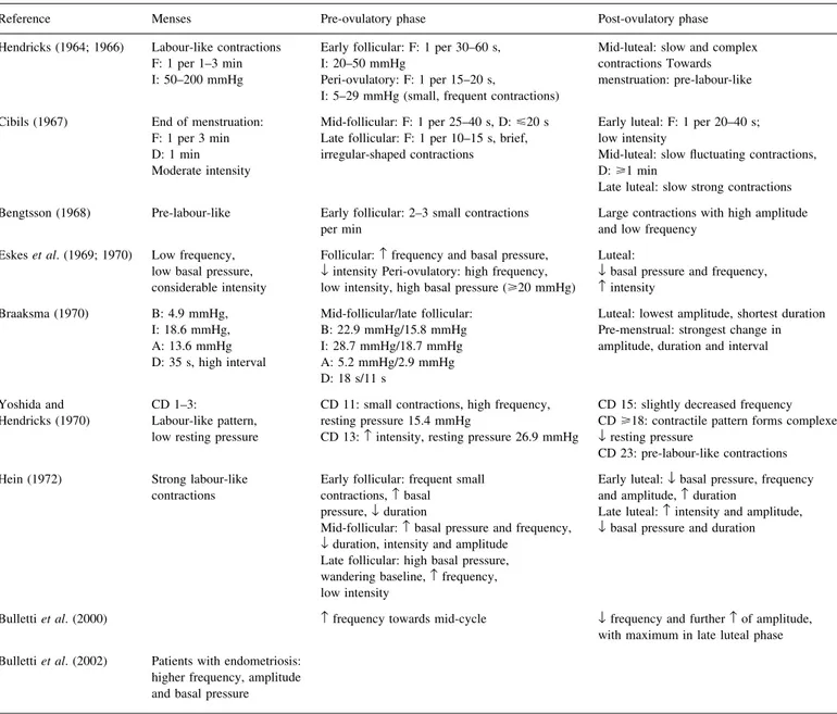
Table II.Ultrasound studies of movements in the non-pregnant human uterus. [Adapted from IJland (2000), with permission.]
Reference Year US
abd/vag Patients(n) Observations(n) Patients Moments in cycle Record time/video/replay3normal speed Birnholz (1984) 1984 + / ± 26 32 Spont, clomid ? 30 s/ ±
Oikeet al. (1990) 1990 ± / + 21 ? Spont ?
de Vrieset al. (1990) 1990 + / + 35 35 Spont 1st and 2nd half cycle 120 s / + / 53
Abramowics and Archer
(1990) 1990 ± / + a. 15b. 10 c. 18
21 cycles 15 cycles 18 cycles
Spont Oac IVF
a+b. CD 5/6, 10/11, 13/15, 20/22
c. CD 5/6, hCG, ET+2 ? / + and ±/ ? Lyonset al. (1991) 1991 ± / + 18 328 Spont Menstrual, follicular
Peri-ovulatory, luteal 120 s / + / 53 Chalubinskiet al. (1993) 1993 ± / + 53 a. 14
b. 9 c. 20 d. 10
Spont a. CD 2 b. CD 7/8 c. Ovulatory phase
d. CD 20/22 ? / +/ ? Fukuda and Fufuda (1994) 1994 ± / + a. 72
b. 18 c. 11
180 cycles 75 cycles 18 cycles
Spont Clomid Gn
Mid-follicular (1±2 times) Late follicular; daily Luteal (3±4 times)
2 min / + / ?
Salamanca and Beltran
(1995) 1995 ± / + 37 37 EndometriosisSpont CD 1/2 5 min / + / ? Kunzet al. (1996) 1996 ± / + 36 a. 11
b. 17 c. 8
Spont a. Early follicular b. Mid-follicular c. Late follicular
5 min / + / ?
Leyendeckeret al. (1996) 1996 ± / + 66 a. 15 b. 27 c. 39 d. 19 e. 18
Endometriosis
Spont a. Menstruationb. Early follicular c. Mid-follicular d. Late follicular e. Luteal
5 min / + / 53
IJlandet al. 1996 1997 1998 1999
± / + ± / + ± / + ± / +
16 47 19 21
23 cycles 59 cycles 35 cycles 21 cycles
a. Spont b. Spont c. Gn d. IVF
a+b. Early, mid- and late follicular; early, mid- and late luteal c. Start, 23stim period, hCG, hCG+2, hCG+6, hCG+9 d. Start, 33stim period, hCG, OPU, ET, hCG+7 (d)
3±15 min / + / 43
Fanchinet al. 1998
2001 ± / +± / + 20943 22043 IVFIVF Before ETHCG, hCG+4, hCG+7 5 min / + / 102 min / ± / ?3
abd = abdominal; CD = cycle day; clomid = clomiphene citrate; ET = embryo transfer; Gn = gonadotrophins; Oac = oral contraceptives; OPU = ovum pick-up; spont = spontaneous cycles; US = ultrasound; vag = vaginal.
by guest on October 6, 2012
http://humupd.oxfordjournals.org/
In spontaneous menstrual cycles and stimulation cycles wave frequency can be analysed according to the presence of unidirectional waves (fundus-to-cervix and cervix-to-fundus). There is an increase in the frequency of fundus-to-cervix waves from the mid-follicular to the late follicular phase, while the frequency of cervix-to-fundus waves also increases from the mid-follicular phase towards the early luteal phase (IJlandet al., 1996;
Kunz et al., 1996). After ovulation, there is a reduction in the
frequency of contractions, which might optimize the contact between the blastocyst and the endometrium to facilitate implantation (Fanchinet al., 1998a; 2001b). Kunz and colleagues reported that during the luteal phase, the upper fundal part of the uterus shows a relative quiescence facilitating embryo implanta-tion (Kunzet al., 2000b).
Wave velocity can be calculated for the unidirectional wave types. There is a trend towards increasing wave velocity in fundus-to-cervix waves from the mid-follicular to the late follicular phase (IJland et al., 1997b). Cervix-to-fundus waves seem to attain their highest velocity in the peri-ovulatory phase, underlining their putative role in rapid sperm transport, and these ®ndings corroborate the results of other studies (Abramowics and Archer, 1990; Lyons et al., 1991). Because of the low inter-observer reproducibility and the complexity of measurements, the wave velocity measurements do not as yet appear to be appropriate for clinical application (IJlandet al., 1997b).
Endometrial waves and fecundability
The contractility pattern in the non-pregnant uterus appears to play a role in the reproductive process. In spontaneous cycles, the endometrial wave type is related to fecundability (IJland et al., 1997a). Conception cycles show a predominance of waves from cervix to fundus in the peri-ovulatory phase, whereas waves from fundus to cervix are seen only during the late follicular phase and have never been detected after ovulation (IJland et al., 1997a; Fanchin et al., 1998b). Conception cycles show an overall dampening of endometrial wave activity during the consecutive phases of the cycle. Both dampening of activity and ®ne-tuning of the activity patterns at mid-cycle appear to be prerequisites for successful embryo implantation (Fanchinet al., 1998a).
The endometrial wave pattern is also related to the occurrence of pregnancy in IVF cycles (IJland et al., 1999). The relative distribution of the different endometrial wave types in the stimulation period showed more fundus-to-cervix waves in conception cycles than in non-conception cycles. After adminis-tration of hCG, fundus-to-cervix waves were no longer detected in either group. Most of the IVF cycles also showed a switch from fundus-to-cervix to cervix-to-fundus waves, referred to as the wave direction switch (WDS). A premature switch from fundus-to-cervix to cervix-to-fundus wavesÐthat is, an early WDSÐis associated with a reduced pregnancy prognosis (29%), while a
Table IV.Aspects of movements in the non-pregnant human uterus studied by ultrasound. [Adapted from IJland (2000), with permission.]
Reference Direction Frequency Amplitude Strength (intensity) Symmetry
Birnholz (1984) ± + ± ± ±
Oikeet al. (1990) + + + + ±
de Vrieset al. (1990) + + + ± +
Abramowics and Archer (1990) ± + + + ±
Lyonset al. (1991) + + + ± ±
Chalubinskiet al. (1993) + + ± ± +
Fukuda and Fufuda (1994) + ± ± ± ±
Salamanca and Beltran (1995) + ± ± ± ±
Kunzet al. (1996) + + ± + ±
Leyendeckeret al. (1996) + + ± ± ±
IJlandet al. (1996) + + ± ± ±
Fanchinet al. (1998) + + ± ± ±
Table III.Description of uterine movements in the non-pregnant human uterus by ultrasound. [Adapted from IJland (2000), with permission.]
Reference Description of uterine movement Direction Birnholz (1984) Stripping movements endometrium ±
Oikeet al. (1990) Endometrial movements Towards fundus
de Vrieset al. (1990) Inner one-third myometrium contractions Antegrade/retrograde Abramowics and Archer (1990) Endometrial peristalsis ±
Lyonset al. (1991) Subendometrial myometrial contractions ±
Chalubinskiet al. (1993) Myometrial contractions Towards fundus and cervix Fukuda and Fufuda (1994) Endometrial echo free space Horizontal, vertical Salamanca and Beltran (1995) Subendometrial contractility Antegrade, retrograde Kunzet al. (1996)/Leyendeckeret al. (1996) Uterine peristalsis Fundo-cervical, cervico-fundal
IJlandet al. (1996) Endometrial waves Cervix to fundus, fundus to cervix, opposing, random Fanchinet al. (1998a) Uterine contractions Antegrade, retrograde, antagonistic, non-propagated
by guest on October 6, 2012
http://humupd.oxfordjournals.org/
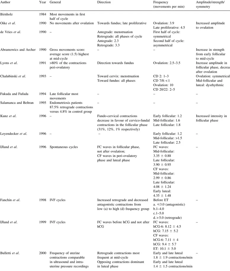
Table V.Results of ultrasound studies of movements in the non-pregnant uterus. (Adapted from IJland, 2000, with permission)
Author Year General Direction Frequency
(movements per min) Amplitude/strength/symmetry Birnholz 1984 Most movements in ®rst
half of cycle
Oikeet al. 1990 No movements after ovulation Towards fundus; late proliferative Ovulation: 3.9
Late proliferative: 4.5 Increased amplitudeto ovulation de Vrieset al. 1990 ± Antegrade: menstruation
Retrograde: all phases of cycle Antegrade: 2.3
Retrograde: 3.3
First half of cycle: symmetrical Second half of cycle: asymmetrical Abramowics and Archer 1990 Gross movements score:
average score (1.5) highest at mid-cycle
± ± Increase in strength from early follicular to mid-cycle Lyonset al. 1991 >80% of the contractions
peri-ovulatory Direction towards fundus Ovulation: 2.5±3.5 Increase amplitude infollicular phase, decrease after ovulation
Chalubinskiet al. 1993 ± Toward cervix: menstruation
Toward fundus: all phases CD 2: 1±3CD 7/8: < 1 Ovulation: 10 CD 20/22: 2±5
Ovulation: symmetrical Mid-follicular and luteal: dysrhythmic Fukuda and Fufuda 1994 Late follicular most
movements ± ± ±
Salamanca and Beltran 1995 Endometriosis patients 87.5% retrograde contractions versus 4.8% in control group
± ± ±
Kunzet al. 1996 ± Fundo-cervical contractions
decrease in favour of cervico-fundal contractions in the follicular phase (31%, 12%, 1% respectively)
Early follicular: 1.2 Mid-follicular: 1.6 Late follicular: 1.8
Increased intensity in follicular phase
Leyendeckeret al. 1996 ± ± Early follicular: 1.2 Mid-follicular: >1.5 Late follicular: 2.5
±
IJlandet al. 1996 Spontaneous cycles FC waves in follicular phase, not after ovulation; CF waves in peri-ovulatory phase and luteal phase
FC waves: Mid-follicular: 3.3560.88 Late follicular: 3.9060.95 CF waves: Mid-follicular: 2.9960.86 Late follicular: 4.0861.24 Early luteal: 4.3561.48
±
Fanchinet al. 1998 IVF cycles Increased retrograde and decreased antagonistic contractions from low (a) to high (d) frequency group
Before ET
a.<3.0 (antagonistic) b.1±4.0
c.1±5.0
d. > 5.0 (retrograde)
±
IJlandet al. 1999 IVF cycles FC waves before hCG and not after
hCG FC waves:hCG-6: 8.1264.5 hCG: 7.1565.2 CF waves: hCG-6: 7.1164 hCG: 9.465.7 ET: 10.165.0
±
Bullettiet al. 2000 Frequency of uterine contractions comparable in ultrasound and intra-uterine pressure recordings
Retrograde contractions most frequent at mid-cycle
Opposing contractions dominant in luteal phase
Early and late luteal 1.861.9 contractions/min Early and late luteal 1.461.5 contractions/min
±
CD = cycle day; CF = cervix±fundus; ET = embryo transfer; FC = fundus±cervix.
by guest on October 6, 2012
http://humupd.oxfordjournals.org/
late WDS is accompanied by a very favourable prognosis of pregnancy (88%). The persistence of fundus-to-cervix waves until the day of hCG administration appears to help a good quality embryo to implant (IJlandet al., 1999).
As was the case in controlled ovarian stimulation (COS) cycles, one group (IJland et al., 1998) showed that endometrial wave activity was more pronounced in IVF cycles. A statistically signi®cant difference was found in wave frequency, which was higher in IVF cycles than in spontaneous cycles (Abramowics and Archer, 1990; IJlandet al., 1999). Although the latter group could not ®nd any relationship between the frequency of endometrial waves and the occurrence of pregnancy in IVF cycles, others (Fanchinet al., 1998b; 2001b) demonstrated a negative correla-tion between the frequency of endometrial waves on the day of embryo transfer and pregnancy outcome. High-frequency en-dometrial waves on the day of embryo transfer appear to affect IVF-embryo transfer outcome in a negative manner, perhaps by expelling embryos from the uterine cavity.
Effect of steroid hormones on endometrial waves
The cycle-dependent pattern of endometrial waves presumes a role for steroid hormones. First, endometrial activity is most pronounced in the peri-ovulatory phase, with high estrogen levels. Also, more endometrial activity is seen in IVF cycles and COS cycles with high estrogen levels, compared with spontaneous cycles. The WDS during the course of the cycle appears to be governed by estrogens and progesterone.
To study this effect of steroids, spontaneous cycles were mimicked in post-menopausal women by giving the patients estradiol (days 1±28) and progesterone (days 14±28) (IJland, 2000). Ultrasound examinations have revealed that the prevalence of spontaneous endometrial wave-like activity is low or absent after menopause. Endometrial waves increase after estrogen administration, with waves from fundus to cervix being recorded in the estradiol-only phase of the cycle and vanishing immediately after the administration of progesterone. Waves from cervix to fundus are seen in both the only phase and the progesterone phase, with a maximum at the end of the estradiol-only phase.
Besides inducing a WDS, progesterone appears to have a relaxing effect in the non-pregnant uterus (Fanchinet al., 1998b; 2000). The contention that the endometrial wave-like activity is predominantly governed by ovarian steroid hormones has also been supported by others (Knaus, 1933; Bengtsson and Theobald, 1965; Oikeet al., 1990; Bullettiet al., 1993; Batra, 1994; Kunz
et al., 1998)
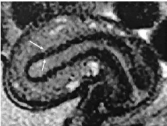
Anatomy
The described endometrial waves are initiated in the subendo-metrial myometrium or junctional zone, and this can be identi®ed using either magnetic resonance imaging (MRI) (Brosens, 1995) (Figure 1) or ultrasound (Kunz et al., 2000b). Like the endometrium, the subendometrial myometrium exhibits a cyclic pattern of estrogen receptor and progesterone receptor expression
(Noeet al., 1999).
The outer layer of the uterus, the non-paramesonephric myometrium, is formed in a later embryological stage and appears to have functions that are predominantly con®ned to parturition (Noe et al., 1999). This outer portion of the uterine wall does not exhibit a cyclic pattern of estrogen and progesterone receptor expression (Noeet al., 1999).
Conclusions
The endometrium in the non-pregnant uterus shows distinct activity patterns throughout the menstrual cycle, which are called endometrial waves. These waves originate in the subendometrial myometrium and are in¯uenced by steroids. The subendometrial activity might be initiated by pacemakers in the uterine muscle, providing the mechanical phenomenon of endometrial waves. A relative quiescence of the uterus and an adequate wave pattern in certain phases of the cycle are related to successful reproduction (Bullettiet al., 1997; IJlandet al., 1997a).
Uterine factors that have so far been implicated in decreased fertility, are the presence of severe intra-uterine adhesions, deformity of the uterine cavity by ®broids, and congenital abnormality of uterus or cervix (Taylor and Collins, 1992). The existence of an abnormal wave pattern in a subfertile patient could be another uterine factor explaining subfertility, and this should be actively sought in future. Other examinations, such as endometrial biopsy, studying the texture of the endometrium by ultrasound, and hysteroscopy to reveal the presence of intra-uterine lesions, contribute little to the understanding of the cause of otherwise unexplained infertility (Taylor and Collins, 1992).
Whilst in assisted reproduction there exists an important role for the pattern and frequency of endometrial waves, more insight
Table VI.Endometrial wave classi®cation system (IJlandet al., 1996)
Fundus-to-cervix waves (FC) Waves from fundus to cervix Cervix-to-fundus waves (CF) Waves from cervix to fundus
Opposing waves (OPP) Starting simultaneously at fundus and cervix Random waves (R) Starting at various foci
No activity
Figure 1.Magnetic resonance image of the uterus in the mid-sagittal plane. The arrows indicate the subendometrial myometrium or junctional zone.
by guest on October 6, 2012
http://humupd.oxfordjournals.org/
is required into the phenomenon of these endometrial waves and the factors which in¯uence them, especially those of a hormonal nature. This might provide an opportunity to correct an abnormal wave pattern or to modulate wave frequency to improve pregnancy rates, for example by exogenously changing hormone levels (Fanchinet al., 2001a). Uterine contractility might also be stimulated by the use of prostaglandins (PG), for example the vaginal administration of a PGE1 analogue (misoprostol) in women undergoing intra-uterine insemination (Brown et al., 2001). If a premature WDS should occur in IVF cycles, it could be hypothesized that it would perhaps be better to freeze the embryos and postpone the embryo transfer to another cycle.
Abnormal wave patterns or dysperistalsis have been described by two groups (Salamanca and Beltran, 1995; Bulletti et al., 2002), both of which detected abnormal activity patterns in women with endometriosis. Others (Leyendecker et al., 1996) identi®ed a relationship between this abnormal activity, endome-triosis and impairment of directed sperm transport, resulting in lower pregnancy rates. Furthermore, documentation of the endometrial wave patterns might contribute to an understanding of conditions such as dysmenorrhoea, endometriosis and habitual abortion.
In the near future, MRI techniques might offer another means of detecting endometrial activity and comparing the results with transvaginal ultrasound ®ndings.
Further investigations are important to gain more insight into the phenomenon of endometrial waves. Moreover, improving the uniformity of recording these waves, as well as gathering details of their description and activity, might add to an understanding, diagnosis and treatment of unexplained fertility disorders.
References
Abramowics, J.S. and Archer, D.F. (1990) Uterine endometrial peristalsis: a transvaginal ultrasound study.Fertil. Steril.,54, 451±454.
Batra, S. (1994) Hormonal control of myometrial function. In Chard, T. and Grudzinskas, J.G. (eds)The Uterus. Press Syndicate of the University of Cambridge, Cambridge, United Kingdom.
Bengtsson, L.P. (1968) The sponge catheter; a modi®cation of the open end catheter for recording of myometrial activityin vivo.J. Reprod. Fertil.,
16, 115.
Bengtsson, L.P. and Theobald, G.W. (1968)J. Physiol.(London),178, 46. Birnholz, J.C. (1984) Ultrasonic visualisation of endometrial movements.
Fertil. Steril.,41, 157±8.
Braaksma, J.T. (1970)Drukregistratie in de niet zwangere uterusin vivo. Thesis, Amsterdam.
Brosens, J.S. (1995) Uterine junctional zone: function and disease.Lancet,
346, 558±560.
Brown, S.E., Toner, J.P., Schnorr, J.A., Williams, S.C., Gibbons, W.E., de Ziegler, D. and Oehninger, S. (2001) Vaginal misoprostol enhances intrauterine insemination.Hum. Reprod.,16, 96±101.
Bulletti, C., Prefetto, R.A., Bazzocchi, G., Romero, R., Mimmi, P., Polli, V., Assuero Lanfranchi, G., Labate, A. and Flamingi, C. (1993) Electromechanical activities of human uteri during extra-corporeal perfusion with ovarian steroids.Hum. Reprod.,8, 1558±1563.
Bulletti, C., DeZiegler, D., Rossi, S., Polli, V., Massoneau, M., Rossi, E., Albonetti, A., Negrini, V. and Flamigni, C. (1997) Abnormal uterine contractility in non-pregnant women.Ann. N. Y. Acad. Sci.,828, 223±229. Bulletti, C., DeZiegler, D., Polli, V., Diotallevi, L., Del Ferro, E. and Flamigni, C. (2000) Uterine contractility during the menstrual cycle.Hum. Reprod.,
15(Suppl. 1), 81±89.
Bulletti, C., DeZiegler, D., Polli, V., Del Ferro, E., Palini, S. and Flamingi, C. (2002) Characteristics of uterine contractility during menses in women with mild to moderate endometriosis.Fertil. Steril.,77, 1156±1161. Chalubinski, K., Deutinger, J. and Bernaschek, G. (1993) Vaginosonography
for recording of cycle-related myometrial contractions.Fertil. Steril.,59, 225±228.
Cibils, L.A. (1967) Contractility of the nonpregnant uterus.Obstet. Gynecol.,
30, 441±459.
Coutinho, E.M. and Maia, H.S. (1971) The contractile response of human uterus, fallopian tubes, and ovary to prostaglandinsin vivo.Fertil. Steril.,
22, 539±543.
Csapo, A.I. and Pino-Pantas, C.R. (1966) The cyclic activity of the nonpregnant human uterus. A new method for recording intrauterine pressure.Fertil. Steril.,17, 34±38.
De Vries, K., Lyons, E.A., Ballard, G., Levi, C.S. and Lindsay, D. (1990) Contractions of the inner third of the myometrium. Am. J. Obstet. Gynecol.,162, 679±682.
Dickinson, R.L. (1937) The technique of timing human ovulation by palpable changes in ovary, tube and uterus. Am. J. Obstet. Gynecol.,33, 1027± 1033.
Eskes, T.K.A.B., Hein, P.R., Kars-Villanueva, E.B., Braaksma, J.T., Janssens, J. and Kollerie, A. (1969) The in¯uence of steroids on the motility of the non-pregnant human uterus in vivo. Arch. Int. Pharmacodyn.,182, 409.
Eskes, T.K.A.B., Hein, P.R., Stolte, L.A.M., Kars-Villanueva, E.B., Crone, A., Braaksma, J.T. and Janssens, J. (1970) In¯uence of dydrogestrone on the activity of the non-pregnant human uterus.Am. J. Obstet. Gynecol.,106, 1235±1241.
Fanchin, R., Righini, C. and Ayoubi, J.M. (1998a) Uterine contractions at the time of embryo transfer: a hind implantation ?Contracept. Fertil. Sex,26, 498±505.
Fanchin, R., Rhigini, C., Olivennes, F., Taylor, S., de Ziegler, D. and Frydman, R. (1998b) Uterine contractions at the time of embryo transfer alter pregnancy rates after in-vitro fertilization.Hum. Reprod.,13, 1968± 1974.
Fanchin, R., Ayoubi, J.M., Olivennes, F., Righini, F., de Ziegler, D. and Frydman, R. (2000) Hormonal in¯uence on the uterine contractility during ovarian stimulation.Hum. Reprod.,15(Suppl. 1), 90±100.
Fanchin, R., Rhigini, C., DeZiegler, D., Olivennes, F., Ledee, N. and Frydman, R. (2001a) Effects of vaginal progesterone administration on uterine contractility at the time of embryo transfer.Fertil. Steril., 75, 1136±1140.
Fanchin, R., Ayoubi, J.-M., Rhigini, C., Olivennes, F., Schonauer, L.M. and Frydman, R. (2001b) Uterine contractility decreases at the time of blastocyst transfers.Hum. Reprod.,16, 1115±1119.
Fukuda, M. and Fufuda, K. (1994) Uterine endometrial cavity movements and cervical mucus.Hum. Reprod.,9, 1013±1016.
Hendricks, C.H. (1964) A new technique for the study of motility in the non-pregnant human uterus.J. Obstet. Gynaecol.Br. Commonw.,71, 712±715. Hendricks, C.H. (1965) Activity patterns in the nonpregnant human uterus. In Paul, W.M., Daniel, E.E., Kay, E.M. and Monckton, G. (eds),The Muscle. Pergamon Press, New York.
Hendricks, C.H. (1966) Inherent motility patterns and response characteristics of the nonpregnant uterus.Am. J. Obstet. Gynecol.,96, 824±843. Hein, P.R. (1972)De contractiliteit van de uterus tijdens de menstruele cyclus
en na toediening van geslachtshormonen. Thesis, Amsterdam.
Heinricius, G. (1889) En metod att gra®skt atergiva kontraktioner hos en icke gravid livmoder.Finsk Lakaresalist Handl.,31, 349.
IJland, M.M. (2000)Endometrial wavelike activity in the nonpregnant uterus. Thesis, Maastricht.
IJland, M.M., Evers, J.L.H., Dunselman, G.A.J., van Katwijk, C., Lo, C.R. and Hoogland, H.J. (1996) Endometrial wavelike movements during the menstrual cycle.Fertil. Steril.,65, 746±749.
IJland, M.M., Evers, J.L.H., Dunselman, G.A.J., Volovics, L. and Hoogland, H.J. (1997a) Relation between endometrial wavelike activity and fecundability in spontaneous cycles.Fertil. Steril.,67, 492±496. IJland, M.M., Evers, J.L.H. and Hoogland, H.J. (1997b) Velocity of
endometrial wavelike activity in spontaneous cycles. Fertil. Steril.,68, 72±75.
IJland, M.M., Evers, J.L.H., Dunselman, G.A.J. and Hoogland, H.J. (1998) Endometrial wavelike activity, endometrial thickness, and ultrasound texture in controlled ovarian hyperstimulation cycles.Fertil. Steril.,70, 279±283.
IJland, M.M., Hoogland, H.J., Dunselman, G.A.J., Lo, C.R. and Evers, J.H.L. (1999) Endometrial wave direction switch and the outcome ofin vitro
fertilization.Fertil. Steril.,71, 476±481.
Knaus, H. (1933) Zur technik der Registration von bewegungen der menschlichen gebaÈrmutter.Zentrallblatt fuÈr gynakologie,45, 2658±2662. Kunz, G., Beil, D., Deininger, H., Wildt, L. and Leyendecker, G. (1996) The
by guest on October 6, 2012
http://humupd.oxfordjournals.org/
dynamics of rapid sperm transport through the female genital tract: evidence from vaginal sonography of uterine peristalsis and hysterosalpingography.Hum. Reprod.,11, 627±632.
Kunz, G., Noe, M., Herbertz, M. and Leyendecker, G. (1998) Uterine peristalsis during the follicular phase of the menstrual cycle: effects of oestrogen, antioestrogen and oxytocin. Hum. Reprod. Update, 5, 647±654.
Kunz, G., Beil, D., Huppert, P. and Leyendecker, G. (2000a) Structural abnormalities of the uterine wall in women with endometriosis and infertility visualized by vaginal sonography and magnetic resonance imaging.Hum. Reprod.,15, 76±82.
Kunz, G., Kissler, S., Wildt, L. and Leyendecker, G. (2000b) In Filicori, M. (eds),Endocrine Basis of Reproductive Function.Monduzzi.
Leyendecker, G., Kunz, G., Widt, L., Beil, D. and Deininger, H. (1996) Uterine hyperperistalsis and dysperistalsis as dysfunctions of the mechanism of rapid sperm transport in patients with endometriosis and infertility.Hum. Reprod.,11, 1542±1551.
Lyons, E.A., Taylor, P.J., Zheng, X.H., Ballard, G., Clifford, C.S. and Kredentser, J.V. (1991) Characterization of subendometrial myometrial
contractions throughout the menstrual cycle in normal fertile women.
Fertil. Steril.,55, 771±774.
Moir, J.C. (1944) The effect of posterior lobe pituitary gland fractions on the intact uterus.J. Obstet. Gynaecol. Br. Empire,51, 181±197.
Noe, M., Kunz, G., Herbertz, M., Mall, G. and Leyendecker, G. (1999) The cyclic pattern of the immunocytochemical expression of oestrogen and progesterone receptors in human myometrial and endometrial layers: characterization of the endometrial-myometrial unit.Hum. Reprod.,14, 190±197.
Oike, K., Ishihara, K. and Kikuchi, S. (1990) A study on endometrial movement and serum hormonal level in connection with uterine contraction.Nippon Sanka Fujinka Gakkai Zasshi,1, 86±92.
Salamanca, A. and Beltran, E. (1995) Subendometrial contractility in menstrual phase visualized by transvaginal sonography in patients with endometriosis.Fertil. Steril.,64, 193±195.
Taylor, P.J. and Collins, J.A. (eds) (1992) Unexplained Infertility. Oxford University Press, Oxford, United Kingdom.
Yoshida, T. and Hendricks, C.H. (1970) Resting pressure patterns in the nonpregnant human uterus.Am. J. Obstet. Gynecol.,108, 450±457.
by guest on October 6, 2012
http://humupd.oxfordjournals.org/
![Table II. Ultrasound studies of movements in the non-pregnant human uterus. [Adapted from IJland (2000), with permission.] Reference Year US](https://thumb-us.123doks.com/thumbv2/123dok_es/5549456.121543/3.918.52.837.136.706/ultrasound-studies-movements-pregnant-adapted-ijland-permission-reference.webp)