Introduction
The Ursidae family comprises three lineages: Ursinae, Tremarctinae and Hemicyoninae (a basal subfamily of Ursidae that is recorded in the Miocene and Pliocene of North America and has some mor-phological affinities with tremarctines); Amphi-cyonidae is the sister group of Ursidae (Wyss and Flynn, 1993).
The four genera of the bear subfamily Tremarctinae (Carnivora: Ursidae) are distributed exclusively in America. 1) PlionarctosFrick is record-ed from the Late Miocene to the Early Pliocene of North America with two species: P. edensisFrick and P. harroldorumTedford and Martin. 2) Arctodus Leidy
contains two North American Late Pliocene and Pleistocene species: A. pristinus Leidy and A. simus (Cope). 3) ArctotheriumBravard includes five South American species: A. latidens Bravard, restricted to the Ensenadan (Early to Middle Pleistocene), A. ve-tustumAmeghino registered only in the Bonaerian (Middle Pleistocene), and three Bonaerian - Lujanian (Middle Pleistocene to Early Holocene) species A. brasiliense(Lund), A. bonariense(Gervais) and A. tari-jense Ameghino (Soibelzon, 2002). Finally, 4) Tremarctos Gervais has two species T. floridanus (Gildey) from the Late Pliocene and Pleistocene of North America, and the only living Tremarctinae T. ornatus(Cuvier) of South America, which has not yet been recorded as a fossil. Regarding the origin of the South American fossil bears, it is noteworthy that these bears arrived in South America from North America during the Great American Biotic Interchange (Marshall et al, 1984).
Departamento Científico Paleontología de Vertebrados, Museo de La Plata, Paseo del Bosque, 1900 La Plata, Buenos Aires, Argentina. CONICET. lsoibelzon@museo.fcnym.unlp.edu.ar
Deciduous teeth morphology of some tremarctines
(Ursidae, Tremarctinae). Descriptions, comparissons and
possible evolutive implications
Leopoldo SOIBELZON and Alfredo CARLINI
Key words. Ursidae, Tremarctinae, Arctotherium, deciduous tooth.
Palabras clave. Ursidae, Tremarctinae, Arctotherium, dientes deciduos.
Abstract. The morphology of some deciduous teeth of Arctotherium tarijense and A. latidens (Ursidae: Tremarctinae) is here described and compared for the first time. The crown morphology of dP4 and dp4 is similar to that of M1 and m1. The general morphology of dP/dp4 in A. tarijenseand Ursinae is similar, but their permanent morphology dentition is different. The dP/dp4 of A. tarijense seems to bear a gener-alized set of characters (i.e. crown outline, the protocone and parastyle of dP4, the metaconid and hypoconid of dp4); unfortunately these teeth cannot be compared with their homologues in other tremarctines. Consequently, we are unable to include these characters in the phylogenetic analysis of the Tremarctinae subfamily in order to know whether the phylogenetic relationships are affected or not. It is noteworthy, that if these characters were present in M/m1 of A. tarijense, at least, some of them could be undoubtedly regarded as plesiomorphic features. In this respect, is the deciduous teeth morphology more conservative than that of permanent teeth?
The Tremarctinae are a monophyletic and sister taxon to the Ursinae (Trajano and Ferrarezzi, 1994; Talbot and Shields, 1996). Plionarctos and Tremarctos constitute the basal clade of the Tremarctinae (spec-tacled bears), and Arctodus and Arctotherium (short-faced bears) later diverging taxa. Within the clade formed by the five Arctotheriumspecies, A. vetustum and A. brasilienseare more basal than A. latidens, and A. bonariense and A. tarijense are later diverging species (Soibelzon, 2002).
During the taxonomic and phylogenetic revision of the South American fossil bears (Soibelzon, 2002), one us (LS) found an incomplete juvenile specimen of A. tarijensewith deciduous teeth, two isolated milk teeth of A. tarijense? and another one of A. latidensin old collections of Museo de La Plata and Museo Argentino de Ciencias Naturales “Bernardino Rivadavia”.
Many studies have been made of the morphology of deciduous teeth of ursines such as Ursus arctosand U. spelaeus (see Koby, 1952; Radulescu and Samson, 1959; Terzea, 1969; Torres, 1988 and references therein) but there is no available information for tremarctines.
Kraglievich (1934) reported the discovery of de-ciduous teeth of South American short-faced bears, found in 1923 at Km 150 of “Canal 9” (at 4 m depth), Buenos Aires Province, Argentina. These teeth were associated with a femur and tibiae of an adult speci-men of A. tarijense. The present study includes the re-examination of this material, the first morphological comparison of deciduous teeth of tremarctines and high quality anatomical illustrations. In addition, a radiograph of a juvenile mandible of A. tarijense is shown. Unfortunately, no deciduous teeth of the sin-gle extant Tremarctos ornatus, are known. Since com-monly only low quality photographs of bears decid-uous teeth published in old papers that are really dif-ficult to access, are available, we expect that this study will provide an identification key for tremarc-tine deciduous teeth in other collections and, useful characters for phylogeny. Our results suggest that some characters of the milk dentition of A. tarijense are significant to understanding the evolution of cheek teeth patterns and as source of characters for future phylogenetic analyses.
Abbreviations.MACN: Museo Argentino de Ciencias Naturales “Bernardino Rivadavia”; MHJ: Museo Histórico de Junín, Junín, Argentina; MLP: Museo de La Plata, La Plata, Argentina. MNHN AC Museum National d’Histoire Naturelle, Anatomie Comparée, Paris, France. MDL: maximum mesiodistal length; BLL: maximum buccolingual length. d: deciduous tooth.
Material and methods
A. tarijenseAmeghino. MACN 8582 left premaxil-la fragment with dI2 and I1-2 in their alveoli; left
maxilla fragment with dP3 and dP4; upper dC; left mandible fragment with dp4, and i1-3, lower C, p1, p4, m1 and m2 in their alveoli; right mandible frag-ment; one metapode and two phalanges, from Km 150 of “Canal 9”, Buenos Aires Province, Argentina; Lujanian (Late Pleistocene).
A. tarijense ? MLP 92-XI-28-1, right dP4, from Camet Norte, Mar Chiquita, Buenos Aires Province, Argentina; Lujanian (Late Pleistocene) (24.450 ( 150 14C yr BP, at 37º 49´S / 57º 29´ W; see Pardiñas et al., 1998). MLP 92-XI-27-1, left dP4, from Centinela del Mar 38° 26´ S / 58° 14´ W, General Alvarado, Buenos Aires Province, Argentina; Belgranian (Last Interglacial ca. 130 Ka, Isla et al., 2000).
A. latidens MACN 6132, right upper dC, from Miramar, Buenos Aires Province, Ensenadan (Early-Middle Pleistocene).
The specimen MACN 8582 is identified as A. tari-jensebecause it has also the m1-2, but the other two (MLP 92-XI-28-1 and MLP 92-XI-27-1) are only isolat-ed dP4 and it is impossible to make a secure specific determination, for this reason we identified them as A. tarijense?
Specimens of Ursinae used for comparisons: Melursus ursinusMNHN AC 10998
Ursus arctosMNHN AC 1896-346 and those de-scribed in Koby (1952), Terzea (1969) and Torres (1988).
Morphological terms and measurements defini-tions follow Koby (1952) and Torres (1988), but we numbered the deciduous premolars mesiodistally as Terzea (1969) and all recent authors (ie. the last de-ciduous premolar is dP/dp 4) to facilitate compar-isons. Measurements were taken with dial calipers to the nearest mm.
Description
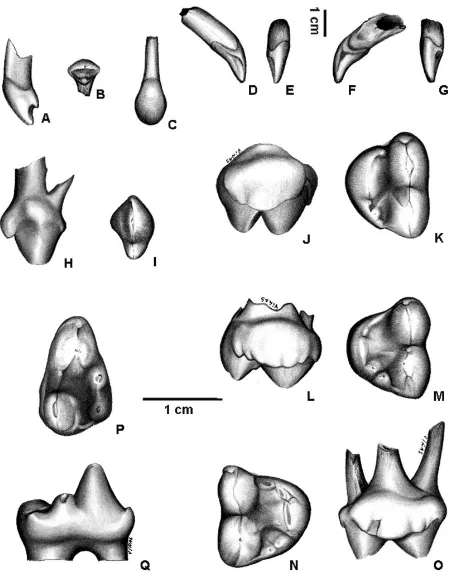
The morphology of the second upper deciduous incisor (dI2) (MACN 8582) is quite different from that of the corresponding permanent tooth (figures 1A-C, table I). In labial view, the upper margin of the crown is rounded, and the mesial and distal margins join toward the base of the crown. In occlusal view, the tooth is sub-triangular in section. An enamel crest crosses the lingual margin mesio-distally, and in the middle of this crest a low cusp is developed. The crown is implanted obliquely and slightly rotated mesially. The root is mesio-distally compressed and sub-triangular in section, as well as the crown.
and the mesial side is slightly flattened. The crown basal margin on the mesial side descends sharply and then ascends forming a V-shaped notch. At the lowest point of this notch there is a protuberance with two enamel crests obliquely diverging, one de-scending toward the apex of the crown, and the oth-er rising through the tooth neck. The uppoth-er decidu-ous canine MACN 6132 (figures 1F-G) of A. latidensis morphologically similar to that of A. tarijense but more robust (table I). The edge that crosses the lin-gual side is much less conspicuous in A. latidens than in A. tarijense. This specimen also shows more wear on the apex of the crown and on the mesial pro-tuberance, the latter produced by abrasion against the lower canine.
The third upper deciduous premolar crown (dP3) (MACN 8582) is laterally compressed, elongated mesio-distally (figures 1H-I, table I), and
mesio-dis-tally rotated. Two conspicuous cusps are aligned, the mesial one (paracone?) is much higher and has a wider base than the distal one (metacone?); a mesiodistally oriented enamel ridge connects both cusps. This ridge is thickest on the mesial margin of the crown base of the anterior cusp, where a small cuspule is developed. The dP3 has two roots, the mesial one being thinner.
The fourth upper deciduous premolar crown (dP4) is slightly wider than long in MACN 8582, but longer than wide in MLP 28-1 and MLP 92-XI-27-1 (Table I). The labial margin is almost straight and longer than the lingual margin, which is very convex (figures 1J-O). The mesial and distal margins are almost straight and converge toward the lingual side. In oclusal view the crown is triangle-shaped. The two labial cusps are elongated mesiodistally, and both are much larger than the lingual cusps. The mesial cusp (paracone) is larger and slightly com-pressed than the distal cusp (metacone). The para-cone is lingually inclined, while the metapara-cone is ver-tical. A small but well differentiated cusp, the parastyle, arises in front of the paracone. Between the paracone and the metacone a very small cusp is de-veloped in MLP 92-XI-27-1. A deep furrow runs mesiodistally between the labial and lingual cusps. On the lingual margin of the crown there are four or five relatively low cusps arranged in a mesiodistal series. The first cusp of the series is small and lies at the mesio-lingual angle. This cusp forms a continu-ous crest with the central cusp (protocone), which is larger. In two of the three specimens studied (MLP 92-XI-28-1 and MLP 92-XI-27-1) the protocone has two apices nearly indistinguishable one from each other (figures 1M-N). Two small cusps are behind the protocone, in front of the metacone, the distal cusp somewhat lower than the proximal. Two of the three specimens show on the lingual side of the metacone, an enamel crest descending toward the center of the crown. In one specimen (MACN 8582) it merges with another crest that comes from the two distal cusps of the lingual margin (Figure 1K), and in the other (MLP 92-XI-27-1) it is in direct contact with both dis-tal cusps (Figure 1M). No cingulum is observed, oth-er than a slight widening of the enamel running along the labial side of the metacone in one of the three specimens (MLP 92-XI-27-1). Deciduous P4 is the only deciduous tooth with three roots, two labial roots that belong to the paracone and the metacone, and one lingual root that corresponds to the series of cusps of the lingual margin.
[image:3.595.62.289.80.368.2]The crown of the fourth deciduous premolar (dp4) (MACN 8582) is transversely compressed, the labial and distal margins are almost straight, the lin-gual margin is slightly convex and the mesial margin rounded (figures 1P-Q, table I). The trigonid shows Figure 1.Arctothenium tarijense, MACN 8582: left dI2. A,lateral
view; B, occlusal view; C,labial view; left dC; D,mesial view; E,
labial view. A. latidens, MACN 6132: rigth dC; F, mesial view; G,
labial view. A. tarijense, MACN 8582: left dP3; H,lingual view; I,
occlusal view; left dP4 (J) lingual view, (K) occlusal view. A. tari-jense?, MLP 92-XI-27-1: left dP4; L,lingual view; M,occlusal view; MLP 92-XI-28-1: rigth dP4; N,occlusal view; O,lingual view. A. tarijense, MACN 8582: left dp4;P,occlusal view; Q,lingual view.
three main cusps, paraconid, protoconid and meta-conid, and an accessory cusp (always present in Tremarctinae) on the posterolabial side of the proto-conid near its base. The dp4 paraproto-conid lies on the mesial margin of the crown and is relatively smaller than in m1. The protoconid is the most conspicuous cusp; it occupies all the width of the crown and com-pletely separates the paraconid from the metaconid, as in m1. Three ridges descend from the apex of the protoconid, one on the antero-labial side toward the paraconid, another on the postero-labial side toward a small accessory cusp, and the third on the postero-lingual side extending toward the metaconid. The metaconid is placed on the lingual side of the crown just behind the protoconid and its size is similar to that of the metaconid of the m1.
Two cusps are present on the talonid area, hypoconid and entoconid, which are separated from the protoconid and metaconid by a deep notch. The hypoconid, larger than the entoconid, is placed on the labial margin. On the base of the cusp, on the lin-gual-mesial angle of the crown, there is a small enamel thickening. Also on the postero-lingual cor-ner at the base of the hypoconid there is an enamel shelf, which together with a thin enamel crest run-ning along the distal margin of the talonid toward the entoconid, closes the distal margin of the talonid. The entoconid is relatively small, formed by a single cusp, and is placed on the postero-lingual angle of the crown. An enamel crest connects the entoconid with the metaconid. A thin cingulum runs along the labial side of the talonid at the level of the hypoconid. The dp4, as well as the m1 has two roots, with the distal root larger than the mesial one.
A fragment of the left premaxilla (MACN 8582) preserves, in addition to the dI2, the I1 and I2 en-crypted at the bottom of their alveoli. The enamel of the main cusp of both incisors is mineralized, while the enamel of the lingual margin and the area close to the neck of the crown is in a relatively early stage of development. The alveolus of the dI1 lies
mesio-lin-gually to the dI2 and is approximately of the same size of the latter. The alveolus of the dI3 lies distal to the dI2.
The left hemimandible (MACN 8582, Figures 2.A-C) preserves most of the horizontal ramus, but it lacks the articular condyle and the ascending ramus. Three alveoli for the deciduous incisors are pre-served labially. The alveolus of the di2 is very close to that of the di1 and both are similar in size. The alveolus of the di3 is larger than the other two, and it is located behind the di2. Below this area, the three permanent incisors remain inside the mandibular body. Externally only the apical end of the i1 and i3 are exposed to view. The i2 crown, which can only be seen by X-Ray, is located both distally and below the i1. The radiograph shows that the i1 has the most de-veloped crown, and the most mineralized enamel.
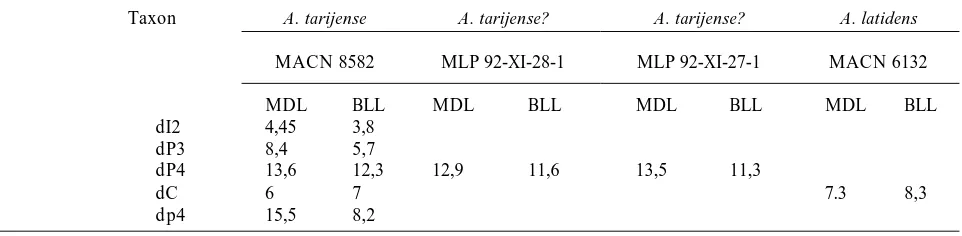
[image:4.595.60.541.100.218.2]On the alveolar margin, there is the alveolus for dp1, followed by one alveolus for dp2, two alveoli for the two roots of dp3, and the implanted dp4. The p1 is seen below the alveolus for the dp2, with a large portion of its apex mineralized (figures 2B-C). The p4 is observed in the radiograph between the two roots of dp4 (figures 2B-C). It is in a relatively earlier stage of development than p1. Labially to the alveoli of the dp1 and dp2, is the alveolus of the large deciduous canine. The permanent canine is seen below this alve-olus in the radiograph (figures 2,B-C). Along the alveolar margin behind the dp4, the first stage in the development of the m1 alveolus may be observed. This area is damaged; consequently the alveoli of the m2 and m3 cannot be confidently reconstructed. During early preparation, the m1 and m2 (figures 2.D-G) were found below this area of the mandible and they were unfortunately removed from its nat-ural place and kept separately in the collection. The m1 (figures 2.D-E) has all cusps fully developed, but the enamel is only mineralized on the paraconid, protoconid, metaconid and hypoconid. The enamel of the entoconid, the floor and the distal margin of the talonid, and all the lower margins of the crown Table 1.Comparison of sizes of all known Tremarctinae deciduous teeth / Comparación de tamaños de todos los dientes deciduos conocidos para los Tremarctinae.
Taxon A. tarijense A. tarijense? A. tarijense? A. latidens
MACN 8582 MLP 92-XI-28-1 MLP 92-XI-27-1 MACN 6132
MDL BLL MDL BLL MDL BLL MDL BLL
dI2 4,45 3,8
dP3 8,4 5,7
dP4 13,6 12,3 12,9 11,6 13,5 11,3
dC 6 7 7.3 8,3
dp4 15,5 8,2
MDL: maximum mesiodistal length (mm) BLL: maximum buccolingual length (mm)
are at a primary stage of maturation. The m2 (figures 2.F-G) shows a still earlier stage of development than the m1. The protoconid and metaconid are the most developed cusps and the hypoconid the least devel-oped in accordance with the description of Tonge (1976). The remaining occlusal surface and the mar-gin of the crown are at an early stage of development. On the talonid lingual margin, both cusps of the en-toconid may be seen.
Comparisons and discussion
The general dental formula of adult Ursidae is I 3/3 C1/1 P 4/4 M 2/3. This is the dental formula of tremarctines, but ursines usually lack the P 2-3/2-3. The deciduous dental formula of U. arctos and U. spelaeusis dI 3/3 dC 1/1 dP 2-4/2-4, and some spec-imens of U. spelaeus have dP/dp 2 completely re-duced (Koby, 1952; Radulescu and Samson, 1959; Terzea, 1969; Torres, 1988). Regarding the deciduous dental formula of A. tarijense, although there is not enough material to allow a definitive determination, it may be tentatively established as dI 3/3 dC 1/1 dP 1-4/1-4. This is coincident with the deciduous dental formula of T. ornatus deduced from the notes of Saporiti (1949).
In most mammals the milk dentition is replaced once, a condition called diphyodonty (hemiphyo-dontism by Mones, 1982), the first set is called pri-mary and the next set secondary (Jernvall, 1995); then deciduous teeth and molars are both primary teeth. As Jernvall (1995) pointed out, the last deciduous premolars always resemble the first permanent mo-lars. In this sense, the crown morphology of dP/dp4 in ursids is quite similar to that of M/m 1 respective-ly and, the dP4 occlude on dp4 in the juvenile in much the same way as the M1 occlude on the m1 in the adult. On the other hand, their corresponding permanent premolars (except for P4) are simpler.
Although the general morphology of dP/dp4 in A. tarijenseresembles that of Ursinae (see Koby, 1952 for details) considered -as previously mentioned- the sister group of Tremarctinae, the adult dentition of this taxon is quite different (see Torres, 1988 and Kurtén, 1967 for more details).
When dP4 and dp4 of A. tarijense are compared with M1 and m1 respectively, significant morpholog-ical differences are seen. Moreover, these morpho-logical features can be seen in the M1 and m1 of re-lated tremarctine taxa, and in Ursinae, Hemicyo-ninae and Amphicyonidae.
Comparisons between dP4 and M1
[image:5.595.58.291.78.519.2]The lingual side of the crown of dP4 of A. tarijense is very convex, but that of the M1 is slightly convex as in all other Arctotheriumspecies (A. bonariense, A. vetustum, A. latidens and A. brasiliense). In the three other tremarctine genera (Arctodus, Plionarctos and Tremarctos) the lingual side of M1 is straight, as in most Ursinae. In dP4 of U. spelaeusthe lingual side of the crown is slightly convex as in M1 of Arctotherium. The fourth upper deciduous premolar of A. tarijense is triangle-shaped in occlusal view, but M1 of all Arctotheriumspecies is sub-quadrate and in the other three genera of Tremarctinae (Tremarctos, Arctodus Figure 2.Arctothenium tarijense, MACN 8582: left hemimandible.
A,external view; B,composed radiograph and C,hemimandible and teeth outlined from the radiograph with the addition of those of m1 and m2 in their assumed position. A. tarijense, MACN 8582: left m1; D,lingual view andE,occlusal view; MACN 8582: left m2; F,lingual view andG,occlusal view /
and Plionarctos) and in all ursines the M1 is sub-rec-tangular. First upper molars with very convex lin-gual sides and triangle-shaped in occlusal view were found only in Amphicyonidae (Carnivora) and in hemicyonines. The protocone of dP4 of A. tarijenseis the largest cusp of the lingual row, this condition is only observed in the M1 of A. latidens (among Tremarctinae), in Hemicyoninae and in the dP4 of Ursinae. In the M1 of all other Tremarctinae and Ursinae, the protocone has the same size as the other lingual cusps. The dP4 of A. tarijensehas a well dif-ferentiated parastyle. This cusp is reduced or absent in the M1 as well as in A. latidensand A. bonariense, but conspicuous in the most basal South American taxa (A. vetustum and A. brasiliense), in all other Tremarctinae, Ursinae and in most Hemicyoninae.
Comparisons between dp4 and m1
The entoconid of dp4 of A. tarijenseis placed on the postero-lingual angle of the crown and formed by a single cusp. In all tremarctines (except for Tremarctos) the entoconid of m1 is placed forward on the lingual side of the talonid and has two or three apices. In some ursines such as U. americanusand all Hemicyoninae the morphology of the entoconid of m1 is the same as that of dp4 of A. tarijenseand m1 of Tremarctos. In others such as U. arctosand U. spelaeus, the condition observed in m1 is that of most tremarctines. The hypoconid of dp4 of A. tarijenseis large and consists of a single conical cusp. In all tremarctines (except for A. latidens), the hypoconid of m1 is mesiodistally elongated and has two apices separated by a shallow line in the enamel. In dp4 of U. spelaeus the hypoconid is represented by one or more cusps (Koby, 1952). In m1 of all Ursinae (except for Ursus spelaeus) and Hemicyoninae the hypoconid has the same morphology as that of dp4 of A. tarijense and m1 of A. latidens. It is noteworthy that the multi-plication of main cusps on the molars is a common feature of U. spelaeus(Torres, 1988).
In addition, it is noteworthy that A. tarijensedI2 (MACN 8582) has a similar morphology than that of the ursine I2 but very different from that of tremarc-tine I2.
Summarizing, the dP4/dp4 of A. tarijenseseem to bear a generalized set of characters (concerning the crown shape, the protocone and parastyle of dP4 and, the metaconid and hypoconid of dp4). Unfortunately these teeth cannot be compared with their homologues in other tremarctines. For this rea-son we cannot include these characters in the phylo-genetic analysis of the Tremarctinae.
In this respect, is the deciduous teeth morphology more conservative than that of permanent teeth? Mottl (1934, cited in Koby, 1952) compared the dp4 of
U. spelaeus with its m1 and observed that some fea-tures of dp4 may be considered primitive. Koby (1952) did not agree with this idea and expressed that the inferences of the author were not well supported. Flower (1871, cited in Woodward, 1892) considered that “the milk teeth of the Eutheria invariably show a more primitive pattern and shape than those of the permanent or second series which replace them”. Von Koenigswald (1967: 779) considered that “this observation is not absolutely correct” and proposed two possibilities: mammalian posterior milk teeth could either preserve dental features characteristic of their ancestors (be “conservative”) or precede the molars and replacement premolars in adapting to changing conditions (be “progressive”). This author considered that carnivores provided the best illustra-tion about how conservative the last deciduous mo-lars could be; and after a discussion of the evidence found in Canis, Meles, Ursus, Hyaena and Felis con-cluded that “That means in some carnivores we find preserved in last deciduous molars elements typical for the Miocene-Pliocene species some 10 million to 12 million years ago.” (Von Koenigswald, 1967: 780).
As it can be seen, there is some evidence found by other authors in related taxa that support our hy-pothesis. Unfortunately, due to the scarcity of decid-uous premolars of short-faced bears in the fossil record we cannot arrive at a definitive conclusion. But the observations on the morphology of the fourth upper and lower deciduous teeth of A. tarijense seems to be a good starting point. Detailed compari-son with ursine deciduous and permanent teeth is planed as future work.
In addition, we can estimate the age of MACN 8582. In T. ornatusi1 is the first permanent tooth that erupts during the fifth month of life (see Saporiti, 1949). If we assume that the sequence of appearance of the permanent teeth in A. tarijenseis similar to that of T. ornatus, MACN 8582 may have been approxi-mately four to five months of age, as the i1 had not yet emerged. The large size of the permanent teeth involves a rebuilding of the jaw in this process; that may have been an important mortality factor (Ehrenberg, in Kurtén, 1958). On this sense, it is note-worthy the great difference in size between the de-ciduous and permanent teeth, for example M1 of A. tarijenseis approximately twice the size of dP4.
Acknowledgments
References
Isla, F.I., Rutter, N. W., Schnack, E. J., and Zárate, M. A. 2000. La transgresión belgranense en Buenos Aires. Una revisión a cien años de su definición. Asociación Geológica Argentina, Serie D, Publicación EspecialNº4: 3-14.
Flower, W.J. 1871. On the milkdentition of Mammalia. Trans. Odont. Soc.
Jernvall, J. 1995. Mammalian molar cusp patterns: Developmental mechanisms of diversity. Acta Zoológica Fennica198: 1-61. Koby, F.E. 1952. La dentition lactele de l´Ursus spelaeus. Revue
Suisse de Zoologie59: 511-541.
Kraglievich, L. 1934. La antigüedad pliocena de las faunas de Monte Hermoso y Chapadmalal, deducidas de su comparación con las que le precedieron y sucedieron.El Siglo Ilustrado, Montevideo, 136 p.
Kurtén, B. 1958. Life and death of the Pleistocene cave bear. Acta Zoologica Fennica95: 1-59.
Kurtén, B. 1967. Pleistocene bears of North America: 2 Genus Arctodus, short faced bears. Acta Zoologica Fennica117: 1-60. Mones, A. 1982. An equivocal nomenclature: what means
hyp-sodonty? Palaeontologische Zeitschrift, 56:107-111.
Pardiñas, U.F.J., Tonni, E.P. and Figini, A. 1998. Camet Norte: di-versidad faunística próxima al último máximo glacial en el sudeste de la Provincia de Buenos Aires (Argentina). 10º Congreso Latinoamericano de Geología y 6º Congreso Nacional de Geología Económica, Actas1: 257-262.
Radulescu, C. and Samson, P. 1959. Contribution à la connais-sance de la dentition lactéale d´Usus spelaeus. Eiszeitalter und Gegenwart 10: 205-216.
Saporiti, E.J. 1949. Contribución al conocimiento de la biología del oso de lentes. Anales de la Sociedad Científica Argentina147: 3-12.
Soibelzon, L H. 2002. [Los Ursidae (Carnivora, Fissipedia) fósiles de la República Argentina. Aspectos Sistemáticos y Paleoecológicos. Tesis Doctoral inédita, Facultad de Ciencias Naturales y Museo, Universidad Nacional de La Plata. 239 pp. Unpublished].
Talbot, S.L. and Shields, G.F. 1996. A Phylogeny of the Bears (Ursidae) inferred from complete sequences of three mitocon-drial genes. Molecular Phylogenetics and Evolution5: 567-575. Terzea, E. 1969. Nouvelles donnés sur la dentition lactele de
l´Ursus spelaeus. Proceedings of the 4º International Congress of Sdpeleology in Yugoslavia4-5: 383-389.
Tongue, C.H. 1976. Morphogenesis and development of teeth. In: B. Cohen and I.R.H. Kramer (eds.), Scientific Foundations of Dentistry, William Heinsmann Medical Books, London, pp. 325-334.
Torres, T. 1988. Osos (Mammalia, Carnivora, Ursidae) del Pleistoceno de la Península Ibérica. Boletín Geológico y Minero 99: 1-316.
Trajano, E. and Ferrarezzi, H. 1994. A fossil bear from northeast-ern Brazil, with a phylogenetic analysis of the South American extinct Tremarctinae (Ursidae). Journal of Vertebrate Paleontology 14: 552-561.
Von Koenigswald, G. H. R. (1967). Evolutionary trends in the de-ciduous molars of the Hominoidea. Journal of Dental Research 46: 779-786.
Woodward, M.D. 1892. On the milkdentition of Provacin (Hyrax) capensis and of Rabbit (Lepus cuniculus) with remarks on the relation of the milk and permanent dentition of the Mammalia. Proceeding of the Zoological Society of London,38-49. Wyss A.R. and Flynn, J.J. 1993. A Phylogenetic Analysis and Definition of the Carnivora. In: F.S. Szalay, M.J. NovaceK and M. C. McKenna (eds.), Mammal Phylogeny: Placentals, Springer-Verlag, New York, pp. 32-52.