www.elsevier.com.mx
medicina
universitaria
* Corresponding author: Anillo Periférico 715-4 Avenue, Colinas de San Jerónimo, Z.P. 64630, Monterrey, N. L., Mexico (O. F. López-Lugo).
OrigiNAL ArtiCLe
Terminal interruption of relux source technique in the treatment
of active venous ulcers
O. F. López-Lugo
*, R. Salinas-Domínguez, J. A. Tamez-del Bosque, G. E.
Muñoz-Maldonado
Service of General Surgery, “Dr. José Eleuterio González” University Hospital, Universidad Autónoma de Nuevo León, Monterrey, N. L., Mexico
received: September 2013; Accepted: March 2014
KEYWORDS
Venous ulcers; Venous insuficiency; treatment; Mexico.
Abstract
Introduction: the treatment for venous ulcers in most cases is unsatisfactory, with recurrences and poor healing.
Objective: to evaluate adjuvant therapy in the treatment of active venous ulcers.
Methods: We analyzed 20 patients with active venous ulcers attending the general Surgery out-patient clinic at the “Dr. José eleuterio gonzález” University Hospital from October 2012 to January 2013. they were randomly divided into 2 groups: group A (11 patients) underwent compression therapy and group B (9 patients) underwent compression therapy plus removal of the vein that gives terminal relux to the ulcer, guided by ultrasound (microphlebectomy). Pa -tients were evaluated weekly (8 weeks). At each assessment, photographs and lesion measure-ments were taken and pain was evaluated using the visual analog scale.
Results: No significant differences were found between the study groups in terms of age,
weight, height, body mass index (BMi), ankle-brachial index, and baseline measurement of the ulcer (p>0.05). Group B showed a greater reduction in ulcer size and a statistically signiicant lower score on the visual analog pain scale (p<0.05) from the second and third week of treatment, respectively.
Conclusions: the results obtained in patients with surgical procedure (group B) are consistent with the reported eficacy of chronic venous ulcer treatment with saphenectomy (conventional surgery), the difference is that in this study we used a minimally invasive procedure (microphle-bectomy).
in pigmentation or eczema in 2.9% and a postphlebitic ul- cer in 0.7%.2 A multicenter study conducted in Poland with 40,085 participants found skin changes in 4.6% of the patients, a healed ulcer in 1% and active ulcers in 0.5%.7 Heit et al. found a venous ulcer incidence of 18 for every 100,000 people a year and a yearly Billion-dollar expenditure on public health.3 Current theories about the etiology of severe chronic
ve-nous insuficiency are related to chronic veve-nous hyperten -sion.4,5 When weakness develops in the walls of the veins, dilatation of the valve ring is produced, impeding valve coaptation.6 recent data indicates that venous hypertension causes extravasation of macromolecules and erythrocytes inside the interstitium.7,8 erythrocytes and protein intersti-tial degradation are a powerful leucocyte chemoattractant. this suggests a role in cytokines, demonstrated by the
pre-sence of beta growth factor, which may cause ibrosis by
stimulation of the extracellular matrix, reducing oxygen and nutrient circulation thus resulting in changes in the color and consistency of the skin.5
Advanced isolated venous insuficiency includes a sensa -tion of excessive weight on the legs as well as edema. Signs include a phlebostatic crown (spider-shaped outbreak con-sisting of small varicose intradermal veins on the medial as-pect of the ankle and foot), and changes in skin such as lipodermatosclerosis, which in its chronic phase develops as shiny, hardened and pigmented skin.9,10 Ulcers have irre-gular edges with neo-epithelization with a pinkish base with
granulation tissue, on occasions with a ibrin coating. Pulse
palpation and the ankle-brachial index test are necessary in
order to rule out an arterial insuficiency.11,12 the Doppler
ultrasound is currently the gold standard in venous insufi -ciency diagnosis.13,14 The American Venous Forum classiies active venous ulcers as C6.15,16
treatment includes general measures: patient education, weight loss, elevation of the extremity and exercise.17-20 Factors like immunosuppression, malnutrition, diabetes
me-llitus, arterial insuficiency, local infection, and cardiac in
-suficiency may affect ulcer healing. A compressive therapy
is the base in venous ulcer treatment, and this has been validated in random studies.21
O’Meara et al. and Ukat et al., proved a statistically
signi-icant decrease in time in ulcer healing (p=0.005) with the use of multiple elastic bandages compared to the inelastic systems.22-24 Myers et al. found supericial venous insufi -ciency with Doppler ultrasound in 38% of the patients with
venous ulcers, and a combination of supericial and deep ve-nous insuficiency in 48% of the patients with ulcers.25 A stu-dy showed venous ulcer healing in 77% of the cases 12
months after supericial venous relux surgical treatment.26 Bush included 14 patients with chronic venous ulcers (6 to 24 months) treated with compressive therapy. the patients
Materials and methods
We performed a prospective, comparative longitudinal stu-dy. A non-blind, experimental, controlled clinical trial.
Inclusion criteria: A C6 classiication by The American Ve -nous Forum (active ve-nous ulcers), age of at least 18 years,
venous insuficiency diagnosed by a Doppler ultrasound, BMI
between 18.5 and 34.9, no local infection, a signed infor-med consent, having no previous venous surgery and an ankle-brachial index over 0.85.
We analyzed 20 patients with a diagnosed venous ulcer in their lower limbs, with a previously signed consent form, attending the general Surgery outpatient clinic at the “Dr. José eleuterio gonzález” University Hospital, and meeting the inclusion criteria, from October 2012 to January 2013. the patients were randomly divided into 2 groups: group A (control) with compression therapy and group B with surgi- cal treatment in addition to compressive therapy (expe- rimental). Patients were evaluated weekly (8 weeks). At each assessment, photographs and lesion measurements were taken and pain was evaluated using the visual analog scale. We indicated rest and elevation of lower extremities for 30 minutes in the morning and 30 minutes in the afternoon, as
well as sleeping with the affected limb elevated. Patients we- re discharged on an outpatient basis with analgesic (para-cetamol 500 mg orally every 8 hours) in case of pain, and treating the wound every 24 hours with a super-oxidized antiseptic solution (Microdacyn®), non-adherent dressings and mild compression elastic socks (20-30 mmHg).
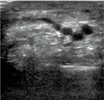
in group B, using an ultrasound with lineal transducer of 7.5 MHz, with the patient standing, the researcher looked for and marked the responsible vein(s) of the terminal blood
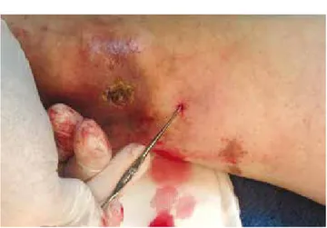
relux to the ulcer (Fig. 1). Once we identiied the vein(s), we proceeded with: iniltration with local anesthesia (lido -caine 1%), a puncture with a Jelco® catheter #14 adjacent to the marked vein, dermis dissection and vein extraction with a hook (Fig. 2), avulsion of the vein with hemostat clamps (Fig. 3), compression in order to achieve hemostasis, application of a super-oxidized antiseptic solution (Microda-cyn®), non-adherent dressings and mild compression elastic socks (20-30 mmHg).
Statistical analysis
the information was gathered in a database using excel®. We performed the Kolmogorov-Smirnov test to evaluate the variable distribution, and we determined that they all showed a normal distribution (gaussian). the other test used to compare both groups was the Student’s t-test with a
95% conidence, and we considered it to be statistically sig
-niicant p<0.05. We used the SPSS® Statistics version 20.0 (iBM Company). the study was approved by the institution’s Committee of ethics.
Results
A total of 20 patients with active venous ulcers were inclu-ded, 11 of them -9 female and 2 male- in group A (compres-sive therapy), and 9 of them -8 female and 1 male- in group
B (surgical and compressive therapy). No signiicant diffe -rences were found between the study groups in terms of age, weight, height, BMi, ankle-brachial index, and baseline measurement of the ulcer (table 1). Venous ulcer mean diame- ter in week 1 was 4.04 cm (± 2.44) for group A and 4.75 cm
(± 3.75) for group B with a p=0.616 (table 2). group B showed a greater reduction in ulcer size and a statistically
signiicant lower score on the visual analog pain scale com -pared to group A (p<0.05) with full healing (scarring) in
3 patients. This difference was signiicant from the second
and third week of treatment, respectively (table 3). the average reduction in ulcer size during 8 weeks of fo-llow-up for group A (compressive therapy) was 1.9 cm and for group B (compressive therapy and surgical treatment)
was 3.4 cm with a conidence interval of 95% (Fig. 4).
Discussion
Based on this study, we were able to ind that the medical-surgical technique shown is better than the more conserva -tive medical option. Favoring the results is the fact that no
signiicant differences were found between the study groups
in terms of age, weight, height, BMi, ankle-brachial index, and baseline measurement of the ulcer. Although patients in group B were younger (median age 58.44 years) than those
in group A (median age 70.27 years), age wasn’t signiicantly
distinct. All this makes the possibility of distractor variables
that could alter the inal result less likely. Group B (medical and surgical therapy) showed a statistically signiicant re -duction in ulcer size and complete healing (scarring) in 3
Figure 2 image showing the extraction with a hook of the vein with terminal relux to the venous ulcer.
Figure 3 image showing the avulsion of the vein with terminal relux to the venous ulcer with a hemostatic clamp.
Table 1 Demographic data
Variable group A (n=11) group B (n=9) p
Age, years,
average (± SD) 70.27 (± 16.3) 58.44 (± 14.2) 0.113 Weight, kg,
MeDiAN (± SD) 76.36 (± 12.57) 77.77 (± 6.74) 0.766 Height, m,
average (± SD) 1.62 (± 0.057) 1.62 (± 0.073) 0.900 BMi, kg/m2,
average (± SD) 28.98 (± 4.46) 29.44 (± 2.14) 0.780 ABi, mmHg,
average (± SD) 0.98 (± 0.087) 0.958 (± 0.06) 0.549
patients compared with group A. As Bush proves in his study with ulcer treatment at 8 weeks (11 patients out of the 14 initially included), group B showed a lower score in the vi-sual analog scale of pain compared with group A, which was
statistically signiicant from the third week of treatment.27 Considering the satisfactory evolution in group B with a
statistically signiicant decrease in pain and ulcer size, it
would be convenient to extend follow-up in order to evalua-te compleevalua-te healing in every patient within this group in particular. in addition, it is important to determine venous ulcer recurrence between both groups in order to
demons-trate if there is a signiicant difference, as mentioned by
the Landmark study eSCHAr (effect of Surgery and Com-pression on Healing and recurrence in patients with venous ulcers), which randomized 500 patients with advanced
chronic venous insuficiency. The irst group included 258
patients treated with compression, a second group with 242 patients was treated with compression and surgery; a
Measurement week 1-week 4
Difference in cm, average (± SD) 1.054 (± 0.75) 1.4 (± 0.82) 0.028
Measurement week 1-week 5
Difference in cm, average (± SD) 1.29 (± 0.88) 2.3 (± 0.97) 0.026
Measurement week 1-week 6 Difference in cm, average (± SD) Measurement week 1-week 7 Difference in cm, average (± SD) Measurement week 1-week 8 Difference in cm, average (± SD)
1.56 (± 0.93)
1.7 (± 0.97)
1.87 (± 1.02)
2.64 (± 1.13)
2.97 (±1.32)
3.4 (± 1.71)
0.025
0.023
0.023
SD: standard deviation.
Table 3 Values on the visual analog scale of the pain during follow-up weeks.
Variable group A (n=11) group B (n=9) p
VAP week, 1, average (± SD) 7 (± 1.18) 7.66 (± 1.32) 0.250
VAP week, 2, average (± SD) 5 (± 1.5) 4.77 (± 1.85) 0.774
VAP week, 3, average (± SD) 4.36 (± 1.2) 2.55 (± 1.23) 0.004
VAP week, 4, average (± SD) 3.81 (± 1.47) 1.33 (± 1.41) 0.001
VAP week, 5, average (± SD) 3.0 (± 1.73) 0.77 (± 1.2) 0.004
VAP week, 6, average (± SD) 2.9 (± 1.75) 0.55 (± 0.08) 0.002
VAP week, 7, average (± SD) 2.36 (± 1.5) 0.33 (± 0.70) 0.002
VAP week, 8, average (± SD) 2.27 (± 1.55) 0.33 (± 0.70) 0.003
VAP: visual analog pain; SD: standard deviation.
Mean reduction
Study group
Group A Group B
Error bars 95% confidence interval 5.00
4.00
3.00
2.00
1.00
0.00
recurrence after 12 months of 28% for the irst group com -pared to 12% for the second group was found.28,29 the obtai- ned results in this study of patients who underwent surgical
procedure (group B) concur with the eficacy reported by
Zamboni et al. in a randomized study of 45 patients with venous ulcers, 24 patients with compressive therapy and 21 patients with postsurgical use of elastic socks, with healing
of 96% in a period of 63 days for the irst group compared
with 100% healing in 31 days for the second group,30 with the difference that in our study we utilized a minimally in-vasive procedure (microphlebectomy). even though a sam-ple of 20 patients is small (this being the study’s main weakness), there is a clear tendency in favor of our experi-mental group. Another limitation of this study is a relatively short follow-up period. Terminal interruption of relux sou-rce technique guided by ultrasound proved with statistically
signiicant results to be more effective than the more con -ventional medical treatment to reduce pain and size of
ve-nous ulcers. The use of this surgical technique may be a
promising line of minimal invasion in venous ulcer treatment
in the immediate future. Further studies are required with this surgical technique and a longer follow-up period in or -der to evaluate recurrences.
Conlicts of interest
The authors have no conlicts of interest to declare.
Funding
No inancial support was provided.
References
1. Callam MJ. epidemiology of varicose veins. Br J Surg 1994;81:167-173.
2. rabe e, Pannier-Fisher F, Bromen K, et al. Bonner Venenstudie der Deutschen gesellschaft fur Phlebologic epidemiologigische Untersuchung zur Frage der Hauigkeit und Avspragung von chronischen Venenkrakheiten inder stadtischen un landlichen wohnbevolkerung. Phlebologic 2003;32:1-14.
3. Heit JA, rooke tW, Silverstein MD, et al. trends in the inciden-ce of venous stasis syndrome and venous ulinciden-cer: a 25 year popu-lation-based study. J Vasc Surg 2001;33:1022-1027.
4. thomas Pr, Nash gB, Dormandy JA. White cell accumulation in the dependent legs of patients with venous hypertension: a possible mechanism for trophic changes in the skin. Br Med J (Clin res ed) 1988;296:1693-1695.
5. Hisley Hr, Ksander gA, gerhardt CO, et al. extravasation of macromolecules and possible trapping of transforming growth factor beta in venous ulceration. Br J Dematol 1995;132:79-85. 6. Alexander CJ. the theoretical basis of varicose vein formation.
Med J Aust 972;1:258-261.
7. Burnand KG, Whimster I, Naidoo A, et al. Pericapillary ibrin in the ulcer bearing skin of the leg. Br Med J 1982;2:243-245. 8. Browse NL, Burnand Kg. the cause of venous ulceration.
Lan-cet 1982;2:243-245.
9. Geyer MJ, Brienza DM, Chib V, et al. Quantifying ibrosis in ve -nous disease: mechanical properties of lipodermatosclerotic and healthy tissue. Adv Skin Wound Care 2004;17:131-142. 10. Navarro tP, Delis Kt, ribeiro AP. Clinical and hemodynamic
sig-niicance of the greater saphenous vein diameter in chronic ve -nous insuficiency. Arch Surg 2002;137:1233-1237.
11. Kjaer ML, Mainz J, Soernsen LT, et al. Clinical quality indicators of venous leg ulcers: development, feasibility, and reliability. Ostomy Wound Manage 2005;51:64-74.
12. Bjellerup M. Does dorsal pedal pulse palpation predict hand-held Doppler measurement of ankle-brachial index in leg ulcer patients? Wounds 2003;15:237-240.
13. Lee YM, ting AC, Cheng SW. Diagnosing deep vein thrombosis in the lower extremity: correlation of clinical and duplex scan in -dings. Hong Kong Med J 2002;8:9-11.
14. Arseculeratne YM, Walton J, Hofman D, et al. A comparison of light relection rheography and duplex scanning in the diagnosis of chronic venous insuficiency. Wounds 2003;15(8).
15. Carpentier PH, Cornu-thenard A, Uhl JF, et al. Appraisal of the information content of the C classes of CEAP clinical classiica -tion of chronic venous disorders: a multicenter evalua-tion of 872 patients. J Vasc Surg 2003;37:827-833.
16. eklof B, rutherford rB, Bergan JJ, et al. American Venous Fo-rum international Ad Hoc Committee for revision of the CeAP. Revision of the CEAP classiication for chronic venous disorders: consensus statement. J Vasc Surg 2004;40:1248-1252.
17. Northeast ADr, Layer gt, Wilson NM, et al. increased compres-sion expedites venous ulcer healing. royal Society of Medicine Venous Forum, 1990.
18. Lorimer Kr, Harrison MB, graham iD, et al. Venous leg ulcer care: how evidence-based is nursing practice? J Wound Ostomy Continence Nurs 2003;30:132-142.
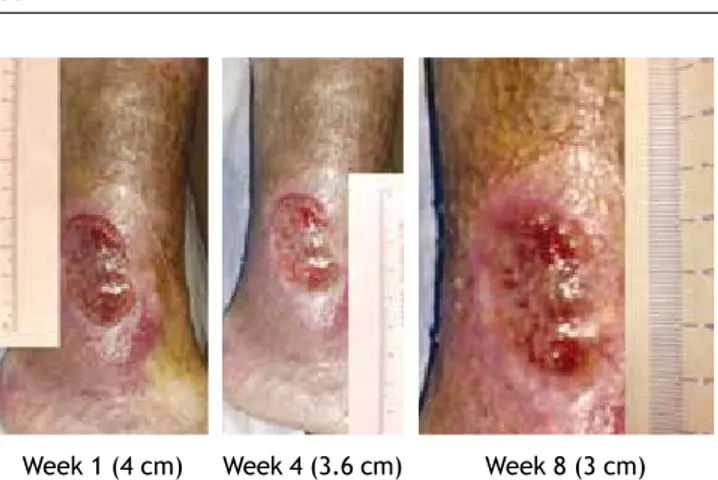
Figure 5 images of the follow-up of a patient from group A (compressive therapy).
Week 1 (4 cm) Week 4 (3.6 cm) Week 8 (3 cm)
Figure 6 images from the follow-up of a patient from group B (surgical and compressive therapy).
ulcers. Cochrane Database Syst rev 2009;CD000265.
24. Ukat A, Konig M, Vanscheidt W, et al. Short-stretch versus mul-tilayer compression for venous leg ulcers: a comparison of hea-ling rates. J Wound Care 2003;12:139-143.
25. Myers KA, Ziegenbein rW, Zeng gH, et al. Duplex ultrasonogra-phy scanning for chronic venous disease: patterns of venous relux. J Vasc Surg 1995;21:605-612.
compression therapy alone versus compression plus surgery in chronic venous ulceration (eSCHAr): randomised controlled trial. BMJ 2007;335:383.