Proteomics. 2011; 11(12): 2555-2559
A novel procedure for protein extraction from formalin-fixed
paraffin-embedded tissues
Teresa Rodríguez-Rigueiro
1, Manuel Valladares-Ayerbes
1,2, Mar Haz-Conde1,
Moisés Blanco
1, Guadalupe Aparicio
1, Patricia Fernández-Puente
3, Francisco J.
Blanco
3, Mª José Lorenzo
2, Luis A. Aparicio
1,2and Angélica Figueroa
11. Translational Cancer Research Group, Proteo-Red-ISCIII, Instituto de Investigación Biomédica A Coruña (INIBIC), Coruña, Spain
2. Medical Oncology Unit, Complejo Hospitalario Universitario A Coruña (CHUAC), Coruña, Spain 3. Proteomics Unit, Proteo-Red-ISCIII, Instituto de Investigación Biomédica A Coruña (INIBIC), Coruña, Spain
Abstract
Most of the archived pathological specimens in hospitals are kept as formalin-fixed paraffin-embedded tissues (FFPE) for long-term preservation. Up to now, these samples are only used for immunohistochemistry in a clinical routine as it is difficult to recover intact protein from these FFPE tissues. Here, we report a novel, short time-consuming and cost-effective method to extract full-length, non-degraded proteins from FFPE tissues. This procedure is combined with an effective and non-toxic deparaffinisation process and an extraction method based on antigen-retrieval, high concentration of SDS and high temperature. We have obtained enough intact protein to be detected by Western blotting analysis. This technique will allow utilising these stored FFPE tissues in several applications for protein analysis helping to advance the translational studies in cancer and other diseases.
Keywords:
In the last few years, biomedical investigations have been focussing on the improvement of the translational research which tries to integrate the findings on basic science more quickly and efficiently into medical practice. At present, the barriers between molecular biology and clinical trials are narrower, thanks to the multi-disciplinary collaboration between scientists and clinicians. For instance, the establishment of a huge number of tissue banks in hospitals for more than a century is one of the most important benefits that scientist can take for their biomedical research. Human tissues obtained from biopsies are generally fixed in formalin and embedded in paraffin (FFPE) to be further analysed by a histopathology expert. FFPE is a standard method for long-term preservation of most archived pathological specimens. Moreover, these FFPE tissue collections are also important resources to develop studies on which DNA and RNA are the starting materials 1. However, research work is still limited on the extraction of intact protein from FFPE tissues as it is believed that their processing with formalin and paraffin destroys proteins and makes their analysis difficult by Western blotting, immunoprecipitation or protein arrays. Western blotting and immunohistochemistry (IHC) are the most frequent techniques used to identify and study protein expression. Although IHC gives information regarding the localisation and distribution of proteins in cells and tissues, it is not adequate for protein quantification as there is an important risk of cross-reacting proteins.
In 1998, Ikeda et al. 2 published the first work on which they obtained intact protein from FFPE tissues; however, they failed to obtain comparable extracts from FFPE and fresh-frozen tissues. Since then, not many works have been published describing approaches to obtain full-length protein for expression analysis 3–6. It is well known that not all the proteins can be extracted under the same conditions. The extraction of individual proteins can be proceeded in several manners considering that the solubilisation of some proteins require conditions that destabilise others. In this article, we describe a novel standardised and reproducible method for full-length protein extraction from FFPE tissues. By Western blotting, we have shown that we obtain comparable yields to that seen from fresh-frozen tissue samples. This method can be used to compare protein expression from different tissues, individuals and pathological conditions.
3% skim milk for 1 h at RT with constant shaking. They were incubated with the indicated antibodies at RT for 1 h, washed three times with PBS plus 0.05% Tween-20 (PBS-T) and incubated for 1 h with the specific secondary mouse or rabbit peroxidase-conjugated anti-IgG antibody. After three washes with PBS-T, the proteins were visualised for immunological detection by enhanced chemiluminescence (ECL+; GE Healthcare, Sweden). Antibodies used in this work were as follows: mouse α-tubulin antibody (Sigma-Aldrich), rabbit Hakai antibody kindly provided by Yasuyuki Fujita (LMCB, UK) 11, E-cadherin (Invitrogen Laboratories, CA) and HRP-rabbit and mouse polyclonal antibodies (GE Healthcare, UK). Primary and secondary antibodies were used at dilutions 1:1000 and 1:5000, respectively.
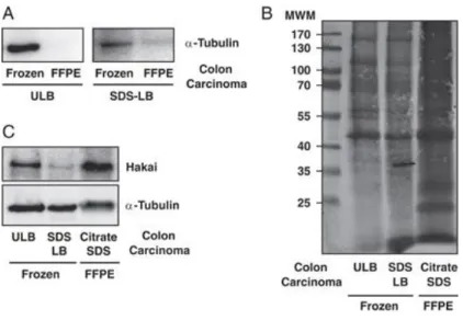
Figure 1. Comparison of the protein extraction protocols from fresh-frozen and FFPE colon carcinoma tissues. (A) Western blotting against α-tubulin antibody by extracting protein following the procedure explained at RT by using ULB and SDS-LB buffers. (B) and (C) Coomassie staining and Western blotting against α-tubulin and Hakai antibodies by using ULB and SDS-LB at RT and citrate-SDS buffer under the high temperature and conditions proposed. All FFPE samples were used <1 year of storage and frozen samples after 5 years of storage. Results are representative of at least three independent experiments.
Figure 2. Western blotting against E-cadherin, Hakai and α-tubulin from different FFPE tissues. (A) FFPE tissues from colon, myoma, kidney and lymph node. (B) FFPE cerebellum tissues from three different blocks (C1, C2 and C3). (C) FFPE colon carcinoma tissues from different patients (T1, T2, T3 and T4). The procedure was performed by using the explained high-temperature extraction in citrate-SDS buffer. Colon, myoma, lymph node samples in (A), and T3 and T4 colon carcinoma samples in (C) were used after 1 year of storage; the rest of the samples were used after 2 years of storage. Results are representative of at least three independent experiments.
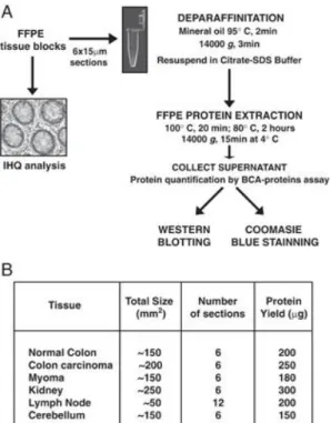
Figure 3. Schematic of the workflow (A) and protein yield (B) obtained from protein extraction from FFPE tissues using citrate-SDS buffer.
Acknowledgements
We thank M. Mayan for critical reading of the manuscript and Y. Fujita for providing Hakai antibody. We also thank Cristina Ruiz-Romero and Valentina Calamia for helpful discussions. T. R. R is the recipient of a grant from Secretaria Xeral I+D+I, Xunta de Galicia, Spain (10CSA916023PR). M. H. C and M. B. are supported by Instituto de Salud Carlos III, Spain (CA09/00116 and CA07/00232). A. F. is supported by Secretaria Xeral I+D+I, Xunta de Galicia, Spain (IPP.08-07). This work is supported by a grant from Conselleria de Sanidade (PS09/24) and from Secretaria Xeral I+D+I (10CSA916023PR) both from Xunta de Galicia, Spain.
References
[1] Gilbert, M. T., Haselkom T., Bunce, M., Sanchez, J. J., et al., The isolation of nucleic acids from fixed, paraffin-embedded tissuew-which methods are useful when?. PLoS One 2007, 2, e537.
[2] Ikeda, K., Monden, T., Kanoh, T., Tsujie, M. et al., Extraction and analysis of diagnostically useful proteins from formalin-fixed, paraffin-embedded tissue sections. J. Histochem. Cytochem. 1998, 46, 397–403.
[3] Becker, K. F., Schott, C., Hipp, S., Metzger, V. et al., Quantitative protein analysis from formalin-fixed tissues: implications for translational clinical research and nanoscale molecular diagnosis. J. Pathol. 2007, 211, 370–378.
[4] Nirmalan, N. J., Harnden, P., Selby, P. J., Banks, R. E., Development and validation of a novel protein extraction methodology for quantitation of protein expression in formalin-fixed paraffin-embedded tissues using Western blotting. J. Pathol. 2009, 217, 497–506.
[5] Addis, M. F., Tanca, A., Pagnozzi, D., Crobu, S. et al., Generation of high-quality protein extracts from formalin-fixed, paraffin-embedded tissues. Proteomics 2009, 9, 3815–3823.
[6] Berg, D., Hipp, S., Malinowsky, K., Böllner, C., Becker, K. F., Molecular profiling of signalling pathways in formalin-fixed and paraffin-embedded cancer tissues. Eur. J. Cancer 2010, 46, 47–55.
[7] Shi, S. R., Liu, C., Balgley, B. M., Lee, C., Taylor, C. R., Protein extraction from formalin-fixed, paraffin-embedded tissue sections: quality evaluation by mass spectrometry. J. Histochem. Cytochem. 2006, 54, 739–743.
[8] Lin, J., Kennedy, S. H., Svarovsky, T., Rogers, J. et al., High-quality genomic DNA extraction from formalin-fixed and paraffin-embedded samples deparaffinized using mineral oil. Anal. Biochem. 2009, 395, 265–267.
[9] Bradford, M. M., A rapid and sensitive method for the quantification of mg quantities of protein. Anal. Chem. 1976, 72, 248–254.
[10] Laemmli, U. K., Cleavage of structural proteins during the assembly of the head of bacteriophage T4. Nature 1970, 227, 680–685.
[11] Calamia, V., Ruiz-Romero, C., Rocha, B., Fernández-Puente, P. et al.,
Pharmacoproteomic study of the effects of chondroitin and glucosamine sulfate on human articular chondrocytes. Arthritis Res. Ther. 2010, 12, R138.
[12] Fujita, Y., Krause, G., Scheffner, M., Zechner, D. et al., Hakai, a c-Cbl-like protein, ubiquitinates and induces endocytosis of the E-cadherin complex. Nat. Cell Biol. 2002, 4, 222–231.
[13] Figueroa, A., Kotani, H., Toda, Y., Mazan-Mamczarz, K. et al., Novel roles of Hakai in cell proliferation and oncogenesis. Mol. Biol. Cell 2009, 20, 3533–3542.