Rodríguez-Lara V, Ramírez-Tirado LA, Barrón F, Zatarain-Barrón ZL, Flores-Estrada D, Arrieta O. Characteristics of non-small cell lung cancer: Differences by sex and hormonal status in a Mexican population. Salud Publica Mex. 2019;61:265-275.
https://doi.org/10.21149/10094
Rodríguez-Lara V, Ramírez-Tirado LA, Barrón F, Zatarain-Barrón ZL, Flores-Estrada D, Arrieta O. Características del cáncer de pulmón de células no pequeñas: diferencias por sexo y estado hormonal en una población mexicana. Salud Publica Mex. 2019;61:265-275.
https://doi.org/10.21149/10094
Characteristics of non-small cell lung
cancer: differences by sex and hormonal
status in a Mexican population
Vianey Rodríguez-Lara, PhD,(1) Laura Alejandra Ramírez-Tirado, MSc,(2) FelicianoBarrón, MD,(2)
Zyanya Lucía Zatarain-Barrón, MSc,(2) Diana Flores-Estrada, SW,(2) Oscar Arrieta, MSc.(2)
(1) Departamento de Biología Celular y Tisular, Facultad de Medicina, Universidad Nacional Autónoma de México. Mexico City, Mexico. (2) Unidad Funcional de Oncología Torácica y Laboratorio de Medicina Personalizada, Instituto Nacional de Cancerología. Mexico City, Mexico.
Received on: October 13, 2018 • Accepted on: February 22, 2019
Corresponding author: Dr. Oscar Arrieta. Instituto Nacional de Cancerología. San Fernando 22, Sección XVI. 14080 Tlalpan, Mexico City, Mexico. E-mail: [email protected]
Resumen
Objetivo. Analizar las diferencias en las características clínico-patológicas, moleculares y en la evolución del cáncer de pulmón de células no pequeñas (CPCNP) por sexo y estadio hormonal. Material y métodos. Estudio retro-spectivo (N=1 104) en pacientes con CPCNP. Se recabaron datos clínico-patológicos y desenlaces de sobrevida y se compararon entre hombres y mujeres, y entre mujeres pre y postmenopáusicas. Resultados. Las mujeres de este estudio tuvieron significativamente mayor probabilidad de ser no fumadoras (p<0.001), tener exposición a humo de leña (p<0.001), mutaciones en EGFR (p<0.001), mejor estado funcional (p=0.020), y una mejor sobrevida global (SG) en comparación con los hombres (p=0.021). Estas diferencias también se encontraron en cuestión al estatus hormonal, con las mujeres postmenopáusicas presentando una mayor sobrevida en comparación con las premenopáusicas (31.1 vs. 19.4 meses; p=0.046). Conclusión. Los presentes resulta-dos apoyan las diferencias en la presentación del CPCNP de acuerdo con el sexo y estatus hormonal.
Palabras clave: cáncer de pulmón de células no pequeñas; hombres; mujeres; premenopausia; postmenopausia
Abstract
Objective. To analyze the differences in the clinico-patho-logical and molecular characteristics of non-small cell lung cancer (NSCLC) as well as the clinical outcome of patients by sex and hormonal status. Materials and methods. We performed a retrospective study among 1 104 NSCLC patients. Clinic-pathologic data was recorded and survival outcomes were compared between male and female sex patients, and further by pre and postmenopausal status in fe-males. Results. Women were significantly more likely to be non-smokers (p<0.001), had higher frequency of wood-smoke exposure (p<0.001), EGFR-sensitizing mutations (p<0.001), had better performance status (p=0.020) and had a better overall survival (OS) compared to men (p=0.021). Differences were found also by hormonal status, postmenopausal women had a longer OS compared to premenopausal women (31.1 vs. 19.4 months p=0.046). Conclusion. Our results support the differences in lung cancer presentation by sex and also by hormonal status.
L
ung cancer remains the leading cause of cancer-related deaths among males and has increased in women in the last decades.1 Lung cancer in womenhas become an important health problem, surpassing mortality from breast cancer and being the first cause of cancer-related death in EEUU and in some countries from Europe.1-5 Interestingly, a higher incidence among
young women compared to men has been observed in the last decades in EEUU, however, this increase in incidence among women cannot be attributed solely to an increase in tobacco use, due to the fact that young women and men show a similar pattern in terms of smoking behavior, nonetheless the increased incidence of lung cancer in women is notorious.6 Moreover, an
increased incidence of lung cancer has also been repor-ted in Latin American women, who present a lower smoking index compared to European and American women.7,8 These data suggest that even though tobacco
use remains an important risk factor for developing lung cancer, women face other risk factors, which also appear to play an important role in lung carcinogenesis. Furthermore, a higher percentage of women who deve-lop lung cancer are never smokers compared to the low percentage of non-smoking men who present with this pathology (53 vs. 15%).9,10 Other risk factors that might
partially explain the increased incidence of lung cancer in women include: wood smoke exposure,11 cooking oil
fumes exposure12 or second hand exposure to cigarette
smoke,13 however exposure to these risk factors does
not fully explain the increased incidence of lung cancer in women. In the last years, it has been reported that estrogen and progesterone play an important role in lung cancer in women, mainly in the adenocarcinoma subtype, activating carcinogenic pathways.14,15 Also,
several studies have shown that hormone replacement therapy based on estrogen and progestin can increase lung cancer incidence and mortality.16,17
Additionally, lung cancer presentation, behavior and response to treatment seem to differ between women and men. For instance, the median age for lung cancer diagnosis in women is lower compared to men. Women with lung cancer more often have no history of smoking and the predominant subtype is adenocarcinoma, frequently associated with mutations in epidermal growth factor receptor gene (EGFR). In addition, women show better response to chemotherapy and longer overall survival, regardless of the clinical stage at diagnosis, compared to men.18,19 Recently,
it has been observed that non-small cell lung cancer (NSCLC) tumors are more immunogenic in men and consequently male patients respond better to immu-notherapy compared to women.20 All these differences
in lung cancer by sex suggest a role of sexual hormones
in lung cancer; however, differences by hormonal status in women (premenopausal/postmenopausal) have been poorly studied. Nevertheless, when characteristics of premenopausal women have been analyzed, they show that these patients are diagnosed in advanced stages, with less differentiated tumors, distant metastases and worse prognosis compared to postmenopausal women. This information suggests that lung cancer is not only influenced by sex, but also by hormonal status.21,22
Several studies have focused on reporting the differences by sex and despite this information, these differences are poorly understood, and the results of studies are contradictory. The controversy in these stu-dies might be due to the inherent characteristics of the population analyzed, differences in the main risk factors, mutational tumor profile, genetic characteristics of pa-tients and hormonal status. With a better understanding about the role of sexual hormones in lung cancer, it is important to investigate differences by hormonal status in Latin America population.23
Nowadays, it is recognized that sex and sexual hormones influence lung cancer. Due to the impact that these differences have in future treatment strategies, it is important to comprehensively characterize them. In this study, we sought to investigate the differences in clinical features and survival of NSCLC patients by sex and hormonal status, in a Mexican population.
Materials and methods
Patient selection
We retrospectively reviewed demographic, clinical, molecular and pathological data of 1 104 NSCLC who attended at the National Cancer Institute of Mexico (In-can) between May 2008 through March 2017. All included patients had a histologically confirmed diagnosis of NS-CLC and molecular genotype available in their clinical file (EGFR and Kirsten rat sarcoma virus oncogene[KRAS] mutation status). Information including sex, age, and me-nopausal status at diagnosis was collected. Menopause was defined according to the international menopause guideline24 as the permanent cessation of menses for 12
or more months in the absence of chemotherapy. Patients who had undergone oophorectomy were excluded from this study. According to standardized guidelines for smoking measurement,25 we defined any patient who
Smoking index was obtained by multiplying the number of cigarettes smoked per day by the number of years the patient reported that he/she had smoked [(# cigarettes per day)(years smoking)/20) and reported as pack-year]. Self-reported wood-smoke exposure while cooking was recorded and the wood smoke exposure index was calcu-lated by multiplying the number of daily hours exposed by the number of years’ exposure.11
Outcome measurement
Clinical baseline characteristics included age, sex, hormonal status (premenopausal/postmenopausal), weight, histologic tumor type, disease stage, smoking status, wood-smoke exposure, exposure to asbestos, ECOG performance status, metastatic sites, and muta-tion profile for EGFR and KRAS. Overall survival (OS) was defined as the time from histological diagnosis until death or last follow-up.
Statistical analysis
For descriptive purposes, continuous variables were summarized as arithmetic means with standard devia-tions, while categorical variables were summarized as frequencies and percentages. The chi2 or Fisher’s exact
tests were used to assess the significance among catego-rical variables. The primary endpoint was OS, defined as the time from histological diagnosis until death or last
follow-up. Median OS was estimated using the Kaplan-Meier’s method, whereas the log-rank test was used for making comparisons among subgroups. A multivariate Cox regression model was used to adjust for potential confounders and hazard ratios (HR) were calculated along with their corresponding 95%CI as a measure of association. Statistical significance was determined as
p≤0.05 using a two-tailed test. SPSS software version 21 (SPSS Inc., Chicago, IL) was used for all statistical analysis.
Results
Characteristics for the entire population of NSCLC patients
Out of 1 104 patient records reviewed for this study, 582 (52.7%) were men and 522 (47.3%) women. The median age was 60.6 years (+12.9). According to risk factors, 55.7% were ever smokers, while 38.5% of the patients had history of wood smoke exposure, and 10.5% had exposure to asbestos; 72.9 % of all patients have an ECOG <1. Most patients had adenocarcinoma histology (84.3%) and 98.9% presented with advanced or metastatic disease (stages IIIB or IV) at diagnosis. Nearly thirty percent of patients (29.6%) had EGFR mutations and 10.2% had KRAS alterations (table I).
Table I
CliniC-pathologiCalCharaCteristiCsofpatientstreatedatthe national CanCer institute
(inCan) in MexiCo CityfroM 2008-2017 (n=1 104)
All patients % (n/N)
Sex
p-Value Female
(n=522) % (n/N) (n=582) % (n/N)Male
Age (years)
Mean (+SD) 60.6 (12.9) 59.8 (13.4) 61.2 (12.5) 0.068
BMI (Kg/m2)
Mean (+SD) 24.8 (4.6) 25.1 (4.9) 24.6 (4.2) 0.036
BMI groups
Normal (<25 kg/m2) 55.2 (609/1 104) 51.6 (269/522) 58.5 (340/582) Overweight (25 - 29.9 kg/m2) 32.4 (358/1 104) 32.4 (169/522) 32.4 (189/582)
Obese (30+ kg/m2) 12.4 (137/1 104) 16.0 (84/522) 9.1 (53/582) 0.003
Tobacco exposure
Absent 44.3 (489/1 104) 68.0 (355/522) 23.0 (134/582)
Present 55.7 (615/1 104) 32.0 (167/522) 77.0 (448/582) <0.001
Tobacco index
Mean (+SD) 34.4 (167.9) 17.7 (21.8) 40.4 (195.3) 0.133
Characteristics of NSCLC patients according to sex
The relative frequency of obese patients was subs-tantially higher in women (16%) than in men (9%),
p=0.003, likewise, female patients showed a higher body mass index (BMI) in comparison with their male counterparts (24.6 vs. 25.1; p=0.036). In addi-tion, women had higher frequencies of wood-smoke
(continuation)
Wood-smoke exposure
Absent 61.5 (679/1 104) 50.0 (261/522) 71.8 (418/582)
Present 38.5 (425/1 104) 50.0 (261/522) 28.2 (164/582) <0.001
Exposure to asbestos
Absent 89.5 (988/1 104) 90.8 (474/522) 88.3 (514/582)
Present 10.5 (116/1 104) 9.2 (48/522) 11.7 (68/582) 0.178
Tumor histologic type
Adenocarcinoma 84.3 (931/582) 88.9 (464/522) 80.2 (467/582)
Squamous 15.7(173/1 104) 11.1 (58/522) 19.8 (115/582) <0.001
Disease stage
II - IIIA 1.1 (12/1 104) 1.2 (6/522) 1.1 (6/582)
IIIB - IV 98.9 (1 092/1 104) 98.8 (516/522) 98.9 (576/582) 0.866
ECOG performance status
0-1 72.9 (805/1 104) 76.2 (398/522) 69.9 (407/582)
>2 27.1 (299/1 104) 23.8 (124/522) 30.1 (175/582) 0.020
Brain metastases at diagnosis*
Absent 53.6 (535/999) 54.7 (262/479) 52.5 (273/520)
Present 46.4 (464/999) 45.3 (217/479) 47.5 (247/520) 0.487
Lymphatic nodes metastases at diagnosis*
Absent 81.8 (817/999) 79.1 (379/479) 84.2 (438/520)
Present 18.2 (182/999) 20.9 (100/479) 15.8 (82/520) 0.037
Adrenal glands metastases at diagnosis*
Absent 95.5 (954/999) 96.0 (460/479) 95.0 (494/520)
Present 4.5 (45/999) 4.0 (19/479) 5.0 (26/520) 0.431
EGFR mutation status‡
WT EGFR 70.4 (286/406) 61.2 (134/219) 81.3 (152/187)
EGFR sensitizing mutation 29.6 (120/406) 38.8 (85/219) 18.7 (35/187) <0.001
KRAS mutation status‡
KRAS (-) 89.8 (256/285) 89.8 (132/147) 89.9 (124/138)
KRAS (+) 10.2 (20/285) 10.2 (15/147) 10.1 (14/138) 0.987
SD: standard deviation, BMI: body mass index, ECOG: eastern cooperative oncology group, WT: wild-type, EGFR: epidermal growth factor receptor, KRAS: Kirsten rat sarcoma virus oncogene.
*Estimation over 999 patients with stage IV disease and known metastases at diagnosis.
‡ Estimations over the number of patients tested for each molecular status.
exposure (50 vs. 28.2%; p=<0.001), EGFR-sensitizing mutations (38.8 vs. 18.7%; p=<0.001) and better ECOG performance status (≤1) (76.2 vs. 69.9%; p=0.020). By contrast, men showed higher frequencies of tobacco smoking exposure compared to women (70 vs. 32%;
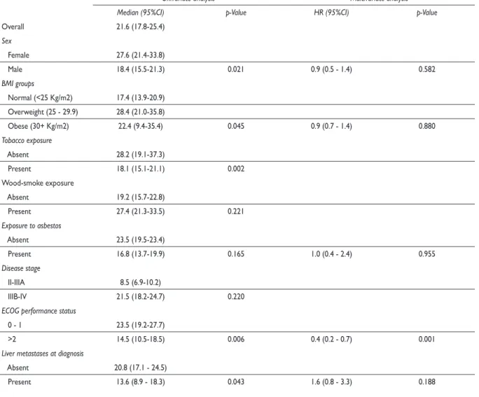
On the other hand, patients with an ECOG <2 (23.5 vs. 14.5 months; p=0.006), absence of liver metastases, and wild type KRAS (38.9 vs. 15.7 months; p<0.001) had better OS. The multivariate analysis showed that ECOG performance status (HR: 0.4, 95%CI (0.2–0.7); p=0.001) and KRAS mutation (HR: 2.1, 95%CI (1.1–3.8); p=0.017) were independently associated with OS (table II).
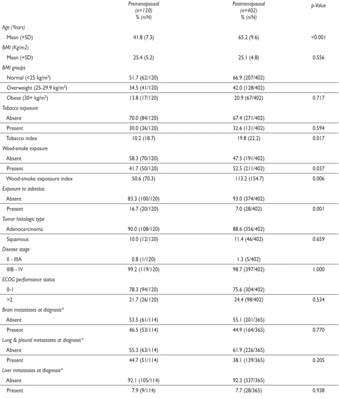
Characteristics of women with NSCLC by hormonal status
Among 522 female patients with NSCLC analyzed, 120 were premenopausal (23%) and 402 postmenopausal (77%). Table III shows the characteristics the female sex
Factors associated with the overall survival of patients
The median OS for the entire cohort was 21.6 months. The univariate analysis showed that the factors associa-ted with OS were: sex, weight, smoking history, ECOG performance status, liver metastases and alterations in KRAS. The OS was prolonged in women compared to men (27.6 vs. 18.4 months, respectively [p=0.021]) (figure 1). Similarly, overweight patients had better results in terms of OS compared with normal weight or obesity (28.4 vs. 17.4 vs. 22.4 months, respectively; p=0.045). Pa-tients with tobacco exposure had a shorter OS compared to never smoker patients (18.1 vs. 28.2 months; p=0.002).
Table II
univariateandMultivariateanalysisofthe os ofpatientstreatedatthe national CanCer
institute (inCan) in MexiCo CityfroM 2008-2017 (n=1 104)
Univariate analysis Multivariate analysis
Median (95%CI) p-Value HR (95%CI) p-Value
Overall 21.6 (17.8-25.4)
Sex
Female 27.6 (21.4-33.8)
Male 18.4 (15.5-21.3) 0.021 0.9 (0.5 - 1.4) 0.582
BMI groups
Normal (<25 Kg/m2) 17.4 (13.9-20.9) Overweight (25 - 29.9) 28.4 (21.0-35.8)
Obese (30+ Kg/m2) 22.4 (9.4-35.4) 0.045 0.9 (0.7 - 1.4) 0.880
Tobacco exposure
Absent 28.2 (19.1-37.3)
Present 18.1 (15.1-21.1) 0.002
Wood-smoke exposure
Absent 19.2 (15.7-22.8)
Present 27.4 (21.3-33.5) 0.221
Exposure to asbestos
Absent 23.5 (19.5-23.4)
Present 16.8 (13.7-19.9) 0.165 1.0 (0.4 - 2.4) 0.955
Disease stage
II-IIIA 8.5 (6.9-10.2)
IIIB-IV 21.5 (18.2-24.7) 0.220
ECOG performance status
0 - 1 23.5 (19.2-27.7)
>2 14.5 (10.5-18.5) 0.006 0.4 (0.2 - 0.7) 0.001
Liver metastases at diagnosis
Absent 20.8 (17.1 - 24.5)
Present 13.6 (8.9 - 18.3) 0.043 1.6 (0.8 - 3.3) 0.188
population according to hormonal status. The relative frequency of wood-smoke exposure was substantially higher in postmenopausal than premenopausal women (52.5 vs. 41.7%, p=0.037). Likewise, postmenopausal patients showed a higher wood-smoke exposure index (113.2 vs. 50.6; p=0.006) and tobacco smoking index (19.8 vs. 10.2; p=0.017) compared to premenopausal women. By contrast, premenopausal women showed higher frequencies of exposure to asbestos in comparison to postmenopausal patients (16.7 vs. 7.0%; p=0.001). We did not find differences among women according to their hormonal status in terms of BMI, tobacco exposure, histology, disease stage, ECOG performance status, sites of metastases (brain, lung, liver, bone, lymphatic nodes, and adrenal glands) nor by EGFR or KRAS mutation status (table III).
Factors associated with overall survival among women NSCLC patients
Median OS for women was 27.6 months; in the univariate analysis the factors associated with a better OS were hor-monal status (postmenopausal vs. premenopausal) (31.1 vs. 19.4 months; p=0.046) (figure 2), tobacco exposure (never vs. ever) (32.1 vs. 18.5 months; p=0.037), ECOG performance status (<2 vs. 2+) (27.9 vs. 18.5 months;
p=0.022), adrenal glands metastases (no vs. yes) (27.6 vs. 10.7; p=0.023) and KRAS mutation status (absent vs. present) (37.6 vs. 15.8 months; p=0.007). In the multiva-riate analysis, the independently associated factors with OS were tobacco exposure (HR: 1.4, 95%CI (1.0 – 1.9);
p=0.031), ECOG performance status (HR: 01.7, 95%CI (1.2-2.4); p=0.005) and KRAS mutation status (HR: 2.1, 95%CI (1.1-4.0); p=0.022) (supplementary table I).26
EGFR mutation status
WT EGFR 27.9 (16.5 - 39.2)
EGFR sensitizing mutation 38.0 (28.9 - 47.1) 0.215 KRAS mutation status
KRAS (-) 38.9 (30.4 - 47.4)
KRAS (+) 15.7 (12.3 - 19.2) <0.001 2.1 (1.1 - 3.8) 0.017
CI: confidence interval, HR: hazard ratio, BMI: body mass index, ECOG: eastern cooperative oncology group, EGFR: epidermal growth factor receptor, WT: wild-type, KRAS: Kirsten rat sarcoma virus oncogene
(continuation)
figura 1. overallsurvivalinMonthsaCCording to gender, in MexiCan population with non -sMallCelllungCanCer. 2008-2017
figura 2. overallsurvivalinMonthsaCCording to horMonal status in MexiCan woMan with non-sMallCelllungCanCer. 2008-2017
Overall Survival
100
80
60
40
20
0
0 10 20 30 40 50 60 70 80 90 100 120 Time (months)
Ov
erall sur
vival (%)
Median (95%CI) p-value Gender
Female: 27.6(21.4-33.8) Male: 18.4(15.5-21.3) 0.021
Overall Survival
100
80
60
40
20
0
Time (months)
Ov
erall sur
vival (%)
Median (95%CI) p-value Hormonal status
Postmenopausal: 31.1(22.4-39.9) Premenopausal: 19.4(11.8-26.9) 0.046
Table III
BaselineCharaCteristiCsofwoMenpatientstreatedatthe national CanCer institute (inCan) in
MexiCo CityfroM 2008-2017 (n=522)
Hormonal status
p-Value Premenopausal
(n=120) % (n/N)
Postmenopausal (n=402) % (n/N) Age (Years)
Mean (+SD) 41.8 (7.3) 65.2 (9.6) <0.001
BMI (Kg/m2)
Mean (+SD) 25.4 (5.2) 25.1 (4.8) 0.556
BMI groups
Normal (<25 kg/m2) 51.7 (62/120) 66.9 (207/402)
Overweight (25-29.9 kg/m2) 34.5 (41/120) 42.0 (128/402)
Obese (30+ kg/m2) 13.8 (17/120) 20.9 (67/402) 0.717
Tobacco exposure
Absent 70.0 (84/120) 67.4 (271/402)
Present 30.0 (36/120) 32.6 (131/402) 0.594
Tobacco index 10.2 (18.7) 19.8 (22.2) 0.017
Wood-smoke exposure
Absent 58.3 (70/120) 47.5 (191/402)
Present 41.7 (50/120) 52.5 (211/402) 0.037
Wood-smoke expossure index 50.6 (70.3) 113.2 (154.7) 0.006
Exposure to asbestos
Absent 83.3 (100/120) 93.0 (374/402)
Present 16.7 (20/120) 7.0 (28/402) 0.001
Tumor histologic type
Adenocarcinoma 90.0 (108/120) 88.6 (356/402)
Squamous 10.0 (12/120) 11.4 (46/402) 0.659
Disease stage
II - IIIA 0.8 (1/120) 1.3 (5/402)
IIIB - IV 99.2 (119/120) 98.7 (397/402) 1.000
ECOG performance status
0-1 78.3 (94/120) 75.6 (304/402)
>2 21.7 (26/120) 24.4 (98/402) 0.534
Brain metastases at diagnosis*
Absent 53.5 (61/114) 55.1 (201/365)
Present 46.5 (53/114) 44.9 (164/365) 0.770
Lung & pleural metastases at diagnosis*
Absent 55.3 (63/114) 61.9 (226/365)
Present 44.7 (51/114) 38.1 (139/365) 0.205
Liver metastases at diagnosis*
Absent 92.1 (105/114) 92.3 (337/365)
Present 7.9 (9/114) 7.7 (28/365) 0.938
NSCLC among EGFR (+) women by hormonal status
Among 522 female patients, only 219 patients were tested for EGFR mutation status, among them only 85 (38.8%) patients harbored an EGFR mutation (17 [20%] were premenopausal and 68 [80%] were postmenopau-sal women). We did not find any differences between premenopausal and postmenopausal EGFR-mutated women in terms of BMI, tobacco, wood-smoke and, asbestos exposure, histology, disease stage, ECOG performance status, sites of metastases (brain, lung, liver, bone, lymphatic nodes, and adrenal glands) nor by KRAS mutation status (supplementary table II).26 Factors associated with the overall survival among EGFR- mutated women
Median OS for women harboring EGFR mutations was 32.4 months; in the univariate analysis the factors associated with a better OS included a good ECOG performance status (<2 vs. 2+) (38.9 vs. 12.9 months;
p=0.012) and absence of adrenal glands metastases (no vs. yes) (32.4 vs. 8.3; p=0.009). We did not find di-fferences in OS when evaluating by hormonal status, age, BMI, tobacco exposure, wood-smoke exposure, exposure to asbestos, disease stage, sites of metastases (brain, lung, liver, bone, lymphatic nodes, and adrenal
glands) nor by KRAS mutation status. None of the previously mentioned characteristics were indepen-dently associated with OS in the multivariate analysis (supplementary table III).26
NSCLC among wt-EGFR women by hormonal status
Among 522 female patients, only 219 were tested for EGFR mutation status; among them, 134 had wt-EGFR (32 [23.9%] premenopausal and 102 [76.1%] postmeno-pausal). Premenopausal wt-EGFR women were more likely to have a history of exposure to asbestos (25% vs. 3%; p<0.001). We did not find differences among premenopausal and postmenopausal wt-EGFR wo-men by the other analyzed variables (supplewo-mentary table IV).26
Factors associated with the overall survival among wt-EGFR women with NSCLC
Median OS for women without an EGFR mutation was 32.1 months. In the univariate analysis the factors asso-ciated with OS were liver metastases (no vs. yes) (27.9 vs. 18.7 months; p=0.044) and KRAS mutation status (absent vs. present) (37.6 vs. 15.8 months; p=0.002). We did not find any significant differences in OS for the
Bone metastases at diagnosis*
Absent 74.6 (85/114) 71.8 (262/365)
Present 25.4 (29/114) 28.2 (103/365) 0.562
Lymphatic nodes metastases at diagnosis*
Absent 74.6 (85/114) 80.5 (294/365)
Present 25.4 (29/114) 19.5 (71/365) 0.170
Adrenal glands metastases at diagnosis*
Absent 95.6 (109/114) 96.2 (351/114)
Present 4.4 (5/114) 3.8 (14/365) 0.793
EGFR mutation status‡
WT EGFR 65.3 (32/49) 60.0 (102/170)
EGFR sensitizing mutation 34.7 (17/49) 40.0 (68/170) 0.502
KRAS mutation status‡
KRAS (-) 86.2 (25/29) 90.7 (107/118)
KRAS (+) 13.8 (4/29) 9.3 (11/118) 0.476
SD: standard deviation, ECOG: eastern oncology group, EGFR: epidermal growth factor receptor, KRAS: Kirsten rat sarcoma virus oncogene. *Estimation over 479 patients with stage IV disease and known metastases at diagnosis.
‡ Estimations over the number of patients tested for each molecular status
other analyzed factors. None of the characteristics were independently associated with OS in the multivariate analysis (supplementary table V).26
Discussion
Lung cancer presentation, behavior and response to treatment depend on several factors including grading and staging of the disease, molecular and histological tumor features, and recently it have been proposed that sex and hormonal status are also associated with tumor behavior and survival of NSCLC patients, however, this information is still controversial. We reported differen-ces in lung cancer presentation and OS according to the sex and hormonal status in a Mexican population of NSCLC.
We observed that women presented higher fre-quencies of wood-smoke exposure, EGFR sensitizing mutations, better ECOG performance, had a higher frequency of obesity and, as a result, higher BMI com-pared to men who instead exhibited a higher smoking index compared to women. OS was also higher in women compared to men, as well as in patients who were overweight, never smokers, had a good ECOG performance status, a wt-KRAS molecular status and were free of liver metastases.
In Mexico, as in other Latin-American countries, smoking habit does not appear to be the main risk factor for the development of lung cancer in women, since a hig-her percentage of women with NSCLC are never smokers. Previously it had been reported that wood-smoke expo-sure was an important risk factor to develop lung cancer in non-smoking Mexican women,27,28 we observed that
this factor remains relevant in the etiology of lung can-cer in women to this day. In addition EGFR-mutations have also been associated with patients who are never smokers and women,29 which was also confirmed by our
results. Nowadays patients with lung adenocarcinomas that exhibit EGFR sensitizing mutation are treated with targeted therapy based on Tyrosine Kinase Inhibitors (TKIs) which produce a high response rate as a first-line treatment.30 Moreover, obesity and high BMI were
re-cently correlated with a reduced risk of death from lung cancer31 consistent with the data reported in the present
study. The characteristics exhibited by women with lung cancer in our population, such as lower smoking index, higher frequency of EGFR sensitizing mutations, better ECOG performance status and higher frequency of obe-sity as well as high BMI, could explain the better OS we observe in women compared to men.
Analysis by hormonal status showed that postme-nopausal women exhibited a higher wood smoke exposure and wood smoke exposure index as well as
tobacco-smoking index compared to premenopausal women, who exhibited higher asbestos exposure. No differences were observed in tobacco exposure, BMI, histology, disease stage, ECOG performance status, sites of metastases and mutation in KRAS and EGFR by hormonal status. Nonetheless an interesting finding was the fact that postmenopausal women presented a better OS compared to their premenopausal counterparts. Older postmenopausal women survived a median of 31.1 months while younger premenopausal women survived only 19.4 months. The lower OS observed in premenopausal women could be explained by the differences in estrogen levels between premenopausal and postmenopausal women and the influence of this hormone in lung carcinogenesis.
Previous studies which have considered hormonal status among women with lung cancer have reported that premenopausal women presented with more advanced stage-disease at the time of diagnosis, less differentiated tumors, distant metastases and had a worse prognosis 21,22
compared to postmenopausal women and men. Although our results did not show significant differences in disease stage, ECOG performance status and metastatic site by hormonal status, premenopausal women exhibited a statistically significant lower OS compared to postme-nopausal. In Mexico as in other countries, lung cancer continues to be diagnosed at advanced stages both in women and men; it is likely that this delay in diagnosis and therefore treatment, as well as other problems con-cerning hospital admission, as well as the low percentage of premenopausal women analyzed in this study could conceal the probable differences in lung cancer metastases and stage at diagnosis by hormonal status. However, our study supports that there are differences in lung cancer behavior, presentation and prognosis not only by sex but also by hormonal status, which can be explained by the response of lung cancer to steroid sexual hormones.14,32,33
Recently lung cancer is being considered as a hormone-dependent cancer, since NSCLC tissues and cell lines exhibited strong estrogen (ER) and aromatase enzyme expression;14,32 the estrogen pathway has been related
to carcinogenic pathway activation and lung cancer progression.34,35 Probably the lower circulating estradiol
levels present in postmenopausal women compared to premenopausal, explain the differences in terms of OS, however it is important to conduct a prospective study where hormonal biochemical characteristics (e.g. circulating estrogen level, exogenous estrogen intake, expression of estrogen receptor and aromatase in tumor) can be evaluated.
postmenopausal women or by BMI, tobacco exposure, wood-smoke exposure, exposure to asbestos, histology, disease stage, ECOG performance status, sites of me-tastases and KRAS mutation status. Also no difference was observed in terms of the OS among EGFR-mutated women. Recently a functional relationship and a crossta-lk between the estrogen and EGFR pathways in lung adenocarcinoma have been observed.36,37 NSCLC cells
stimulated with estradiol resulted in EGFR pathway activation and EGFR activation also increased the ex-pression and activity of the aromatase enzyme in NS-CLC cells.14,38 Although premenopausal women usually
exhibit higher circulating estrogen level compared to postmenopausal women and men, it is probably that the level of estrogen in tumor microenvironment as well as the ER tumor expression are important factors to consider in terms of stimulating EGFR expression as previously reported.38,39 Accordingly, it is important to
conduct a prospective study to evaluate the hormonal characteristics of each patient as well as hormone tumor status (aromatase and ER tumor expression, levels of estrogen in tumor microenvironment, etc.), in relation to EGRF expression, activity and mutation profile.
Finally, this study supports the differences by sex and also by hormonal status in lung cancer presentation and sustains the relevance that sexual hormones have in the course and prognosis of lung cancer, since women exhibited higher OS compared to men and premenopau-sal women showed a significantly lower OS compared to postmenopausal women. Due to the differences that lung cancer exhibited by sex and hormonal status it is important to consider not only women and men as it has previously been done, but also premenopausal and postmenopausal women in futures studies. Moreover the identification of hormonal markers in tumors from patients with lung adenocarcinoma would be relevant in order to design new treatment schemes based on anti-hormone therapy as has been recently proposed.40-42 Declaration of conflict of interests. The authors declare that they have no conflict of interests.
References
1. Torre LA, Bray F, Siegel RL, Ferlay J, Lortet-Tieulent J, Jemal A. Global cancer statistics, 2012. CA Cancer J Clin. 2015;65(2):87-108. https://doi. org/10.3322/caac.21262
2. Siegel RL, Miller KD, Fedewa SA, Ahnen DJ, Meester RGS, Barzi A, Jemal A. Colorectal cancer statistics, 2017. CA Cancer J Clin. 2017;67(3):177-93. https://doi.org/10.3322/caac.21395
3. Islami F, Torre LA, Jemal A. Global trends of lung cancer mortality and smoking prevalence. Transl Lung Cancer Res. 2015;4(4):327-38. https://doi. org/10.3978/j.issn.2218-6751.2015.08.04
4. Eilstein D, Eshai K. Lung and breast cancer mortality among women in France: future trends. Cancer Epidemiol. 2012;36:(6)e341-8. https://doi. org/10.1016/j.canep.2012.07.008
5. Malvezzi M, Bertuccio P, Levi F, La Vecchia C, Negri E. European cancer mortality predictions for the year 2012. Ann Oncol. 2012;23:(4)1044-52. https://doi.org/10.1093/annonc/mds024
6. Jemal A, Miller KD, Ma J, Siegel RL, Fedewa SA, Islami F, et al. Higher lung cancer incidence in young women than young men in the United States. N Engl J Med. 2018;378:1999-2009. https://doi.org/10.1056/NEJMoa1715907 7. Pineros M, Sierra MS, Forman D. Descriptive epidemiology of lung cancer and current status of tobacco control measures in Central and South America. Cancer Epidemiol. 2016;44(suppl 1): S90-9. https://doi. org/10.1016/j.canep.2016.03.002
8. Raez LE, Cardona AF, Santos ES, Catoe H, Rolfo C, Lopes G, et al. The burden of lung cancer in Latin-America and challenges in the access to genomic profiling, immunotherapy and targeted treatments. Lung Cancer. 2018;119:7-13. https://doi.org/10.1016/j.lungcan.2018.02.014
9. Jenks S. Is lung cancer incidence increasing in never-smokers? J Natl Cancer Inst. 2016;108(1). https://doi.org/10.1093/jnci/djv418
10. Jemal A, Bray F, Center MM, Ferlay J, Ward E, Forman D. Global cancer statistics. CA Cancer J Clin. 2011;61:69-90. https://doi.org/10.3322/ caac.20107
11. Arrieta O, Campos-Parra AD, Zuloaga C, Aviles A, Sanchez-Reyes R, Manriquez ME, et al. Clinical and pathological characteristics, outcome and mutational profiles regarding non-small-cell lung cancer related to wood-smoke exposure. J Thorac Oncol. 2012;7:(8)1228-34. https://doi. org/10.1097/JTO.0b013e3182582a93
12. Kim C, Gao YT, Xiang YB, Barone-Adesi F, Zhang Y, Hosgood HD, et al. Home kitchen ventilation, cooking fuels, and lung cancer risk in a pros-pective cohort of never smoking women in Shanghai, China. Int J Cancer. 2015;136:632-8. https://doi.org/10.1002/ijc.29020
13. Kim CH, Lee YC, Hung RJ, McNallan SR, Cote ML, Lim WY, et al. Expo-sure to secondhand tobacco smoke and lung cancer by histological type: a pooled analysis of the International Lung Cancer Consortium (ILCCO). Int J Cancer. 2014;135:1918-30. https://doi.org/10.1002/ijc.28835 14. Rodriguez-Lara V, Hernandez-Martinez JM, Arrieta O. Influence of estrogen in non-small cell lung cancer and its clinical implications. J Thorac Dis. 2018;10(1):482-97. https://doi.org/10.21037/jtd.2017.12.61
15. Marquez-Garban DC, Mah V, Alavi M, Maresh EL, Chen HW, Bagryano-va L, et al. Progesterone and estrogen receptor expression and activity in human non-small cell lung cancer. Steroids. 2011;76(9):910-20. https://doi. org/10.1016/j.steroids.2011.04.015
16. Greiser CM, Greiser EM, Doren M. Menopausal hormone therapy and risk of lung cancer-Systematic review and meta-analysis. Maturitas. 2010;65(3):198-204. https://doi.org/10.1016/j.maturitas.2009.11.027 17. Chlebowski RT, Wakelee H, Pettinger M, Rohan T, Liu J, Simon M, et al. Estrogen plus progestin and lung cancer: follow-up of the women’s health initiative randomized trial. Clin Lung Cancer. 2016;17(1):10-7e1. https:// doi.org/10.1016/j.cllc.2015.09.004
18. De Matteis S, Consonni D, Pesatori AC, Bergen AW, Bertazzi PA, Caporaso NE, et al. Are women who smoke at higher risk for lung cancer than men who smoke? Am J Epidemiol. 2013;177(7):601-12. https://doi. org/10.1093/aje/kws445
19. Rosell R, Moran T, Queralt C, Porta R, Cardenal F, Camps C, et al. Screening for epidermal growth factor receptor mutations in lung cancer. N Engl J Med. 2009;361:958-67. https://doi.org/10.1056/NEJMoa0904554 20. Conforti F, Pala L, Bagnardi V, De Pas T, Martinetti M, Viale G, et al. Cancer immunotherapy efficacy and patients’ sex: a systematic review and meta-analysis. Lancet Oncol. 2018;19(6):737-46. https://doi.org/10.1016/ S1470-2045(18)30261-4
trials. J Clin Oncol. 2007;25(suppl 18):7549. https://doi.org/10.1200/ jco.2007.25.18_suppl.7549
22. Rodriguez-Lara V, Pena-Mirabal E, Baez-Saldana R, Esparza-Silva AL, Garcia-Zepeda E, Cerbon Cervantes MA, et al. Estrogen receptor beta and CXCR4/CXCL12 expression: differences by sex and hormonal status in lung adenocarcinoma. Arch Med Res. 2014;45(2):158-69. https://doi. org/10.1016/j.arcmed.2014.01.001
23. Wong MCS, Lao XQ, Ho KF, Goggins WB, Tse SLA. Incidence and mor-tality of lung cancer: global trends and association with socioeconomic status. Sci Rep. 2017;7:14300. https://doi.org/10.1038/s41598-017-14513-7 24. Lumsden MA, Davies M, Sarri G, for the Guideline Development Group for Menopause: Diagnosis and Management (NICE Clinical Guide-line No. 23). Diagnosis and Management of Menopause: The National Ins-titute of Health and Care Excellence (NICE) Guideline. JAMA Intern Med. 2016;176(8):1205-6. https://doi.org/10.1001/jamainternmed.2016.2761 25. World Health Organization. Guidelines for Controlling and Monitoring the Tobacco Epidemic. Geneva: WHO, 1998.
26. Arrieta O. Suppl files characteristics of non-small cell lung cancer. Di-fferences by sex and hormonal status in a Mexican population. Dataverse 2019. https://doi.org/10.7910/DVN/PAVQ3T
27. Arrieta O, Martinez-Barrera L, Trevino S, Guzman E, Castillo-Gonzalez P, Rios-Trejo MA, et al. Wood-smoke exposure as a response and survival predictor in erlotinib-treated non-small cell lung cancer patients: an open label phase II study. J Thorac Oncol. 2008;3(8):887-93. https://doi. org/10.1097/JTO.0b013e31818026f6
28. Hernandez-Garduno E, Brauer M, Perez-Neria J, Vedal S. Wood smoke exposure and lung adenocarcinoma in non-smoking Mexican women. Int J Tuberc Lung Dis. 2004;8(3):377-83.
29. Tseng CH, Chiang CJ, Tseng JS, Yang TY, Hsu KH, Chen KC, et al. EGFR mutation, smoking, and gender in advanced lung adenocarcinoma. Onco-target. 2017;8:98384-93. https://doi.org/10.18632/oncoOnco-target.21842 30. Nan X, Xie C, Yu X, Liu J. EGFR TKI as first-line treatment for patients with advanced EGFR mutation-positive non-small-cell lung cancer. Onco-target. 2017;8:75712-26. https://doi.org/10.18632/oncoOnco-target.20095 31. Sepesi B, Gold KA, Correa AM, Heymach JV, Vaporciyan AA, Roszik J, et
al. The influence of body mass index on overall survival following surgical resection of non-small cell lung cancer. J Thorac Oncol. 2017;12(8):1280-7. https://doi.org/10.1016/j.jtho.2017.05.010
32. Niikawa H, Suzuki T, Miki Y, Suzuki S, Nagasaki S, Akahira J, et al. Intratumoral estrogens and estrogen receptors in human non-small cell lung carcinoma. Clin Cancer Res. 2008;14(14):4417-26. https://doi. org/10.1158/1078-0432.CCR-07-1950
33. Mah V, Marquez D, Alavi M, Maresh EL, Zhang L, Yoon N, et al. Expres-sion levels of estrogen receptor beta in conjunction with aromatase pre-dict survival in non-small cell lung cancer. Lung Cancer. 2011;74(2):318-25. https://doi.org/10.1016/j.lungcan.2011.03.009
34. Deng F, Li M, Shan WL, Qian LT, Meng SP, Zhang XL, Wang BL. Corre-lation between epidermal growth factor receptor mutations and the ex-pression of estrogen receptor-beta in advanced non-small cell lung cancer. Oncol Lett. 2017;13(4):2359-65. https://doi.org/10.3892/ol.2017.5711 35. Chen XQ, Zheng LX, Li ZY, Lin TY. Clinicopathological significance of oestrogen receptor expression in non-small cell lung cancer. J Int Med Res. 2017;45(1):51-8. https://doi.org/10.1177/0300060516666229 36. Stabile LP, Lyker JS, Gubish CT, Zhang W, Grandis JR, Siegfried JM. Com-bined targeting of the estrogen receptor and the epidermal growth factor receptor in non-small cell lung cancer shows enhanced antiproliferative effects. Cancer Res. 2005;65(4):1459-70. https://doi.org/10.1158/0008-5472.CAN-04-1872
37. Giovannini M, Belli C, Villa E, Gregorc V. Estrogen receptor (ER) and epidermal growth factor receptor (EGFR) as targets for dual lung cancer therapy: not just a case? J Thorac Oncol. 2008;3(3):684-5. https://doi. org/10.1097/JTO.0b013e3181757aec
38. Marquez-Garban DC, Chen HW, Goodglick L, Fishbein MC, Pietras RJ. Targeting aromatase and estrogen signaling in human non-small cell lung cancer. Ann N Y Acad Sci. 2009;1155(1):194-205. https://doi.org/10.1111/ j.1749-6632.2009.04116.x
39. Nose N, Sugio K, Oyama T, Nozoe T, Uramoto H, Iwata T, et al. Associa-tion between estrogen receptor-beta expression and epidermal growth factor receptor mutation in the postoperative prognosis of adenocarci-noma of the lung. J Clin Oncol. 2009;27(3):411-7. https://doi.org/10.1200/ JCO.2008.18.3251
40. Tang H, Liao Y, Zhang C, Chen G, Xu L, Liu Z, et al. Fulvestrant-mediated inhibition of estrogen receptor signaling slows lung cancer progression. Oncol Res. 2014;22(1):13-20. https://doi.org/10.3727/096504 014X14077751730315