Otras secciones de este sitio:
☞ ☞ ☞ ☞
☞ Índice de este número
☞ ☞ ☞ ☞
☞ Más revistas
☞ ☞ ☞ ☞
☞ Búsqueda
Others sections in this web site:
☞ ☞ ☞ ☞
☞ Contents of this number ☞
☞ ☞ ☞
☞ More journals ☞
☞ ☞ ☞ ☞ Search Artículo:
Molecular bases of the excretion of fetal bile acids and pigments through the fetal liver-placenta-maternal liver pathway
Copyright © 2005: Mexican Association of Hepatology
ANNALS OF HEPATOLOGY
Number 2 April-June 2 0 0 5
Volume 4
Annals of Hepatology 4(2) 2005: 70-76
MG
70
edigraphic.com
Annals of Hepatology 2005; 4(2): April-June: 70-76
Annals of Hepatology
Abstract
Since the excretion of potentially toxic cholephilic or-ganic anions (COAs) produced by the fetus, such as bile acids and biliary pigments, cannot be performed by the fetal liver alone, the placenta and the maternal liver must play a key role collaborating in this function. COAs are transported across the plasma membranes of fetal and maternal hepatocytes and trophoblastic cells via similar carrier proteins. OATPs (organic anion-transporting polypeptides), mainly OATP1B1 and OATP1B3 are involved in COA uptake across the basal membrane of adult hepatocytes and trophoblastic cells. Certain OATPs may also play a role in COA efflux from fetal hepatocytes toward the fetal blood and from the trophoblast to the maternal blood. Either unmodi-fied or biotransformed during their transit across the placenta, COAs are transferred to the maternal blood by MRPs (multidrug resistance-associated proteins),
such as MRP1, MRP2 and MRP3. BCRP (breast cancer resistance protein) may also be involved in this step. Under physiological circumstances, fetal COAs are tak-en up by the maternal liver, which eliminates them across the canalicular membrane via MRP2 and BSEP (bile salt export pump). However, when normal biliary excretion is not possible, the accumulation of COAs, in particular in the fetal liver, placenta and maternal liver trio, induces oxidative stress and apoptosis, which has noxious repercussions on normal fetal development and even challenges pregnancy outcome. Treatment of pregnant rats with ursodeoxycholic acid, even though maternal hypercholanemia is not corrected, prevents oxidative damage and the subsequent deleterious effects on the placenta and fetal liver.
Key words: Bilirubin, cholephilic Organic Anions, Cholestasis, Oxidative Stress, Pregnancy, Ursodeoxy-cholic Acid.
A large variety of structurally unrelated compounds can be secreted into bile by the adult liver. For some of them, the hepatobiliary pathway is by far the major one for their elimi-nation from the body. These are commonly termed as chole-philic compounds, which include several endogenous an-ions, such as bile acids (BAs) and biliary pigments, mainly biliverdin and bilirubin. The present review focus on the molecular bases of the mechanisms responsible for the elim-ination of these cholephilic organic anions (COAs) during the fetal life through the excretory pathway constituted by the fetal liver, placenta, and maternal liver trio (Figure 1).
Some COAs are taken up by the liver and excreted into bile without undergoing major biotransformation. Detoxifi-cation is therefore carried out only by transport mechanisms: i.e., phase 0 (uptake) and phase III (secretion). In contrast, other COAs undergo chemical modifications during their transcellular residence due to detoxification mechanisms in-volving oxidation/reduction reactions (phase I) and/or con-jugation with polyatomic groups (phase II).1
Although some COAs, such as unconjugated biliru-bin, are able to enter cells by simple diffusion,2 most
COAs are taken up mainly via plasma membrane carrier proteins (for a review, see 3). Thus, in adult hepatocytes,
this process is performed in part by members of the fam-1Department of Physiology and Pharmacology.
2Research Unit, University Hospital.
3Department of Biochemistry and Molecular Biology, University of Salamanca, 37007-Salamanca, Spain.
Abbreviations:
BAs, bile acids; BP, biliary pigments; COAs, cholephilic organic anions; ICP, intrahepatic cholestasis of pregnancy; OCP, obstructive cholestasis during pregnancy; UCB, unconjugated bilirubin.
Financial Support
This study was supported in part by the Junta de Castilla y León (Grant SA013/04 and Grant SA017/03) Spain. Fondo de
Investigaciones Sanitarias, Ministerio de Sanidad y Consumo, Spain, alone (CP03/00093) and co-funded by the FEDER-FSE Program of the E.U. (Grant 01/1043). Ministerio de Ciencia y Tecnología, Plan Nacional de Investigación Científica, Desarrollo e Innovación Tecnológica (Grant BFI2003-03208).
The group is member of the Network for Cooperative Research on Hepatitis, Instituto de Salud Carlos III, FIS (Grant G03/015), Spain.
Address for correspondence: José J. G. Marín
Department of Physiology and Pharmacology. Campus Miguel de Unamuno E.D. S-09. 37007-Salamanca, Spain
Telephone: 34-923-294674 Fax: 34-923-294669 E-mail: [email protected]
Manuscript received and accepted: 12 February, 2005.
Concise Review
Molecular bases of the excretion of fetal bile
acids and pigments through the fetal
liver-placenta-maternal liver pathway
JJG Marin et al. Excretion of fetal bile acids and biliary pigments 71
edigraphic.com
ily of organic anion-transporting polypeptides (OATPs), including OATP-A or OATP1A2, according to the new nomenclature recently adopted by the HUGO Gene No-menclature Committee4 (gene symbol SLCO1A2),
OATP-C or OATP1B1 (SLOATP-CO1B1) and OATP-8 or OATP1B3 (SLCO1B3). Although all three isoforms have been shown to be able to transport BAs (for a review, see 5)
and, some of them, also unconjugated bilirubin (UCB).6,7
owing to the low expression of OATP1A2 in normal adult liver cells its role in COAs uptake is probably lower than that of OATP1B1 and OATP1B3.
Another transport system involved in COAs uptake by the liver that is not present in lower vertebrates, and hence is probably phylogenetically more modern than OATPs, is the Na+-taurocholate-cotransporting
polypep-tide (NTCP, gene symbol SLC10A1).8,9 This carrier
be-longs to a family of sodium-dependent co-transporters that also includes the intestinal bile acid transporter (IBAT, gene symbol SLC10A2), which plays a major role in active bile acid uptake by the intestine and is also ex-pressed in cholangiocytes and renal proximal tubular cells.10 A negligible expression of NTCP has been found
in rat11 and human12 placenta, which is consistent with
functional evidence that suggest that the carrier-mediated uptake of BAs13 across the basal plasma membrane of the
trophoblast - as happen for UCB14 - is not sensitive to
so-dium gradients.
Other transporters of the SLC22A family are able to transport organic anions (OATs) or organic cations (OCTs), and they hence participate in the uptake by the liver of a large variety of compounds,15 including some
COAs and their derivatives.16 In this respect, it should be
noted that OCT3 (gene symbol SLC22A3) is particularly abundant in human placenta.17
In adult hepatocytes, phase III processes are performed by efflux pumps, most of them belonging to the superfami-ly of ATP-binding cassette (ABC) proteins. These ATPas-es use the energy of ATP hydrolysis to actively transport a large number of different substrates out of cells. The fol-lowing ABC proteins are located in the canalicular plasma membrane: the P-glycoprotein, also termed multidrug re-sistance protein (MDR1; gene symbol ABCB1), which is probably involved in the secretion of organic and inorgan-ic cations;18 the sister of the P-glycoprotein or bile salt
ex-port pump (BSEP; gene symbol ABCB11), which is the major mechanism of bile acid secretion into bile;19 the
iso-form 2 of the multidrug resistance-associated protein (MRP2; gene symbol ABCC2), which exports conjugated bilirubin, dianionic sulfated or glucuronated metabolites, including BAs, and drugs, such as cisplatin,20,21 and the
breast cancer resistance protein (BCRP; gene symbol
ABCG2), which can transport steroids, including BAs, with
higher efficiency for sulfated derivatives.22,23
The level of expression of MRP1 (ABCC1) and MRP3 (ABCC3) in the basolateral plasma membrane of adult hepatocytes is very low, but this can be markedly
in-creased in response to cholestasis24-26 and endotoxemia.27
Up-regulation of these pumps in pathological conditions might favor the elimination from liver cells of potentially toxic COAs, which would subsequently be excreted by the kidney when they cannot be secreted into bile.28
Handling of cholephilic organic anions by the fetal liver
In healthy adult humans, the major BAs are primary BAs - those synthesized directly by the liver from choles-terol -, and secondary BAs - those resulting of modifica-tion of primary BAs by intestinal bacteria -.29
Surprising-ly, although fetal intestinal function is very poor, and hence BAs seem unnecessary, at least for digestive pur-poses, as from very early on in gestation the fetal liver is able to carry out bile acid synthesis,30 and indeed serum
BA concentrations are higher in fetuses than in their mothers in both rats31 and humans.32-34
However, the fetal hepatobiliary excretory pathway is not yet fully functional.30 Therefore, the fetal liver must
transfer these COAs to the placenta, which would elimi-nate them toward the mother. A minor contribution by the fetal kidney, which can secrete them into the amniotic fluid, also exists.35
Although not only of hepatic origin, bilirubin is also produced by the fetus, and contributes to enhanced con-centrations of this COA in fetal serum.34,36 It is not known
how the COAs produced by the fetal liver exit from hepa-tocytes. However, at least some OATPs are believed to behave as bi-directional transporters37 and their
expres-sion has been detected in fetal liver. Using real-time quantitative RT-PCR the mRNA levels of several trans-porters have been measured recently at different time-points during rat gestation.11 Except for Oatp4a1, the
abundance of mRNA in fetal liver is lower than in adult liver. Thus, although an efficient bile acid transporter, Oatp1b2 (previously named Oatp4), is up-regulated dur-ing the last third of gestation its mRNA levels increase from only 1% to 10% of values found in adult liver. Among the substrates transported by Oatp4a138 and its
human ortholog, OATP4A1 (previously named OATP-E),39 are some BAs.
Regarding the possibility that active export of COAs across the basolateral plasma membrane of fetal hepato-cytes might be mediated by MRPs, it is noteworthy that mRNA levels for Mrp1 in fetal liver are three-fold higher than in adult liver.11
Placental transfer of bile acids
Annals of Hepatology 4(2) 2005: 70-76
MG
72
edigraphic.com
are in anionic form, which are poorly diffusible across cell membranes.40 Moreover, simple diffusion would
per-mit BA transfer in both directions, which would reduce efficiency of the overall process and would not prevent the risk of the increased fluxes of these compounds from the maternal blood that may occur under pathological cir-cumstances accompanied by hypercholanemia.
In fact, BA transfer in the mother-to-fetus direction also exists, because secondary BAs are present in the fetal BA pool, even though the bacteria accounting for their produc-tion are absent in the fetus. The transplacental gradient for secondary BAs is inverse to that of primary species: i.e., secondary BAs are more abundant in maternal than in fetal serum.34 However, this transfer is very low, as
demonstrat-ed by the fact that maternal cholestasis, inducdemonstrat-ed in rats by complete obstruction of common bile duct, which is ac-companied by marked hypercholanemia, results in only a moderate increase in BA concentrations in fetal serum.41
The existence in the trophoblast, as the major mechanism of BA transfer across the placenta, of transport proteins, some of which have unidirectional transport characteris-tics, accounts for the vectorial properties of the overall pro-cess (for a review, see figure 1).42
The first evidence for the existence of carrier proteins involved in BA transport across the human placenta came from functional experiments using plasma mem-brane preparations obtained from the basal, or
fetal-fac-ing,13,43 and apical, or maternal-facing,44-46 poles of the
human trophoblast.
The placental phase 0 for fetal COAs involves the up-take of these substances across the basal plasma mem-brane. For fetal BAs, this step is carried out by sodium-independent anion exchange.13 Since there is a
bicarbon-ate-gradient from maternal-to-fetal blood and since bicarbonate has been shown to activate BA transport across this membrane, it is likely that BA:bicarbonate ex-change would mediate this process,47 which is not
similar-ly effective, and hence partsimilar-ly selective for the different molecular species of BAs.48 This is probably involved in
establishing the differences in the composition of fetal BA pool as compared to that of the mother.34 This
trans-port system is able to transtrans-port COAs other than BAs,49
suggesting that it could play a role in the detoxification of other compounds from the fetal compartment.
Functional characteristics suggest that members of the OATP family could be involved in this transport process. The mRNA for some of these proteins in rat11,50 and
hu-man7 placenta has been detected. In human placenta, the
expression levels of OATP1B3 were found to be higher than those of OATP1B1 and OATP1A1 - all of them able to transport BAs make it smaller.7 Although OATP4A1
(previously OATP-E) is believed to be a thyroid hormone transporter, it also transports certain BAs and is highly expressed in human placenta.39 The ortholog Oatp4a1
(previously Oatp12) is also abundantly expressed in rat placenta.11 However, the overall role of OATP4A1 in
fe-tal BA uptake is not clear, since this carrier has been pre-dominantly detected at the apical surface of the human syncytiotrophoblast.39 The expression levels of the BA
carriers Oatp1a1, Oatp1a4 and Oatp1b2 in rat placenta are very low under physiological conditions,11 but they
are up-regulated during maternal cholestasis and, even more so when pregnant rats are treated with ursodeoxy-cholic acid.50 Moreover a role in this function of other
transporters cannot be ruled out. Thus, interesting candi-dates are OATP2B1 (previously OATP-B), which has been localized at the basal membrane of human tropho-blast,51 and Oatp2b1 (previously Oatp9), whose mRNA is
also detected (although at less than 10% of that found in adult rat liver) in rat placenta.11 However, there is some
controversy regarding substrate-specificity for both orthologs. While rat Oatp2b1 seems to be able to trans-port taurocholate,52 no ability to transport BAs is
ob-served when OATP2B1 (previously OATP-B) is ex-pressed in Xenopus laevis oocytes.51
As shown in functional studies,46,53 phase III or the
ex-port of BAs from the trophoblast toward the maternal blood, is probably mainly carried out by ATP-dependent transport systems. However, a role for ATP-independent mechanisms has been also suggested.44,45 The latter could
involve OATPs, such as OATP4A1.
Regarding ATP-dependent pumping mechanisms, several ABC proteins expressed in placenta may be in-volved (for a review, see 54). Among these transporters,
several multidrug-resistance associated proteins (MRPs) with a known ability to transport BAs whose expression in placenta has been detected include MRP1, MRP2 and MRP3 in human placenta55 and Mrp1, Mrp2 and Mrp3
in rat placenta.50 At least in rats, these three transporters
are markedly up-regulated during maternal cholestasis,50
which could enhance the defensive barrier against the inverted gradient of BAs which may favor the entry of these compounds into the trophoblast and which these transporters may return to the maternal blood. Indeed, whereas rat common bile duct ligation resulted in an in-crease in serum BA concentrations of 220 µM in the mother, these were only approximately 30 µM in their fetuses.50 Another candidate to be involved in this
func-tion is MRP4, which is expressed in basolateral mem-brane of human hepatocytes, and is able to mediate ef-flux of glutathione by cotransport with anionic BAs.56
Moreover, at least in rats, Mrp4 mRNA levels have been found to be about 20-fold higher in placenta than in nor-mal liver, although lower - approximately 30% - than those in kidney.57
Although BSEP mRNA has been detected in
hu-man12,58 and rat placenta,11 its abundance is so low that a
major physiological role for this protein in BA transport across the placenta is unlikely.
In spite of the high expression of BCRP in placenta,59
JJG Marin et al. Excretion of fetal bile acids and biliary pigments 73
edigraphic.com
sustraídode-m.e.d.i.g.r.a.p.h.i.ccihpargidemedodabor
:rop odarobale FDP
VC ed AS, cidemihparG arap
acidémoiB arutaretiL :cihpargideM
sustraídode-m.e.d.i.g.r.a.p.h.i.c
ability to transport BAs,23 its relevance in overall BA
transport across this organ has not been evaluated yet.
Placental transfer of biliary pigments
Fetal hemeoxygenase catalyzes the convertion of pro-toporphyrin IX into biliverdin, mainly biliverdin IXα plus carbon monoxide (CO).60,61 Hemeoxygenase (HO)
con-sists of two isoenzymes: HO-1 (inducible and mainly ex-pressed in liver and spleen) and HO-2 (constitutive and widely distributed throughout the body). In placenta, HO-2 is expressed in syncytiotropoblast and endothelial cells, where CO release may play a role in the control of pla-cental perfusion.62 The activity of biliverdin
reductase-al-pha, which is detectable in several organs63 including the
placenta,64 accounts for the biotransformation of
biliver-din – a non-toxic and water-soluble green pigment – into bilirubin – a potentially toxic and poorly water-soluble yellow pigment –, mainly bilirubin IXα. In the adult, UCB is efficiently taken up by the liver via saturable and concentrative mechanisms that are mediated by transport proteins.65 In hepatocytes, UCB is mono- or
di-conjugat-ed with glucuronic acid by the bilirubin uridine diphos-phate-glucuronosyl transferase-1A166 to generate
water-soluble derivatives that are secreted into bile by MRP2.20
When the excretion of bilirubin into bile is impaired, up-regulation of MRP3 permits the regurgitation of bilirubin glucuronides into the plasma across the basolateral plas-ma membrane of hepatocytes.67
Serum concentrations of UCB are higher in fetal than in maternal blood.34,36 This can be explained in terms of
the following two additive features: on one hand, the fe-tus has a high rate of bilirubin production due to active haemoglobin F turnover during late gestation68 and an
el-evated fragility of blood red cells.69 On the other hand, the
activity of UCB-conjugating enzyme in fetal liver is very poor, as is the hepatobiliary excretory pathway.70 In fact,
in fetal gallbladder bile only appreciable amounts of biliru-bin IXβ have been found, probably due to the fact that this is much more water-soluble than the IXα isomer, and hence conjugation is not mandatory for its secretion into bile.71 Therefore, for fetal bilirubin IXα it is transplacental
elimination that constitutes the major excretory pathway. Owing to the existence of a fetal-to-maternal concen-tration gradient and in view of the physical-chemical li-pophilic characteristics of UCB, it has also long been ac-cepted that one of the advantages of the evolutionary se-lection of the transformation of biliverdin into bilirubin was the need for a route for heme catabolites to cross the placenta, which is easily carried out by UCB via simple diffusion, but not by biliverdin.64 Moreover, when
biliru-bin was administered in utero to fetal guinea pigs72 and
monkeys73-75 the transfer of UCB from the fetal to the
ma-ternal circulation was rapid, while that of the conjugated derivative was almost absent, indicating that only UCB is able to cross the placenta.
Nevertheless, although a diffusional pathway of UCB across plasma membranes does exist2,14 and may
contrib-ute to transplacental UCB transfer, functional in vitro ev-idence suggest that the major pathway for UCB transport across the basal and apical membranes of the human tro-phoblast is carrier-mediated14 (Figure 1). Studies carried
out using in situ perfused rat placenta have found that UCB is transported from the fetal to the maternal com-partment via a process that can be inhibited by several COAs, which is not consistent with simple diffusional transfer.76 Moreover, UCB was not biotransformed during
its transit across the rat trophoblast.
When antipyrine, a highly diffusible compound,77 was
intravenously administered to pregnant rats, serum anti-pyrine concentrations readily became similar in mothers and fetuses.76 However, transfer from the maternal serum
to the fetal compartment of UCB, when co-administered with antipyrine, was very poor.76 The following
addition-al observation suggests that UCB transfer has vectoriaddition-al properties and hence further supports the concept that this process is not mainly due to simple diffusion: as happens with BAs, when complete obstructive cholestasis was in-duced in pregnant rats, this resulted in a marked increase in bilirubin concentrations in maternal serum, whereas those in fetal serum were only moderately affected.50
Moreover, the normal placental capacity for the trans-port of bilirubin in the fetus-to-mother direction is limit-ed. Indeed this is exceeded in situations of enhanced de-struction of fetal blood red cells, where an elevation in fe-tal serum bilirubin concentrations often precedes the development of antenatal anemia, which is associated with fetal hemolytic disease.78
Functional and molecular biology studies have sug-gested that the transport systems responsible for the up-take of UCB from fetal blood are sodium gradient-inde-pendent and ATP-indegradient-inde-pendent mechanisms,14 probably
including members of the OATP family, and in particular OATP1B3.7 In contrast, efflux from the trophoblast
to-ward the maternal blood involves transporters activated by ATP hydrolysis. Owing to the presence of MRPs in the human55 and rat50 trophoblast, it is tempting to suggest
a role for these proteins in UCB transport. Thus, indirect evidence obtained in experiments on BeWo human cho-riocarcinoma cells led the authors to suggest a role for MRP1 in this process.79 However, the actual role of MRPs
in UCB transport across the placenta mechanism remains obscure.
Repercussions of maternal cholestasis on the placenta and fetal liver
Annals of Hepatology 4(2) 2005: 70-76
MG
74
edigraphic.com
cholestasis that may develop during late pregnancy and usually resolves soon after delivery. For the mothers, this condition is usually benign since it is only associated with certain discomfort due to pruritus. However, ICP is fre-quently the cause of premature delivery and increased risk of fetal mortality during the third trimester of preg-nancy in patients suffering this disease (for a review, see 80).
Moreover, the severity of fetal complications is propor-tional to the magnitude of maternal hypercholanemia.81
An inverse relationship between maternal serum concen-trations of BAs and the functional activity of the ATP-de-pendent bile transporter located at the apical membrane of human trophoblast has also been reported.82
To experimentally induce accumulation of COAs in maternal blood of laboratory animals, complete obstruc-tive cholestasis during the last third of pregnancy and the lactation period (OCP) has been imposed on pregnant rats in a series of studies. Elevated serum BA concentrations were detected both in utero41 and at birth31 in offspring
born from OCP rats. Congenital alterations in hepatobil-iary function were detected in young animals,
character-ized by a partial impairment in the ability of the liver to secrete COAs, whereas the BA-induced biliary secretion of phospholipids, but not that of cholesterol, was marked-ly enhanced.31,83 These alterations have been associated in
part with delayed maturation of the mechanisms involved in hepatocyte transcytosis,84 as well as with the presence
of multilamellar bodies in the bile canaliculi, which might act as plugs to hinder bile flow.83 In contrast, no
alter-ations in the expression of basolateral transporters in-volved in COAs uptake85,86 or in the efficiency of
ATP-dependent BA transport across the canalicular mem-brane87 were found in these animals. However, Mrp1 was
markedly up-regulated and cholesterol transporters ABCG5/ABCG8 were down-regulated.86
An interesting issue that has recently been addressed is how the accumulation of COAs can affect fetal and placen-tal tissues. Since the placenta is exposed to high concentra-tions of BAs at the maternal side, it is reasonable to assume that the well-known cytotoxicity associated with the most hydrophobic BAs may become an insult for the tropho-blast.88 Indeed, OCP induces impairment of the placental
antioxidant system and oxidative damage. These alter-ations are accompanied by enhanced activation of the mi-tochondrial pathway of apoptosis. Treatment of pregnant rats with ursodeoxycholic acid has a beneficial effect on the placenta by partly preventing these changes.89
Nevertheless, the placental barrier for COAs is not completely abolished since, as has been commented above, despite the existence of marked maternal hyper-cholanemia, OCP causes only a moderate accumulation of COAs in the fetal compartment. However, this is suffi-cient to induce marked oxidative damage and apoptosis in the fetal liver.90 Treatment of pregnant rats with
ursodeox-ycholic acid has beneficial effects by lowering the expo-sure of the fetus to toxic BAs, restoring the levels of glu-tathione in fetal liver, preventing lipid peroxidation and protein carbonylation, and correcting pro-apoptotic alter-ations in the Bax-α/Bcl-2 ratio.90
OCP-induced alterations in fetuses and placentas may be responsible for both reduction in the number of fetuses per pregnancy and post-natal impairment in hepatobiliary function. Both changes can be prevented by treatment of pregnant rats with cholestasis with ursodeoxycholic acid.86
References
1. Vavricka SR, Van Montfoort J, Ha HR, Meier PJ, Fattinger K. Inter-actions of rifamycin SV and rifampicin with organic anion uptake systems of human liver. Hepatology 2002; 36(1): 164-172. 2. Zucker SD, Goessling W. Mechanism of hepatocellular uptake of
al-bumin-bound bilirubin. Biochim Biophys Acta 2000; 1463(2): 197-208. 3. Ferenci P, Zollner G, Trauner M. Hepatic transport systems. J
Gastroenterol Hepatol 2002; 17(Suppl): S105-S112.
4. Hagenbuch B, Meier PJ. Organic anion transporting polypeptides of the OATP/SLC21 family: phylogenetic classification as OATP/SLCO superfamily, new nomenclature and molecular/functional properties.
Pflugers Arch 2004; 447(5): 653-665.
5. Meier PJ, Stieger B. Bile salt transporters. Annu Rev Physiol 2002; 64: 635-661.
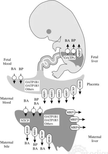
Figure 1. Schematic representation of the plasma membrane carriers involved in the normal uptake/secretion of bile acids (BA) and biliary pigments (BP) by adult and fetal hepatocytes and trophoblastic cells. NTCP, Na+-taurocholate-cotransporting polypeptide; OATPs, organic anion-transporting polypeptides; MDR1, multidrug resistance protein; BSEP, bile salt export pump; MRPs, multidrug-resistance associated proteins; BCRP, breast cancer resistance protein.
Maternal
bile BA BPBA BA
BSEP MRP2
BCRP
NTCP OATP1B1OATP1B3
Others
BA BP BA
MRP1 BCRP
Maternal blood
Maternal liver
MRP2 MRP3 MRP?
MDR1
BA BP Fetal blood
OATP1B1 OATP1B3 Others
Placenta Fetal liver OATPs
BA BP
MRP?
MDR1
MRP1
MRP3
JJG Marin et al. Excretion of fetal bile acids and biliary pigments 75
edigraphic.com
6. Cui Y, Konig J, Leier I, Buchholz U, Keppler D. Hepatic uptake of bilirrubin and its conjugates by the human organic anion-transporting polypeptide SLC21A6. J Biol Chem 2001; 276(13): 9626-9630. 7. Briz O, Serrano MA, Macias RI, Gonzalez-Gallego J, Marin JJG.
Role of organic anion-transporting polypeptides, OATP-A, OATP-C and OATP-8 in the human placenta-maternal liver tandem excretory pathway for foetal bilirubin. Biochem J 2003a; 371(3): 897-905. 8. Hagenbuch B, Stieger B, Foguet M, Lubbert H, Meier PJ. Functional
expression cloning and characterization of the hepatocyte Na+/bile acid cotransport system. Proc Natl Acad Sci USA 1991; 88(23): 10629-10633. 9. Boyer JL, Hagenbuch B, Ananthanarayanan M, Suchy F, Stieger B, Meier PJ. Phylogenic and ontogenic expression of hepatocellular bile acid transport. Proc Natl Acad Sci USA 1993; 90(2): 435-438. 10. Craddock AL, Love MW, Daniel RW, Kirby LC, Walters HC, Wong
MH, Dawson PA. Expression and transport properties of the human ileal and renal sodium-dependent bile acid transporter. Am J Physiol 1998; 37(1): G157-G169.
11. St-Pierre MV, Stallmach T, Grundschober AF, Dufour J-F, Serrano MA, Marin JJG, Sugiyama Y, et al. Temporal expression profiles of organic anion transport proteins in placenta and fetal liver of the rat.
Am J Physiol 2004; 287(6): R1505-R1516.
12. Patel P, Weerasekera N, Hitchins M, Boyd CA, Johnston DG, Williamson C. Semi quantitative expression analysis of MDR3, FIC1, BSEP, OATP-A, OATP-C, OATP-D, OATP-E and NTCP gene transcripts in 1st and 3rd trimester human placenta. Placenta 2003; 24(1): 39-44.
13. Marin JJG, Serrano MA, El-Mir MY, Eleno N, Boyd CA. Bile acid transport by basal membrane vesicles of human term placental tro-phoblast. Gastroenterology 1990; 99(5): 1431-1438.
14. Serrano MA, Bayon JE, Pascolo L, Tiribelli C, Ostrow JD, Gonzalez-Gallego J, Marin JJG. Evidence for carrier-mediated transport of unconjugated bilirubin across plasma membrane vesicles from hu-man placental trophoblast. Placenta 2002; 23(7): 527-535. 15. Suzuki H, Sugiyama Y. Transport of drugs across the hepatic
sinu-soidal membrane: sinusinu-soidal drug influx and efflux in the liver. Semin
Liver Dis 2000; 20(3): 251-263.
16. Briz O, Serrano MA, Rebollo N, Hagenbuch B, Meier PJ, Koepsell H, Marin JJG. Carriers involved in targeting the cytostatic bile acid-cisplatin derivatives cis-diammine-chloro-cholylglycinate-platinum(II) and cis-diammine-bisursodeoxycholate-cis-diammine-chloro-cholylglycinate-platinum(II) to-ward liver cells. Mol Pharmacol 2002; 61(4): 853-860.
17. Kekuda R, Prasad PD, Wu X, Wang H, Fei YJ, Leibach FH, Ganapathy V. Cloning and functional characterization of a potential-sensitive, polyspecific organic cation transporter (OCT3) most abundantly ex-pressed in placenta. J Biol Chem 1998; 273(26): 15971-15979. 18. Muller M, Mayer R, Hero U, Keppler D. ATP-dependent transport of
amphiphilic cations across the hepatocyte canalicular membrane mediated by Mdr1 P-glycoprotein. FEBS Lett 1994; 343(2): 168-172. 19. Gerloff T, Stieger B, Hagenbuch B, Madon J, Landmann L, Roth J, Hofmann AF, et al. The sister of P-glycoprotein represents the canali-cular bile salt export pump of mammalian liver. J Biol Chem 1998; 273(16): 10046-10050.
20. Jedlitschky G, Leier I, Buchholz U, Hummel-Eisenbeiss J, Burchell B, Keppler D. ATP-dependent transport of bilirubin glucuronides by the multidrug resistance protein MRP1 and its hepatocyte canalicular isoform MRP2. Biochem J 1997; 327(1): 305-310.
21. Cui Y, Konig J, Buchholz JK, Spring H, Leier I, Keppler D. Drug resistance and ATP-dependent conjugate transport mediated by the apical multidrug resistance protein, MRP2, permanently expressed in human and canine cells. Mol Pharmacol 1999; 55(5): 929-937. 22. Imai Y, Asada S, Tsukahara S, Ishikawa E, Tsuruo T, Sugimoto Y.
Breast cancer resistance protein exports sulfated estrogens but not free estrogens. Mol Pharmacol. 2003; 64(3): 610-618.
23. Janvilisri T, Shahi S, Venter H, Balakrishnan L, van Veen HW. Argi-nine-482 is not essential for transport of antibiotics, primary bile ac-ids and unconjugated sterols by the human breast cancer resistance protein (ABCG2). Biochem J 2005; 385(2): 419-426.
24. Ogawa K, Suzuki H, Hirohashi T, Ishikawa T, Meier PJ, Hirose K, Akizawa T, et al. Characterization of inducible nature of MRP3 in rat liver. Am J Physiol 2000; 278(3): G438-G446.
25. Soroka CJ, Lee JM, Azzaroli F, Boyer JL. Cellular localization and up-regulation of multidrug resistance-associated protein 3 in
hepato-cytes and cholangiohepato-cytes during obstructive cholestasis in rat liver.
Hepatology 2001; 33(4): 783-791.
26. Donner MG, Keppler D. Up-regulation of basolateral multidrug re-sistance protein 3 (Mrp3) in cholestatic rat liver. Hepatology 2001; 34(2): 351-359.
27. Vos TA, Hooiveld GJ, Koning H, Childs S, Meijer DK, Moshage H, Jansen PL, et al. Up-regulation of the multidrug resistance genes, Mrp1 and Mdr1b, Mrp1 and Mdr1b, and down-regulation of the or-ganic anion transporter, Mrp2, and the bile salt transporter, Spgp, in endotoxemic rat liver. Hepatology 1998; 28(6): 1637-1644. 28. Tanaka Y, Kobayashi Y, Gabazza EC, Higuchi K, Kamisako T,
Kuroda M, Takeuchi K, et al. Increased renal expression of bilirubin glucuronide transporters in a rat model of obstructive jaundice. Am J
Physiol 2002; 282(4): G656-G662.
29. Javitt NB. Cholesterol, hydroxycholesterols, and bile acids. Biochem
Biophys Res Commun 2002; 292(5): 1147-1153.
30. Suchy FJ, Bucuvalas JC, Novak DA. Determinants of bile formation during development: Ontogeny of hepatic bile acid metabolism and transport. Semin Liver Dis 1987; 7(2): 77-84.
31. Monte MJ, Morales AI, Arevalo M, Alvaro I, Macias RI, Marin JJG. Reversible impairment of neonatal hepatobiliary function by mater-nal cholestasis. Hepatology 1996; 23(5): 1208-1217.
32. Colombo C, Roda A, Roda E, Buscaglia M, dell’Agnola CA, Filippetti P, Ronchi M, et al. Correlation between fetal and maternal serum bile acid concentrations. Pediatr Res 1985; 19(2): 227-231.
33. Balistreri WF, A-Kader HH, Setchell KD, Gremse D, Ryckman FC, Schroeder TJ. New methods for assessing liver function in infants and children. Ann Clin Lab Sci 1992; 22(3): 162-174.
34. Monte MJ, Rodriguez-Bravo T, Macias RI, Bravo P, el-Mir MY, Serrano MA, Lopez-Salva A, et al. Relationship between bile acid transplacen-tal gradients and transport across the fetransplacen-tal-facing plasma membrane of the human trophoblast. Pediatr Res 1995; 38(2): 156-163.
35. Nakagawa M, Setchell KD. Bile acid metabolism in early life: Stud-ies of amniotic fluid. J Lipid Res 1990; 31(6): 1089-1098.
36. Knudsen A, Lebech M. Maternal bilirubin, cord bilirubin, and pla-centa function at delivery and the development of jaundice in mature newborns. Acta Obstet Gynecol Scand 1989; 68(8): 719-724. 37. Li L, Meier PJ, Ballatori N. Oatp2 mediates bidirectional organic
solute transport: a role for intracellular glutathione. Mol Pharmacol 2000; 58(2): 335-340.
38. Fujiwara K, Adachi H, Nishio T, Unno M, Tokui T, Okabe M, Onogawa T, et al. Identification of thyroid hormone transporters in humans: different molecules are involved in a tissue-specific man-ner. Endocrinology 2001; 142(5): 2005-2012.
39. Sato K, Sugawara J, Sato T, Mizutamari H, Suzuki T, Ito A, Mikkaichi T, et al. Expression of organic anion transporting polypeptide E (OATP-E) in human placenta. Placenta 2003; 24(2-3): 144-148. 40. Cabral DJ, Small DM, Lilly HS, Hamilton JA. Transbilayer movement
of bile acids in model membranes. Biochemistry 1987; 26(7): 1801-1804. 41. Macias RI, Pascual MJ, Bravo A, Alcalde MP, Larena MG, St-Pierre MV, Serrano MA, et al. Effect of maternal cholestasis on bile acid transfer across the placenta-maternal liver tandem. Hepatology 2000; 31(4): 975-983.
42. Marin JJG, Briz O, Serrano MA. A review on the molecular mecha-nisms involved in the placental barrier for drugs. Current Drug Deliv 2004; 1: 275-289.
43. Iioka H, Moriyama I, Hino K, Ichijo M. A study on the mechanism of bile acid transport in the human placenta (the passive transport sys-tem of taurocholate across microvillous membrane). Nippon Sanka
Fujinka Gakkai Zasshi 1986; 38(6): 837-844.
44. Dumaswala R, Setchell KD, Moyer MS, Suchy FJ. An anion exchanger mediates bile acid transport across the placental microvillous mem-brane. Am J Physiol 1993; 264(6): G1016-G1023.
45. Iioka H, Hisanaga H, Akada S, Shimamoto T, Yamada Y, Sakamoto Y, Moriyama IS, et al. Characterization of human placental activity for transport of taurocholate, using brush border (microvillous) mem-brane vesicles. Placenta 1993; 14(1): 93-102.
Annals of Hepatology 4(2) 2005: 70-76
MG
76
edigraphic.com
47. El-Mir MY, Eleno N, Serrano MA, Bravo P, Marin JJG. Bicarbon-ate-induced activation of taurocholate transport across the basal plasma membrane of the human term trophoblast. Am J Physiol 1991; 260(6): G887-G894.
48. Serrano MA, Bravo P, El-Mir MY, Marin JJ. Influence of hydroxyla-tion and conjugahydroxyla-tion in cross-inhibihydroxyla-tion of bile acid transport across the human trophoblast basal membrane. Biochim Biophys Acta 1993; 1151(1): 28-34.
49. Bravo P, El-Mir MYA, Serrano MA, Boyd R, Marin JJG. Interaction between cholephilic anions and bile acid transport across basal mem-brane of human trophoblast. Am J Physiol 1993; 265(2 Pt 1): G242-G250. 50. Serrano MA, Macias RI, Vallejo M, Briz O, Bravo A, Pascual MJ, St-Pierre MV, et al. Effect of ursodeoxycholic acid on the impair-ment induced by maternal cholestasis in the rat placenta-maternal liver tandem excretory pathway. J Pharmacol Exp Therap 2003; 305(2): 515-524.
51. St-Pierre MV, Hagenbuch B, Ugele B, Meier PJ, Stallmach T. Char-acterization of an organic anion transporting polypeptide OATP B in human placenta. J Clin Endocrin Metab 2002; 87(4): 1856-1863. 52. Nishio T, Adachi H, Nakagomi R, Tokui T, Sato E, Tanemoto M, Fujiwara
K, et al. Molecular identification of a rat novel organic anion transporter moat1, which transports prostaglandin D(2), leukotriene C(4), and tauro-cholate. Biochem Biophys Res Commun 2000; 275(3): 831-838. 53. Bravo P, Marin JJG, Beveridge MJ, Novak DA. Reconstitution and
characterization of ATP-dependent bile acid transport in human and rat placenta. Biochem J 1995; 311(2): 479-485.
54. Young AM, Allen CE, Audus KL. Efflux transporters of the human placenta. Adv Drug Deliv Rev 2003; 55(1): 125-132.
55. St-Pierre MV, Serrano MA, Macias RI, Dubs U, Hoechli M, Lauper U, Meier PJ, et al. Expression of members of the multidrug resistance protein family in human term placenta. Am J Physiol 2000; 279(4): R1495-R1503.
56. Rius M, Nies AT, Hummel-Eisenbeiss J, Jedlitschky G, Keppler D. Cotransport of reduced glutathione with bile salts by MRP4 (ABCC4) localized to the basolateral hepatocyte membrane. Hepatology 2003; 38(2): 374-84.
57. Leazer TM, Klaassen CD. The presence of xenobiotic transporters in rat placenta. Drug Metab Dispos 2003; 31(2): 153-167.
58. St-Pierre MV, Serrano MA, Lauper U, Marin JJG, Meier PJ. Identifi-cation of bile salt transporters in human and rat placenta. (Abstract)
Placenta 1999; 20: A62.
59. Allikmets R, Schriml LM, Hutchinson A, Romano-Spica V, Dean M. A human placenta-specific ATP-binding cassette gene (ABCP) on chromosome 4q22 that is involved in multidrug resistance. Cancer
Res 1998; 58(23): 5337-5339.
60. Galbraith R. Heme oxygenase: who needs it? Proc Soc Exp Biol Med 1999; 222(3): 299-305.
61. Ryter SW, Tyrrell RM. The heme synthesis and degradation path-ways: role in oxidant sensitivity. Heme oxygenase has both pro- and antioxidant properties. Free Radic Biol Med 2000; 28(2): 289-309. 62. Lyall F, Barber A, Myatt L, Bulmer JN, Robson SC. Hemeoxygenase
expression in human placenta and placental bed implies a role in regu-lation of trophoblast invasion and placental function. FASEB J 2000; 14(1): 208-219.
63. McCoubrey WK, Cooklis MA, Maines MD. The structure, organiza-tion and differential expression of the rat gene encoding biliverdin reductase. Gene 1995; 160(2): 235-240.
64. McDonagh AF, Palma LA, Schmid R. Reduction of biliverdin and placental transfer of bilirubin and biliverdin in the pregnant guinea pig. Biochem J 1981; 194(1): 273-282.
65. Stollman YR, Gartner U, Theilmann L, Ohmi N, Wolkoff AW. He-patic bilirubin uptake in the isolated perfused rat liver is not facili-tated by albumin binding. J Clin Inves; 1983 72(2): 718-723. 66. Bosma PJ, Seppen J, Goldhoorn B, Bakker C, Oude Elferink RP,
Chowdhury JR, Chowdhury NR, et al. Bilirubin UDP-glucuronos yltransferase 1 is the only relevant bilirubin glucuronidating isoform in man. J Biol Chem 1994; 269(27): 17960-17964.
67. Keppler D, Kamisako T, Leier I, Cui Y, Nies AT, Tsujii H, Konig J. Local-ization, substrate specificity, and drug resistance conferred by conjugate export pumps of the MRP family. Adv Enzyme Regul 2000; 40: 339-349.
68. Terrenato L, Bertilaccio C, Spinelli P, Colombo B. The switch from haemoglobin F to A: the time course of qualitative and quantitative varia-tions of haemoglobins after birth. Br J Haematol 1981; 47(1): 31-41. 69. Ogita S, Shimamoto T, Ohnishi M, Kamei T, Noma H, Ishiko O,
Ando T, et al. Hemolytic pattern of erythrocytes in the newborn mea-sured by the coil planet centrifuge system and its relationship to neo-natal jaundice. Eur J Pediatr 1978; 127(2): 67-73.
70. Kawade N, Onishi S. The prenatal and postnatal development of UDP-glucuronyltransferase activity towards bilirubin and the effect of pre-mature birth on this activity in the human liver. Biochem J 1981; 196(1): 257-260.
71. McDonagh AF. Turning green to gold. Nat Struct Biol 2001; 8(3): 198-200.
72. Schenker S, Dawber NH, Schmid R. Bilirubin metabolism in the fe-tus. J Clin Invest 1964; 43: 32-39.
73. Bernstein RB, Novy MJ, Piasecki GJ, Lester R, Jackson BT. Biliru-bin metabolism in the fetus. J Clin Invest 1969; 48(9): 1678-1688. 74. Lester R, Behrman RE, Lucey JF. Transfer of bilirubin-C across
mon-key placenta. Pediatrics 1963; 32: 416-419.
75. Bashore RA, Smith F, Schenker S. Placental transfer and disposition of bilirubin in the pregnant monkey. Am J Obstet Gynecol 1969; 103(7): 950-958.
76. Briz O, Macias RI, Serrano MA, Gonzalez-Gallego J, Bayon JE, Marin JJG. Excretion of foetal bilirubin by the rat placenta-maternal liver tandem. Placenta 2003b; 24(5): 462-472.
77. Brodie BB, Axelrod J, Soberman R, Levy BB. The estimation of an-tipyrine in biological materials. J Biol Chem 1949; 179: 25-29. 78. Weiner CP. Human fetal bilirubin levels and fetal hemolytic disease.
Am J Obstet Gynecol 1992; 166(5): 1449-1454.
79. Pascolo L, Fernetti C, Garcia-Mediavilla MV, Ostrow JD, Tiribelli C. Mechanisms for the transport of unconjugated bilirubin in human trophoblastic BeWo cells. FEBS Lett 2001; 495(1-2): 94-99. 80. Lammert F, Marschall HU, Matern S. Intrahepatic cholestasis of
preg-nancy. Curr Treat Options Gastroenterol 2003; 6(2): 123-132. 81. Glantz A, Marschall HU, Mattsson LA. Intrahepatic cholestasis of
pregnancy: Relationships between bile acid levels and fetal compli-cation rates. Hepatology 2004; 40(2): 467-474.
82. Serrano MA, Brites D, Larena MG, Monte MJ, Bravo MP, Oliveira N, Marin JJG. Beneficial effect of ursodeoxycholic acid on alterations induced by cholestasis of pregnancy in bile acid transport across the human placenta. J Hepatol 1998; 28(5): 829-839.
83. El-Mir MY, Monte MJ, Morales AI, Arevalo M, Serrano MA, Marin JJG. Effect of maternal cholestasis on biliary lipid and bile acid se-cretion in the infant rat. Hepatology 1997; 26(3): 527-536. 84. Monte MJ, Villanueva GR, Macias RI, Vazquez DJ, Toledo M,
Dominguez M, Marin JJG. Effect of maternal obstructive cholestasis during pregnancy on the biliary transport of horseradish peroxidase in the rat offspring. Clin Sci 2003; 105(3): 347-353.
85. Arrese M, Trauner M, Ananthanarayanan M, Boyer JL, Suchy FJ. Maternal cholestasis does not affect the ontogenic pattern of expres-sion of the Na+/taurocholate cotransporting polypeptide (ntcp) in the fetal and neonatal rat liver. Hepatology 1998; 28(3): 789-795. 86. Macias RI, Serrano MA, Monte MJ, Jimenez S, Hernandez B, Marin
JJG. Long-term effect of treating pregnant rats with ursodeoxycholic acid on the congenital impairment of bile secretion induced in the pups by maternal cholestasis. J Pharmacol Exp Ther 2005; 312(2): 751-758. 87. Serrano MA, Monte MJ, Martinez-Diez MC, Marin JJG. Effect of maternal cholestasis on the kinetics of bile acid transport across the canalicular membrane of infant rat livers. Int J Exp Pathol 1997; 78(6): 383-390.
88. Attili AF, Angelico M, Cantafora A, Alvaro D, Capocaccia L. Bile acid-induced liver toxicity: relation to the hydrophobic-hydrophilic balance of bile acids. Med Hypotheses 1986; 19(1): 57-69. 89. Perez MJ, Macias RI, Marin JJG. Maternal cholestasis induces
pla-cental oxidative stress and apoptosis. Protective effect of ursodeoxycholic acid. Placenta (in press) 2005a.
90. Perez MJ, Macias RI, Duran C, Monte MJ, Gonzalez-Buitrago JM, Marin JJG. Oxidative stress and apoptosis in fetal rat liver induced by maternal cholestasis. Protective effect of ursodeoxycholic acid. J