Recurrent Hyperammonemia After
Abernethy Malformation Type 2 Closure: a Case Report
Hui Li, Zhi Ma, Ying Xie, Feng Tian
Department of gastroenterology, Shengjing Hospital Affiliated to China Medical University, Shenyang, Liaoning, China May-June, Vol. 16 No. 3, 2017: 460-464
INTRODUCTION
The Abernethy malformation is a rare congenital com-munication between the portal vein and systemic circula-tion; it was first reported by John Abernethy in 1793. According to the type of anastomosis and status of the por-tal vein, it is classified into 2 types.1 Type 1 is
character-ized by a complete absence of the portal vein and is
predominantly (74% of cases) found in females.1-4 Type 1
malformation is further divided into subtypes A and B:5 in
type 1A, the splenic vein and the superior mesenteric vein drain separately into the inferior vena cava; in type 1B, these veins form a common trunk. Partial shunts between the portal vein and the systemic venous circulation are de-fined as type 2; these allow for some potential portal per-fusion of the liver. Various clinical presentations and some congenital malformations are associated with these
abnor-mal shunts:6 hepatic encephalopathy, portopulmonary
syndrome, hepatic masses, cardiac defects, and vascular anomalies have all been reported.
We report a patient with type 2 Abernethy malforma-tion and hepatic encephalopathy. Although she underwent successful interventional shunt closure, her hyperammon-emia recurred 3 months postoperatively. This is the first
reported case of recurrent hyperammonemia after inter-ventional treatment; we discuss herein the therapeutic op-tions for Abernethy malformation.
Case report
A 58-year-old female presented to the gastroenterology department of Shengjing Hospital (affiliated with China Medical University) with an 8-month history of dizziness, sleepiness. She was previously diagnosed, at another insti-tution, with metabolic encephalopathy when she present-ed with elevatpresent-ed alanine transaminase levels without hepatic dysfunction. She was otherwise healthy, with a history significant only for a simple vocal-cord surgery. Physical examination revealed lower-extremity edema but no splenomegaly or evidence of ascites.
Laboratory testing revealed aspartate aminotransferase level of 39 IU/L (normal range, 5-34 IU/L), an alkaline phosphatase level of 180 IU/L (normal range, 40-150 IU/ L), a total bilirubin level of 24 μmol/L (normal range, 3.4-20.5 μmol/L), and an indirect bilirubin level of 15.7 μmol/
L (normal range, 3.4-11.9 μmol/L). Her white blood-cell
count was 3.9 x 10⁹/L (normal range, 3.5-9.7 x 10⁹/L), her
hemoglobin level was 116 g/L (normal range, 110-150 g/L),
The Official Journal of the Mexican Association of Hepatology, the Latin-American Association for Study of the Liver and
the Canadian Association for the Study of the Liver
Manuscript received: Manuscript received: Manuscript received: Manuscript received:
Manuscript received: May 05, 2016. Manuscript accepted:Manuscript accepted:Manuscript accepted: August 29, 2016.Manuscript accepted:Manuscript accepted:
DOI:10.5604/16652681.1235492
A B S T R A C T A B S T R A C T A B S T R A C T A B S T R A C T A B S T R A C T
The Abernethy malformation is a rare congenital malformation defined by the presence of an extrahepatic portosystemic shunt. Al-though most patients are asymptomatic, clinical encephalopathy is present in 15% of cases. We present a patient with type 2 Aber-nethy malformation, hyperammonemia, and encephalopathy. Shunt closure was performed successfully using interventional angiography; however, hyperammonemia recurred 3 months later. The diagnosis of Abernethy malformation can be made easily, but the ideal patient management strategy has not yet been established. This is the first reported patient with recurrence of hyperam-monemia after interventional treatment; we discuss the therapeutic options for Abernethy malformation.
Key words. Key words. Key words. Key words.
her platelet count was 180 x 109/L (normal range, 135-350 x
109/L), and her coagulation profile was normal. The tumor
marker cancer antigen 19-9 (CA19-9) measured 58.47 U/ mL (normal range, 0-37U/mL). Serological evaluation for ceruloplasmin, hepatitis viruses A E, and immunological markers was negative, although her plasma ammonia level
was 147.6 μmol/L (normal range, 9-33 μmol/L).
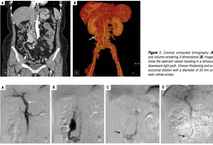
Comput-ed tomography of the abdomen was performComput-ed using a 64-row multidetector. Multiplanar reconstruction and 3-dimensional volume-rendered images confirmed the presence of an abnormal shunt between the inferior vena cava and the superior mesenteric vein; this shunt had a di-ameter of 33 mm (Figure 1).
The patient subsequently underwent shunt closure, us-ing interventional angiography (Figure 2). Her plasma am-monia level returned to normal range, with complete resolution of encephalopathy and the lower-extremity edema. However, about 3 months after surgery, routine follow-up examination revealed an elevated plasma
am-monia level of 155 μmol/L; repeat angiography confirmed
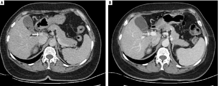
successful shunt closure and did not reveal any other por-tal-venous vessels that could explain the recurrence of hyperammonemia(Figure 2). However, contrast-enhanced CT of the liver revealed that the portal vein was 1.5 cm in diameter, larger than the presurgical value (Figure 3). Since she was asymptomatic, conservative observation was recommended. Over a year of follow-up, the serological evaluation for liver function was normal, while the plasma
ammonia level was in the range of 60-80 μmol/L.
DISCUSSION
Abernethy malformation has a female dominance and is usually diagnosed during childhood.7 Hao, et al.,8 reviewed
101 previously reported patients with Abernethy malfor-mation and found that 65.3% were female, 69.3% were younger than 18 years of age, and less than 10% had type-2 malformations.
Some patients remain asymptomatic throughout life.9
When symptoms do occur, the clinical manifestations are
Figure 1. Figure 1.Figure 1.
Figure 1.Figure 1. Coronal computed tomography (AAAA)A and volume-rendering 3-dimensional (BBBBB) images show the aberrant vessel traveling in a tortuous, downward-right path. Uneven thickening and an-eurysmal dilation with a diameter of 33 mm are seen (white arrow).
Figure 2. Figure 2. Figure 2.
Figure 2. Figure 2. Angiographic images (AAAAA) shows the intrahepatic portal system (white arrow) before shunt closure; (BBBB) shows the aberrant drainage branches of su-B perior mesenteric vein into the inferior vena cava prior to closure (white arrow); (CCCCC) shows the successful closure of the shunt with coil embolization(black ar-row); (DDDDD) repeat angiographic image shows the persistent closure of the shunt.
A AA
AA BBBBB CCCCC DDDDD
A A A A
mainly associated with the hepatic shunt and associated congenital malformations. In the Abernethy malformation, diversion of the portal blood flow into the inferior vena cava leads to systemic hypertension, which can explain the leg edema seen in our patient. Metabolic and vasoactive substances bypass the liver through portosystemic collat-erals and thus cause portopulmonary hypertension,
hyper-ammonemia, and encephalopathy.10 Nodular liver lesions
are seen in up to half of patients: these include benign fo-cal nodular hyperplasia, hepatocellular adenoma, and
re-generative nodules4,11 and may be explained by the absence
of portal flow and compensatory increased hepatic arterial blood flow. Although most of these lesions are benign, there are several case reports in the literature describing the coexistence of hepatocellular carcinoma and hepatob-lastoma. Therefore, long-term follow-up and monitoring
are recommended.12 Congenital hepatic shunts can also
present with metabolic dysregulation, such as hypoglyc-emia, due to metabolic alterations in the liver.13
Despite the existence of a direct connection between the portal and systemic venous systems, hyperammonemia is found in only 26% of patients with the Abernethy
mal-formation,14 and only 15% of all patients experience
hepat-ic encephalopathy.15 A recent review identified 316
published cases and reported that hyperammonemia/neu-rological abnormalities occurred in 35% of patients with
congenital portosystemic shunt.16 Hyperammonemia can
be present without encephalopathy, especially in younger patients; clinical encephalopathy is more common at older ages. Possible explanations for this phenomenon are an age-dependent increase in sensitivity to deleterious me-tabolites and the impact of the extent of shunting, deter-mined by the portal/systemic shunt ratio-a shunt ratio of
more than 60% may predict the age of onset of encepha-lopathy.17,18
Current treatment options, including interventional or surgical shunt closure and liver transplantation, are de-scribed in case reports; however, the therapeutic
experi-ence with Abernethy malformation is still limited.19 The
choice of strategy depends on the type of shunt, symp-toms, complications, and comorbidities. Shunt occlusion is not feasible in patients with type 1, as the shunt is the only drainage route for the mesenteric venous blood. Thus, liver transplantation may be required to provide a patent portal system and to treat the metabolic abnormali-ties and liver disease in these patients.20 Kanamori, et al.21
previously reported on a patient with type 2 Abernethy malformation who required liver transplantation after de-veloping postoperative portal hypertension and another portosystemic shunt. Thus, it is important to ensure ade-quate portal vein capacity and normal portal pressure prior to closure, in order to avoid postprocedure portal
hyper-tension and subsequent mesenteric ischemia.22
The balloon shunt occlusion test shows a visible intra-hepatic portal system (IHPS) in patients with both type 1
and type 2 Abernethy malformation.23 This finding could
represent small portal vein branches which cannot be seen on ultrasonography but can be visualized on shunt angiog-raphy.2,24 Kanazawa H, et al.,23 proposed a new
classifica-tion based on IHPS hypoplasia (mild, moderate, and severe types) under shunt occlusion; this system guides the decision on whether to perform single-stage or 2-stage shunt closure. Single-stage closure is recommended for mild and moderate types with crescent-shaped portal veins and a portal venous pressure below 25 mmHg. This sug-gests that the intrahepatic portal vascular bed is able to
ac-Figure 3. Figure 3. Figure 3. Figure 3.
Figure 3. Axial contrast-enhanced CT images show the change in size of portal vein before (AAAAA) and after (BBBBB) shunt closure (white arrow). A
A A A
cept sufficient blood flow immediately upon shunt clo-sure. Patients with the severe type, with a small portal-tri-ad area and a portal venous pressure over 25 mmHg, are candidates for either 2-stage shunt closure or liver trans-plantation. In the present case, the intrahepatic portal vein system was well displayed under angiography, similar to mild type of IHPS, which indicated the possibility of shunt closure. The balloon shunt occlusion test was not performed prior to operation and a direct shunt closure was carried out in this patient. The treatment was proved effective in both the improvement of symptoms and labo-ratory results. However, her plasma ammonia level was el-evated again just 3 months later. Although repeat angiography showed persistent closure of the shunt, an en-larged portal vein was confirmed; thus, we conjectured that the closure of the original large shunt caused a sudden increased inflow into the portal vein, leading to portal hy-pertension, when then resulted in the development of new collaterals between the portal and venous vessels. As the diameter of shunt vessels grows with age, the difficul-ty of intervention increases. In our case, the large diameter of the longstanding aberrant vessel increased the difficulty of the original operation and the chance of complications. Therefore, it is necessary to carefully assess the severity of the hypoplasia of IHPS as well as the PVP under the oc-clusion test, and for cases with large abnormal vessel, sim-ple shunt closure may not be the first choice.
In conclusion, the diagnosis of Abernethy malforma-tion is challenging, as it is an uncommon congenital mal-formation with nonspecific clinical symptoms. However, patients with unexplainable hyperammonemia and hepatic encephalopathy require evaluation for a potential porto-systemic shunt. Since most significant presentations ap-pear in older patients with large-diameter shunts, as well as in those with severe portal dysplasia, the incidence of postsurgical complications increases. Thus, it is necessary to evaluate the IHPS by angiography, using the shunt-oc-clusion test, and to measure the portal venous pressure un-der shunt occlusion in orun-der to choose the optimal therapeutic approach. Moreover, in the present case, al-though the repeat angiography failed to show the new col-laterals between the portal and venous veins, we couldn't deny the existence of the recurrent shunts that might ex-plain the recurrent hyperammonemia. Additionally, fur-ther tests should be done to explore the possible defects of urea cycle which might result in high ammonia. Post-operative clinical improvement, such as regression of be-nign liver masses and of pulmonary, cardiac, neurological,
and renal complications, has been reported.24 However,
there are patients described in the literature with hepatic lesions that did not improve by 12 to 24 months after shunt closure. These findings suggest that the malforma-tion should be treated before a significant complicamalforma-tion
occurs and that long-term follow-up and monitoring for
complications as well as malignancy are mandatory.23
REFERENCES
1. Morgan G, Superina R. Congenital absence of the portal vein:Two cases and a proposed classification system for portasystemic vascular anomalies. J Pediatr Surg 1994; 29: 1239-41.
2. Howard ER, Davenport M. Congenital extrahepatic porto-caval shunts: The Abernethy malformation. J Pediatr Surg
1997; 32: 494-7.
3. Murray CP, Yoo SJ, Babyn PS. Congenital extrahepatic por-tosystemic shunts. Pediatr Radiol 2003; 33: 614-20. 4. Grazioli L, Alberti D, Olivetti L, Rigamonti W, Codazzi F,
Matri-cardi L, Fugazzola C, et al. Congenital absence of portal vein with nodular regenerative hyperplasia of the liver. Eur Radi-ol 2000; 10: 820-5.
5. Witters P, Maleux G, George C, Delcroix M, Hoffman I, Gewil-lig M, Verslype C, et al. Congenital veno-venous malforma-tions of the liver: widely variable clinical presentamalforma-tions. J Gastroenterol Hepatol 2008; 23: e390-4.
6. Lee WK, Chang SD, Duddalwar VA, Comin JM, Perera W, Lau Wing FE, Bekhit EK, et al. Imaging assessment of con-genital and acquired abnormalities of the portal venous sys-tem. RadioGraphics 2011; 31: 905-26.
7. Asran MK, Loyer EM, Kaur H, Choi H. Case 177: congenital absence of the portal vein with hepatic adenomatosis. Radi-ology 2012; 262: 364-7.
8. Yabin H, Xu H, Xinyan Z. Congenital absence of the portal vein associated with focal nodular hyperplasia of the liver and con-genital heart disease (Abernethy malformation): A case report and literature review. Oncol lett 2015; 9: 695-700.
9. Hoeper MM, Krowka MJ, Strassburg CP. Portopulmonary hy-pertension and hepatopulmonary syndrome. Lancet 2004; 363: 1461-8.
10. Mistinova J, Valacsai F, Varga I. Congenital absence of the portal vein-Case report and a review of literature. Clin Anat
2010; 23: 750-8.
11. Collard B, Maleux G, Heye S, Cool M, Bielen D, George C, Roskams T, et al. Value of carbon dioxide wedged venogra-phy and transvenous liver biopsy in the definitive diagnosis of Abernethy malformation. Abdom Imaging 2006; 31: 315-9. 12. Alonso-Gamarra E, Parrón M, Pérez A, Prieto C, Hierro L, López-Santamaría M. Clinical and radiologic manifestations of congenital extrahepatic portosystemic shunts: a compre-hensive review. RadioGraphics 2011; 31: 702-22.
13. Duprey J, Gouin B, Benazet MF, et al. Glucose intolerance and poststimulatory hypoglycemia secondary to a probably congenital intrahepatic portacaval anastomosis. Ann Med In-terne 1985; 136: 655-8.
14. De Gaetano AM, Gui B, Macis G, Manfredi R, Di Stasi C. Con-genital absence of the portal vein associated with focal nod-ular hyperplasia in the liver in an adult woman: imaging and review of the literature. Abdom Imaging 2004; 29: 455-9. 15. Murray CP, Yoo SJ, Babyn PS. Congenital extrahepatic
por-tosystemic shunts. Pediatr Radiol 2003; 33: 614-20. 16. Sokollik C, Bandsma RH, Gana JC, van den Heuvel M, Ling
SC. Congenital portosystemic shunt: characterization of a multisystem disease. J Pediatr Gastroenterol Nutr 2013; 56: 675-81.
18. Singhal A, Srivastava A, Goyal N, Vij V, Wadhawan M, Bera M, Gupta S. Successful living donor liver transplant in a child with Abernethy malformation with biliary atresia, ventricular septal defect and intrapulmonary shunting. Pediatr Trans-plant 2009; 13: 1041-7.
19. KCS, Matsutani S. Maruyama H. Fukamachi T. Nomoto H. Aki-ike T. Ebara M, et al. Portal systemic encephalopathy in two patients without liver cirrhosis and portal hypertension.
Hepatol Res 2002; 23: 122-9.
20. Orii T, Ohkohchi N, Kato H, Doi H, Hirano T, Sekiguchi S, Aka-matsu Y, et al. Liver transplantation for severe hypoxemia caused by patent ductus venosus. J Pediatr Surg 1997; 32: 1795-7.
21. Kanamori Y, Hashizume K, Kitano Y, Sugiyama M, Motoi T, Tange T. Congenital extrahepatic portocaval shunt (Aber-nethy type 2), huge liver mass, and patent ductus arterio-sus-a case report of its rare clinical presentation in a young girl. J Pediatr Surg 2003; 38: E15.
22. Venkateshwaran S, Krishnamoorthy KM, Sivasankaran S. Percutaneous Device Closure of Abernethy Malformation-A
Treatable Cause of Hepatopulmonary Syndrome. Catheter Cardiovasc Interv 2014; 83: 968-70.
23. Kanazawa H, Nosaka S, Miyazaki O, Sakamoto S, Fukuda A, Shigeta T, Nakazawa A, et al. The classification based on in-trahepatic portal system for congenital portosystemic shunts. J Pediatr Surg 2015; 50: 688-95.
24. Franchi-Abella S, Branchereau S, Lambert V, Fabre M, Ste-imberg C, Losay J, Riou JY, et al. Complications of congenital portosystemic shunts in children: therapeutic options and outcomes. J Pediatr Gastroenterol Nutr 2010, 51: 322-30.
Correspondence and reprint request: Feng Tian, M.D., Ph.D.
Department of Gastroenterology, Shengjing Hospital Affiliated to China Medical University,
No. 36 Sanhao Street, Shenyang, Liaoning 110004, China Tel: +86 - 1894 - 025 - 6110