Annals of Hepatology 5(3) 2006: 150-156
MG
150
edigraphic.com
Annals of Hepatology 2006; 5(3): July-September: 150-156Annals of Hepatology
Original Article
Differential diagnosis of acute
liver failure in India
Amarapurkar Deepak N;1 Nikhil D Patel2
Abstract
Background: Acute liver failure (ALF) is a condition with rapid deterioration of liver function resulting in hepatic encephalopathy and/or coagulopathy in pa-tients with previously normal liver. Complicated forms of certain infectious diseases like falciparum malaria, leptospirosis, dengue fever, ricketsial fever, typhoid fe-ver, haemophagocytosis, herpes simplex virus, cytome-galovirus, tuberculosis or amoebic liver abscess can present with altered mentation and/or bleeding mani-festations in presence of jaundice and mimic ALF due to acute viral hepatitis (AVH). Methods: We describe our experience in last 2 years with 28 patients of ALF due to above mentioned conditions (ALF-ID) and com-pared them with 28 patients with ALF due to AVH (ALF-AVH). Results: In ALF-ID, typhoid fever was
1Head.
2Clinical Assistant, Department Of Gastroenterology; Bombay
Hospital and Medical Research Centre, Mumbai, India.
Address for correspondence: Deepak Amarapurkar
D-401.402 Ameya RBI Employees Co-op Hsg Society Plot No. 947-950
New Prabhadevi Road Prabhadevi
Mumbai 400 025 Fax No. 91 22 4368623 Tel No. 4222432- 4306262 Email : [email protected]
Manuscript received and acepted: 17 June and 20 July 2006.
Abbreviations
Acute liver failure (ALF), international standardized ratio of prothrombin time (INR), hepatitis A virus (HAV), hepatitis B virus (HBV), hepatitis C virus (HCV), hepatitis D virus (HDV), hepatitis E virus (HEV), infectious diseases (ID), falciparum malaria (FM), Leptospirosis (LP), dengue fever (DF), ricketsial fever (RF), typhoid fever (TF), haemophagocytosis (HP), herpes simplex virus (HSV), cytomegalovirus (CMV), amoebic liver abscess (ALA), acute viral hepatitis (AVH), ALF due to infectious diseases other than A-E hepatitis viruses ID), ALF due to acute viral A-E hepatitis (ALF-AVH), alanine aminotransferase (ALT), aspartate aminotransferase (AST), alkaline phosphatase (ALP), prothrombin time (PT), lactate dehydrogenase (LDH), creatine kinase (CK), erythrocyte sedimentation rate (ESR), statistically significant (S), statistically not significant (NS), times upper limit of normal value (xULN),
present in 1, haemophagocytosis in 1, ricketsial infec-tion in 4 (scrub typhus = 2, endemic typhus = 2), amoe-bic liver abscess in 4, leptospirosis in 5, dengue fever in 5 and falciparum malaria in 8 patients. In ALF-AVH, hepatitis E and B co-infection was responsible in 1, hepatitis A and E co-infection in 1 and hepatitis E, B and C co-infection in 1, hepatitis E in 18, hepatitis A in 2 and hepatitis B in 5 patients. Differentiation of vari-ous forms of ALF-ID from ALF-AVH depends on var-ious clinical, haematological and biochemical parame-ters, in addition to specific diagnostic tests. Patients with ALF-AVH had mortality rate of 50% (14/28) and ALF-ID had mortality rate of 25% (7/28). Conclu-sions: In developing countries, ALF-mimicking infec-tions should be looked for in differential diagnosis of ALF. Early identification and treatment of these infec-tions is important in reducing mortality.
Key words: Acute liver failure, severe malaria, dengue fever, leptospirosis, enteric fever, amoebic liver ab-scess, ricketsial infection, haemophagocytosis, infec-tious diseases,
Introduction
edigraphic.com
:rop odarobale FDPVC ed AS, cidemihparG
arap
acidémoiB arutaretiL :cihpargideM
sustraídode-m.e.d.i.g.r.a.p.h.i.c
sustraídode-m.e.d.i.g.r.a.p.h.i.c cihpargidemedodabor
for ALF in 171% cases, HEV in 2.7-25%, HBV in 0-10.7%, HCV in 0-2.5%, HDV in 16.7%, mixed infection in 10-22% and non A-E infections in 17.9%.15 There is no specific treatment available for ALF for most of the caus-es, and mortality due to ALF even with a liver transplan-tation remains high.
In recent years, a subgroup of patients is often looked at, who harbor complicated forms of common infectious diseases (ID) and present as ALF.16-21 These infections are falciparum malaria (FM), Leptospirosis (LP), dengue fe-ver (DF), ricketsial fefe-ver (RF), typhoid fefe-ver (TF), hae-mophagocytosis (HP), herpes simplex virus (HSV), cy-tomegalovirus (CMV), adenovirus, Epstein-Barr virus, Varicella-Zoster virus, tuberculosis or amoebic liver ab-scess (ALA). Complicated forms of all of them can present with altered mentation and/or bleeding manifesta-tions in presence of jaundice and mimic ALF due to acute viral hepatitis (AVH). Early recognition of these condi-tions is essential, as most of them can be completely treat-ed with specific therapies.
In tropical countries like India, where these infections are common; differentiation of ALF due to infectious dis-eases other than A-E hepatitis viruses (ALF-ID) from ALF due to acute viral A-E hepatitis (ALF-AVH) be-comes crucial to provide specific therapy for ALF-ID in addition to supportive treatment for ALF. This study was planned to recognize features that can alert clinicians to suspect these infections in a patient with ALF and differ-entiate ALF-ID from ALF-AVH.
Material and methods
Study design and study population
This prospective case-controlled study was carried out at our institution during the 2-year study period from January 2004 to December 2005. All the consecutive patients pre-senting as ALF were evaluated for presence of various infec-tious diseases. Patients with ALF-ID were included in the study. During the study period, equal numbers of the consec-utive patients with AVH who fulfilled criteria for ALF (ALF-AVH) were included in the study for comparison.
Baseline evaluation
All the patients underwent detailed history taking with special emphasis on recent travel, exposures to drugs or toxins, sexual activities, prodromal symptoms of viral hepatitis, history of fever, psychiatric illness, other con-comitant diseases, previous history suggestive of chronic liver disease and detailed history of alcohol consumption. Clinical examination included assessment and documen-tation of mental status, stigmata of chronic liver disease, jaundice, right upper quadrant tenderness, measurement of liver span, splenomegaly and ascites. Baseline labora-tory evaluation included liver function tests [including
alanine aminotransferase (ALT), aspartate aminotrans-ferase (AST), alkaline phosphatase (ALP), gamma-glutyl transferase, total/conjugated bilirubin, albumin and glob-ulin, prothrombin time (PT) and INR], renal function tests [including creatinine, blood urea nitrogen], serum chem-istry [including sodium, potassium, chloride, bicarbonate, calcium, magnesium, phosphate, lactate dehydrogenase (LDH), creatine kinase (CK)], blood glucose, arterial blood gas analysis, arterial lactate, arterial ammonia, com-plete blood count (including haemoglobin, leukocyte count, platelet count, erythrocyte sedimentation rate (ESR), reticulocyte count), blood group typing, amylase and lipase level and pregnancy test (if female). All the pa-tients underwent ultrasonography of abdomen with spe-cial reference to liver size and echotexture, features of portal hypertension and presence of ascites and pleural ef-fusion. Computerized tomography of head was performed in cases with worsening in mental status to exclude intrac-ranial haemorrhage and other causes. To define aetiology, following tests were performed: serology for viral mark-ers (HIV, IgM anti HAV, HBsAg, IgM anti HBc, anti HCV, IgM anti HEV and anti HDV), serum acetami-nophen level, toxicology screening, autoimmune markers (ANA, ASMA, Anti LKM1, p-ANCA), tests for Wilson disease (serum ceruloplasmin, slit lamp study, urinary copper), liver biopsy (in suspected autoimmune hepatitis, Wilson disease, metastatic/ malignant infiltration, lym-phoma or herpes simplex hepatitis). In addition, all the patients were checked for specific diagnostic tests for in-fectious diseases causing ALF-ID: peripheral smears for malarial parasites and Plasmodium Falciparum antigen (for FM); IgM Dengue virus antibody (for DF); IgM Lep-tospirosis antibody and urine test for LepLep-tospirosis (for LP); IgM herpes simplex virus antibody (for HSV); IgM cytomegalovirus antibody (for CMV); Widal test and blood culture for Salmonella (for TF); Weil-Felix reaction (for RF) and blood culture for bacteria and fungus. IHA test for amoebiasis and culture of the aspirate were per-formed in case of liver abscess on imaging (for ALA). Transjugular liver biopsy and/or bone marrow biopsy were performed as and when necessary to define aetiolo-gy. HP was diagnosed on basis of peripheral smear pic-ture and bone marrow biopsy examination.
Treatment and outcome
All the patients received standard supportive treatment for ALF. Patients with ALF-ID received, in addition, the specific treatment for the infection.
Statistical analysis
MG
edigraphic.com
ResultsIn ALF-AVH, HEV and HBV co-infection was respon-sible in 1, HAV and HEV co-infection in 1 and HEV, HBV and HCV co-infection in 1, HEV in 18, HAV in 2 and HBV in 5 patients. In ALF-ID, TF was present in 1, HP in 1, RF in 4 (scrub typhus = 2, endemic typhus = 2), ALA in 4, LP in 5, DF in 5 and FM in 8 patients.
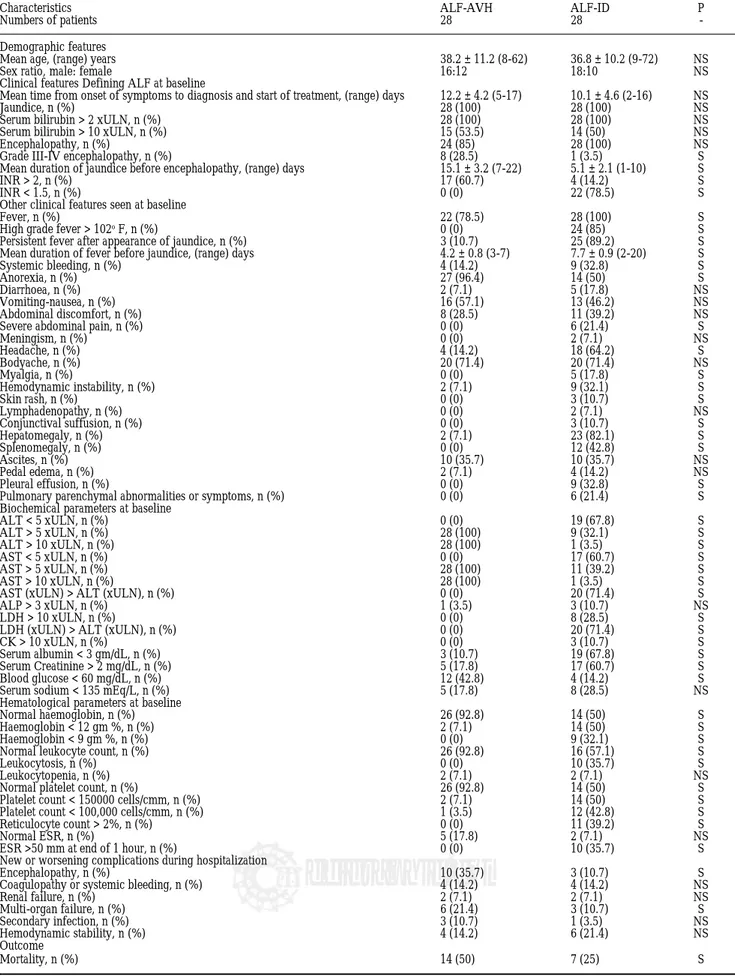
Demographic, clinical, biochemical and haematologi-cal parameters at baseline evaluation of both the groups are tabulated in table I.
Statistically significant features which were exclusive to ALF-ID, were not specific for any single infection pre-senting as ALF-ID and were not seen in ALF-AVH were as follows: high grade fever (> 102o F), splenomegaly, ALT or AST < 5 times upper limit of normal value (xULN), higher AST or LDH than ALT and INR < 1.5 (normal PT).
Other statistically significant exclusive features of ALF-ID were specific to the underlying infection: skin rash, myalgia, conjunctival suffusion, severe abdominal pain, pleural effusion, pulmonary parenchymal involve-ment, CK > 10 xULN, LDH > 10 xULN, severe anaemia (haemoglobin < 9 gm %), leukocytosis (leukocyte count > 11000 cells/cmm), high reticulocyte count (> 2%) and ESR > 50 mm at end of 1 hour.
Non-significant but exclusive features to ALF-ID were: meningism and lymphadenopathy.
Other statistically significant features seen more com-monly in ALF-ID, but were not exclusive to ALF-ID, were as follows: shorter duration of jaundice before en-cephalopathy, persistent fever after appearance of dice, longer duration of fever before appearance of jaun-dice, overt bleeding manifestations (including petechiae), headache, haemodynamic instability (pulse rate > 90 beats/min and systolic blood pressure < 90 mm Hg), hepatomegaly, hypoalbuminaemia (Serum albumin < 3 gm/dL), renal failure (creatinine > 2 mg/dL), anemia (hae-moglobin < 11 gm%), thrombocytopenia (platelet count < 150,000 or < 100,000 cells/cmm).
Statistically significant features seen more commonly in ALF-AVH were as follows: grade III-IV encephalopa-thy, INR > 2, ALT and AST > 5 xULN or > 10 xULN, hypoglycaemia, normal haemoglobin (> 12 gm %), leuko-cyte (4000-11,000 cells/cmm) and platelet (150,000-500,000 cells/cmm) count.
Patients with ALF-AVH had mortality rate of 50% (14/ 28) and ALF-ID had mortality rate of 25% (7/28) (p = S).
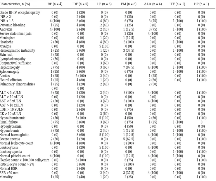
Table II shows characteristics of individual infections
responsible for ALF-ID, taking in account exclusive or significant features in table I.
Discussion
In developing countries like India, ID like FM, TF, LP, and DF can present in a complicated form with febrile
jaundice with encephalopathy features and may mimic ALF; and so they should be looked for in all the cases pre-senting as ALF. Baseline routine clinical and laboratory data help in raising suspicion for presence of such ID. Af-ter reaching specific diagnosis, specific therapy for ID in addition to supportive management of ALF is important in reducing mortality.
In a patient presenting with jaundice and encephalopa-thy (altered mentation) and/or coagulopaencephalopa-thy (deranged coagulation parameters with/without systemic bleeding), primary diagnostic consideration is ALF-AVH, which is the commonest form of ALF in India. But, as seen in our study, presence of high grade fever, splenomegaly, mild ALT and AST elevation (< 5 xULN), AST/ALT ratio > 1, LDH/ALT ratio > 1 and normal PT should make a clini-cian suspicious about presence of ID other than AVH. Additional such features are persistent fever after appear-ance of jaundice, longer duration of fever, presence of hepatomegaly, and abnormal haemoglobin, leukocyte and platelet counts. In patients with suspected ALF-ID, pres-ence of peculiar features to respective ID can help to nar-row down further investigations. Mortality rates were lower for ALF-ID than ALF-AVH in our study, despite statistically insignificant delay in diagnosis and subse-quent start of specific therapy between the two groups.
It is known since long that in the tropics, viral hepatitis needs to be differentiated from other common ID such as typhoid, amoebic or malarial hepatitis.22
edigraphic.com
Table I. Comparison of ALF-AVH and ALF-ID.
Characteristics ALF-AVH ALF-ID P
Numbers of patients 28 28
-Demographic features
Mean age, (range) years 38.2 ± 11.2 (8-62) 36.8 ± 10.2 (9-72) NS
Sex ratio, male: female 16:12 18:10 NS
Clinical features Defining ALF at baseline
Mean time from onset of symptoms to diagnosis and start of treatment, (range) days 12.2 ± 4.2 (5-17) 10.1 ± 4.6 (2-16) NS
Jaundice, n (%) 28 (100) 28 (100) NS
Serum bilirubin > 2 xULN, n (%) 28 (100) 28 (100) NS
Serum bilirubin > 10 xULN, n (%) 15 (53.5) 14 (50) NS
Encephalopathy, n (%) 24 (85) 28 (100) NS
Grade III-IV encephalopathy, n (%) 8 (28.5) 1 (3.5) S
Mean duration of jaundice before encephalopathy, (range) days 15.1 ± 3.2 (7-22) 5.1 ± 2.1 (1-10) S
INR > 2, n (%) 17 (60.7) 4 (14.2) S
INR < 1.5, n (%) 0 (0) 22 (78.5) S
Other clinical features seen at baseline
Fever, n (%) 22 (78.5) 28 (100) S
High grade fever > 102o F, n (%) 0 (0) 24 (85) S
Persistent fever after appearance of jaundice, n (%) 3 (10.7) 25 (89.2) S Mean duration of fever before jaundice, (range) days 4.2 ± 0.8 (3-7) 7.7 ± 0.9 (2-20) S
Systemic bleeding, n (%) 4 (14.2) 9 (32.8) S
Anorexia, n (%) 27 (96.4) 14 (50) S
Diarrhoea, n (%) 2 (7.1) 5 (17.8) NS
Vomiting-nausea, n (%) 16 (57.1) 13 (46.2) NS
Abdominal discomfort, n (%) 8 (28.5) 11 (39.2) NS
Severe abdominal pain, n (%) 0 (0) 6 (21.4) S
Meningism, n (%) 0 (0) 2 (7.1) NS
Headache, n (%) 4 (14.2) 18 (64.2) S
Bodyache, n (%) 20 (71.4) 20 (71.4) NS
Myalgia, n (%) 0 (0) 5 (17.8) S
Hemodynamic instability, n (%) 2 (7.1) 9 (32.1) S
Skin rash, n (%) 0 (0) 3 (10.7) S
Lymphadenopathy, n (%) 0 (0) 2 (7.1) NS
Conjunctival suffusion, n (%) 0 (0) 3 (10.7) S
Hepatomegaly, n (%) 2 (7.1) 23 (82.1) S
Splenomegaly, n (%) 0 (0) 12 (42.8) S
Ascites, n (%) 10 (35.7) 10 (35.7) NS
Pedal edema, n (%) 2 (7.1) 4 (14.2) NS
Pleural effusion, n (%) 0 (0) 9 (32.8) S
Pulmonary parenchymal abnormalities or symptoms, n (%) 0 (0) 6 (21.4) S Biochemical parameters at baseline
ALT < 5 xULN, n (%) 0 (0) 19 (67.8) S
ALT > 5 xULN, n (%) 28 (100) 9 (32.1) S
ALT > 10 xULN, n (%) 28 (100) 1 (3.5) S
AST < 5 xULN, n (%) 0 (0) 17 (60.7) S
AST > 5 xULN, n (%) 28 (100) 11 (39.2) S
AST > 10 xULN, n (%) 28 (100) 1 (3.5) S
AST (xULN) > ALT (xULN), n (%) 0 (0) 20 (71.4) S
ALP > 3 xULN, n (%) 1 (3.5) 3 (10.7) NS
LDH > 10 xULN, n (%) 0 (0) 8 (28.5) S
LDH (xULN) > ALT (xULN), n (%) 0 (0) 20 (71.4) S
CK > 10 xULN, n (%) 0 (0) 3 (10.7) S
Serum albumin < 3 gm/dL, n (%) 3 (10.7) 19 (67.8) S
Serum Creatinine > 2 mg/dL, n (%) 5 (17.8) 17 (60.7) S
Blood glucose < 60 mg/dL, n (%) 12 (42.8) 4 (14.2) S
Serum sodium < 135 mEq/L, n (%) 5 (17.8) 8 (28.5) NS
Hematological parameters at baseline
Normal haemoglobin, n (%) 26 (92.8) 14 (50) S
Haemoglobin < 12 gm %, n (%) 2 (7.1) 14 (50) S
Haemoglobin < 9 gm %, n (%) 0 (0) 9 (32.1) S
Normal leukocyte count, n (%) 26 (92.8) 16 (57.1) S
Leukocytosis, n (%) 0 (0) 10 (35.7) S
Leukocytopenia, n (%) 2 (7.1) 2 (7.1) NS
Normal platelet count, n (%) 26 (92.8) 14 (50) S
Platelet count < 150000 cells/cmm, n (%) 2 (7.1) 14 (50) S
Platelet count < 100,000 cells/cmm, n (%) 1 (3.5) 12 (42.8) S
Reticulocyte count > 2%, n (%) 0 (0) 11 (39.2) S
Normal ESR, n (%) 5 (17.8) 2 (7.1) NS
ESR >50 mm at end of 1 hour, n (%) 0 (0) 10 (35.7) S
New or worsening complications during hospitalization
Encephalopathy, n (%) 10 (35.7) 3 (10.7) S
Coagulopathy or systemic bleeding, n (%) 4 (14.2) 4 (14.2) NS
Renal failure, n (%) 2 (7.1) 2 (7.1) NS
Multi-organ failure, n (%) 6 (21.4) 3 (10.7) S
Secondary infection, n (%) 3 (10.7) 1 (3.5) NS
Hemodynamic stability, n (%) 4 (14.2) 6 (21.4) NS
Outcome
MG
edigraphic.com
Table II. Comparison of different infections causing ALF-ID.
Characteristics, n (%) RF (n = 4) DF (n = 5) LP (n = 5) FM (n = 8) ALA (n = 4) TF (n = 1) HP (n = 1)
Grade III-IV encephalopathy 0 (0) 1 (20) 0 (0) 0 (0) 0 (0) 0 (0) 0 (0)
INR > 2 0 (0) 2 (40) 0 (0) 2 (25) 0 (0) 0 (0) 0 (0)
INR < 1.5 4 (100) 3 (60) 4 (80) 6 (75) 3 (75) 1 (100) 1 (100)
Systemic bleeding 1 (25) 4 (80) 2 (40) 2 (25) 0 (0) 0 (0) 0 (0)
Anorexia 4 (100) 2 (40) 2 (40) 1 (12.5) 3 (75) 1 (100) 1 (100)
Severe abdominal pain 0 (0) 0 (0) 0 (0) 2 (25) 4 (100) 0 (0) 0 (0)
Meningism 0 (0) 0 (0) 1 (20) 1 (12.5) 0 (0) 0 (0) 0 (0)
Headache 2 (50) 4 (80) 4 (80) 8 (100) 0 (0) 1 (100) 0 (0)
Myalgia 0 (0) 0 (0) 5 (100) 0 (0) 0 (0) 0 (0) 0 (0)
Hemodynamic instability 1 (25) 3 (60) 1 (20) 3 (37.5) 0 (0) 1 (100) 0 (0)
Skin rash 3 (75) 0 (0) 0 (0) 0 (0) 0 (0) 0 (0) 0 (0)
Lymphadenopathy 2 (50) 0 (0) 0 (0) 0 (0) 0 (0) 0 (0) 0 (0)
Conjunctival suffusion 0 (0) 0 (0) 3 (60) 0 (0) 0 (0) 0 (0) 0 (0) Hepatomegaly 3 (75) 4 (80) 3 (60) 7 (87.5) 4 (100) 1 (100) 1 (100)
Splenomegaly 1 (25) 3 (60) 0 (0) 6 (75) 0 (0) 1 (100) 1 (100)
Ascites 1 (25) 5 (100) 2 (40) 0 (0) 1 (25) 0 (0) 1 (100)
Pleural effusion 1 (25) 4 (80) 1 (20) 0 (0) 2 (50) 0 (0) 1 (100)
Pulmonary abnormalities 1 (25) 1 (20) 2 (40) 0 (0) 2 (50) 0 (0) 0 (0)
ALT < 5 xULN 3 (75) 1 (20) 2 (40) 8 (100) 4 (100) 0 (0) 1 (100)
ALT > 10 xULN 0 (0) 1 (20) 0 (0) 0 (0) 0 (0) 0 (0) 0 (0)
AST < 5 xULN 2 (50) 0 (0) 3 (60) 8 (100) 4 (100) 0 (0) 0 (0)
AST > 10 xULN 0 (0) 1 (20) 0 (0) 0 (0) 0 (0) 0 (0) 0 (0)
LDH > 10 xULN 0 (0) 2 (40) 0 (0) 6 (75) 0 (0) 0 (0) 0 (0)
CK > 10 xULN 0 (0) 0 (0) 3 (60) 0 (0) 0 (0) 0 (0) 0 (0)
Hypoalbuminemia 2 (50) 5 (100) 5 (100) 4 (50) 2 (50) 0 (0) 1 (100)
Renal failure 3 (75) 3 (60) 3 (60) 6 (75) 1 (25) 1 (100) 0
Hypoglycaemia 0 (0) 0 (0) 0 (0) 4 (50) 0 (0) 0 (0) 0 (0)
Hyponatremia 3 (75) 0 (0) 2 (40) 1 (12.5) 0 (0) 1 (100) 1 (100)
Normal haemoglobin 0 (0) 3 (60) 5 (100) 1 (12.5) 4 (100) 1 (100) 0 (0)
Severe anemia 2 (50) 1 (20) 0 (0) 5 (62.5) 0 (0) 0 (0) 1 (100)
Normal leukocyte count 4 (100) 4 (80) 0 (0) 8 (100) 0 (0) 0 (0) 0 (0)
Leukocytosis 0 (0) 1 (20) 5 (100) 0 (0) 4 (100) 0 (0) 0 (0)
Leukocytopenia 0 (0) 0 (0) 0 (0) 0 (0) 0 (0) 1 (100) 1 (100)
Normal platelet count 4 (100) 0 (0) 5 (100) 1 (12.5) 4 (100) 1 (100) 0 (0) Platelet count < 100,000 cells/cmm 0 (0) 5 (100) 0 (0) 6 (75) 0 (0) 0 (0) 1 (100) Reticulocyte count > 2% 0 (0) 3 (60) 0 (0) 8 (100) 0 (0) 0 (0) 0 (0)
Normal ESR 0 (0) 2 (40) 0 (0) 0 (0) 0 (0) 0 (0) 0 (0)
ESR >50 mm 0 (0) 0 (0) 2 (40) 3 (37.5) 4 (100) 1 (100) 0 (0)
Mortality 1 (25) 2 (40) 1 (20) 2 (25) 0 (0) 0 (0) 1 (100)
Previously from India, ALF due to FM and TF is de-scribed in various series.16-19,29 Few series are reported from India looking at differentiation of complicated FM and TF respectively from ALF-AVH. One such series suggested disproportionate anaemia, renal failure and only mildly elevated liver enzymes should help in differ-entiating FM from ALF-AVH.17 In another study compar-ing 25 patients with complicated FM and 25 patients of ALF-AVH suggested that longer duration of fever, hepatomegaly, splenomegaly, lower haemoglobin, throm-bocytopenia, lower AST and ALT levels and normal PT were commonly present with FM.18 In one prospective se-ries, analysis of 11 patients of complicated TF and 36 ALF-AVH, hepatomegaly, thrombocytopenia, elevated ALP, AST > ALT and only mildly prolonged PT suggest-ed a diagnosis of TF.19 Both these series have achieved survival rate of 100% with TF and 76% with FM as
com-pared to ALF-AVH group (16.6% and 24%, respective-ly).18,19 Our study also showed survival benefit to ALF-ID group as compared to ALF-AVH. In a study on severe FM presenting as multi-organ failure, multiple organ in-volvement was associated with increased mortality and hepatic failure was associated with around 50% mortality rate.30
edigraphic.com
TF is diagnosed in presence of high grade fever, right hypochondrial pain, tender hepatomegaly, relative lym-phocytosis with leucopenia, thrombocytopenia, mild-moderate ALT abnormalities (AST > ALT), elevated ALP, normal PT, presence of biliary complications like cholecystitis or pyogenic abscess, presence of positive culture for salmonella from blood, stool or urine, positive Widal test or rising titers of Widal test and response to Quinolone or Cephalosporin antibiotics.19,34,35
LP should be suspected in the presence of hectic fever, severe myalgia, headaches, severe prostration, manifesta-tions like gastrointestinal bleeding, purpura or epistaxis, exposure to water or soil potentially contaminated with animal waste (recreational activities like hiking or swim-ming; animal contact; work in farms during heavy rain falls), conjunctival suffusion, tender hepatomegaly, pres-ence of meningism, mild anemia, leukocytosis with neu-trophilia, thrombocytopenia, high conjugated bilirubin with mild-moderate elevations of ALT, worsening renal failure, abnormal urine analysis and severely elevated CK. A positive IgM leptospira antibody test or isolation of leptospira from CSF or blood during the first week of fever and from urine during the second week confirms the diagnosis. The disease responds to Penicillin, Ampicillin or Doxycyclin therapy.20,34,36,37
Recently, few case reports are published on occurrence of ALF in DF and dengue hemorrhagic fever, which are endemic in India.38,39 DF is diagnosed in presence of high grade fever of > 3 days duration, gastrointestinal symp-toms, hemorrhagic manifestations, facial flushing, wors-ening drowsiness, shock with narrow pulse pressure, hepatomegaly, circulatory disturbances (restlessness, cold extremities, capillary refill time > 2 sec and tachycardia), positive tourniquet test (> 10 petechiae/2.5 cm2 skin area), profound thrombocytopenia, normal leukocyte count and ESR, evidence of hemoconcentration, evidence of capil-lary leakage (pleural effusion, ascites and/or hypopro-teinemia), mild to severe elevation (> 10 xULN) in AST or ALT levels, absence of hypoglycemia and hyponatrem-ia, and positive test of IgM dengue antibody.21,40-43
Ricketsial fever is rarely associated with liver failure. As seen in our study, these infections are characterized by fever, headache, anorexia, gastrointestinal symptoms, bodyache, skin rash, lymphadenopathy, hepatomegaly, petechiae, hypoalbuminemia, hyponatremia, renal failure, pneumonitis, encephalitis, normal leukocyte and platelet counts, mild-moderate ALT or AST elevation and normal PT.44
ALA rarely presents with hyperbilirubinemia and/or encephalopathy, which are identified as poor prognostic factors and independent risk factors for mortality in a pre-vious Indian series.45 In our study, ALA was associated with fever, hepatomegaly, anorexia, severe right hypo-chondrial pain, pleural effusion and pulmonary parenchy-mal abnorparenchy-malities, norparenchy-mal PT, mild-moderate ALT or AST elevation, leukocytosis, normal haemoglobin and
platelet count and high ESR. All these features are well described in patients with ALA previously.46
Hepatomegaly and deranged liver functions are previ-ously described in fatal HP.47
High index of suspicion and awareness are required to identify various common infections causing symptom complex similar to ALF. This help in identifying a patient with ALF who may have low mortality if specific treat-ment for such infection is given in time.
References
1. Polson J, Lee WM. AASLD Position Paper: The management of acute liver failure. Hepatology 2005; 41: 1179-1197.
2. Ostapowicz J, Lee WM. Acute hepatic failure: a western perspective.
J Gastroenterol Hepatol 2000; 15: 480-488.
3. Acharya SK, Dasarathy S, Kumar TL, Sushma S, Presanna U, Tandon A, et al. Fulminant hepatitis in a tropical population: clinical course, cause and early predictors of outcome. Hepatology 1996; 23: 1448-1455.
4. Dhiman RK, Chawla Y, Jain S. Spontaneous bacterial peritonitis and bacteremia in fulminant hepatic failure. Am J Gastroenterol 2000; 95: 2126-8.
5. Dhiman RK, Seth AK, Jain S, Chawla YK, Dilawari JB. Prognostic evaluation of early indicators in fulminant hepatic failure by multi-variate analysis. Dig Dis Sci 1998; 43: 1311-16.
6. Bendre SV, Bavdekar AR, Bhave SA, Pandit AN, Chitambar SD, Arankalle VA. Fulminant hepatic failure- etiology, viral markers and outcome. Indian Pediatrics 1999; 36: 1107-1112.
7. Acharya SK, Batra Y, Hazari S, Choudhury V, Panda SK, Dattagupta S. Etiopathogenesis of acute hepatic failure: eastern versus western countries. J Gastroenterol Hepatol 2002; 17: S268-S273.
8. Chadha MS, Walimbe A, Chobe LP, Arankalle VA. Comparison of etiology of sporadic acute and fulminant viral hepatitis in hospital-ized patients in Pune, India during 1978-81 and 1994-97. Ind J Gastroenterol 2003; 22: 11-15.
9. Khuroo MS, Kamili S. Etiology and prognostic factors in acute liver failure in India. J Viral Hepatitis 2003; 10: 224-231.
10. Acharya SK, Pande SK, Saxena A, Gupta SD. Acute hepatic failure in India: a perspective from the east. J Gastroenterol Hepatol 2000; 15: 473-479.
11. Bowen DG, Shackel NA, McCaughan GW. East meets west: acute liver failure in the global village. J Gastroenterol Hepatol 2000; 15: 467-469.
12. Ostapowicz G, Fontana RJ, Shiodt FV, et al. Results of a prospective study of acute liver failure at 17 tertiary care centers in the United States Ann Intern Med 2002; 137: 947-954.
13. Riordan SM, Williams R. Fulminant hepatic failure. Clin Liver Dis
2004; 4: 25-45.
14. Bernuau J, Benhamou JP. Fulminant and subfulminant hepatic fail-ure. In Bircher J, Benhamou JP Mclntyre N, Rizzetto M Rodes J, editors Oxford textbook of clinical Hepatology. Oxford University Press 1999: 1341-1372.
15. Arora NK, Mathur P, Ahuja A, Oberoi A. Acute liver failure. Indian J Pediatr 2003; 70: 73-79.
16. Anand AC. Malarial liver failure: myth or reality? Trop Gastroenterol
2001; 22: 55-56.
17. Srivastava A, Khanduri A, Lakhatia S, Pandey R, Choudhuri G. Falciparum malaria with acute liver failure. Trop Gastroenterol 1996; 17: 172-4.
18. Devarbavi H, Alvares JF, Kumar KS. Severe Falciparum malaria simu-lating fulminant hepatic failure. Mayo Clin Proc 2005; 80: 355-8. 19. Kamath PS, Jalihal A, Chakraborty A. Differentiation of typhoid
fe-ver from fulminant hepatic failure in patients presenting with jaun-dice and encephalopathy. Mayo Clin Proc 2000; 75: 462-466. 20. Dutta TK, Christopher M. Leptospirosis- an overview. J Assoc
MG
edigraphic.com
21. John TJ. Dengue fever and dengue hemorrhagic fever. Lancet 2003; 361: 181-182.
22. Khosla SN. Typhoid hepatitis. Postgrad Med J 1990; 66: 923-925. 23. El-Newihi HM, Alamy ME, Reynolds TB. Salmonella hepatitis:
analy-sis of 27 cases and comparison with acute viral hepatitis. Hepatology
1996; 24: 516-519.
24. Durrani AB. Typhoid hepatitis. J Pak Med Assoc 1995; 5: 317-318. 25. Mazumder R, Mishra RK, Mazumder H, Mukherjee P. Jaundice in
falciparum malaria—some prospective observations. J Indian Med Assoc 2002; 100: 312-4.
26. Anand AC, Ramji C, Narula AS, Singh W. Malarial hepatitis: a het-erogeneous syndrome? Natl Med J India 1992; 5: 59-62.
27. Murthy GL, Sahay RK, Sreenivas DV, Sundaram C, Shantaram V. Hepa-titis in falciparum malaria. Trop Gastroenterol 1998; 19: 152-154. 28. Kochar DK, Singh P, Agarwal P, Kochar SK, Pokharna R, Sareen
PK. Malarial hepatitis. J Assoc Physicians India 2003; 51: 1069-1072.
29. Joshi YK, Tandon BN, Acharya SK, Babu S, Tandon M. Acute he-patic failure due to Plasmodium falciparum liver injury. Liver 1986; 6: 357-360.
30. Krishnan A, Karnad DR. Severe falciparum malaria: an important cause of multiple organ failure in Indian intensive care unit patients.
Crit Care Med 2003; 31: 2278-84.
31. Anand AC. Liver in Malaria. Ind J Gastroenterol 2001; 20: C37-C38.
32. Anand AC. Malarial Hepatitis: an entity in search of recognition.
Gastroenterol today 1996; 1: 49-51.
33. Anand AC, Puri P. Jaundice in malaria. J Gastroenterol Hepatol 2005; 20: 1322-1332.
34. Sorabjee JS. The liver in enteric fever and Leptospirosis. Ind J Gastroenterol 2001; 20: C44-C46.
35. Jagadish K, Patwari AK, Sarin SK, Prakash C, Srivastava DK, Anand VK. Hepatic manifestation in typhoid fever. Indian Pediatrics 1994; 31: 807-811.
36. Gayotto L, Silva L, Alves VAF. Leptospirosis. In Oxford textbook of Hepatology, 2nd ed. Oxford press. 2000: 1017-1023.
37. Tripathi K. Clinical aspects of Leptospirosis. In Leptospirosis: round table conference series, Ranbaxy science foundation; 1998: 81-84. 38. Subramanian V, Shenoy S, Joseph AJ. Dengue hemorrhagic fever and
fulminant hepatic failure. Dig Dis Sci 2005; 50: 1146-1147. 39. Vinodh BN, Bammigatti C, Kumar A, Mittal V. Dengue fever with
acute liver failure. J Postgrad Med 2005; 51: 322-323.
40. Solomon, Dung NM, Vaughn DW, Kneen R, Thao LTT, Raengsakulrach B, et al. Neurological manifestations of dengue in-fection. Lancet 2000; 355: 1053-59.
41. Monath TP. Early indicators in acute dengue infection. Lancet 1997; 350: 1719-1720.
42. Lum LC, Lam SK, George R, Devi S. Fulminant hepatitis in dengue infection. Southeast Asian J Trop Med Public Health 1993; 24: 467-471. 43. Nguyen TL, Nguyen TH, Tieu NT. The impact of dengue
hemor-rhagic fever on liver function. Res Virol 1997; 148: 273-277. 44. Walker D, Raoult D, Dumler JS, Marrie T. Ricketsial diseases. In:
Harrison’s Principles of Internal Medicine. Braunwald E, Fauci AS, Kasper DL, Hauser SL, Longo DL, Jameson JL Eds. McGraw-Hill Medical Publishing Division. 2001: 1065-1073.
45. Sharma MP, Dasarathy S, Verma N, Saksena S, Shukla DK. Prognos-tic markers in amoebic liver abscess- a prospective study. Am J Gastroenterol 1996; 91: 2584-2588.
46. Amarapurkar DN, Patel ND, Amarapurkar AD. Amoebic liver ab-scess. J Hepatology 2003; 39: 291-292.