INTRODUCTION
Viruses belonging to Aviadenovirus genus are much diverged and could be classified into 5 (A to E) spe-cies and also into 12 serotypes. Adenoviral infections are widely prevalent among birds including poultry and wild species. Most of these viruses do not produce any clinical signs as the primary pathogens and their role as the opportunistic factors is indicated (Fitzgerald, 2008). However, some fowl adenoviruses (FAdV) directly af-fect health and could cause disease conditions and symptoms such as inclusion body hepatitis, hydroperi-cardium syndrome, respiratory disease, decreased egg production, diarrhea, poor growth, and reduced feed
conversion (McConnell Adair and Fitzgerald, 2008). Gizzard erosions in broiler chickens as a result of FAdV serotype 1 (FAdV-1) infections have been recently re-ported mostly in Japan but also in Europe (Okuda et al., 2001; Ono et al., 2001, 2003a; Marek et al., 2010). Such pathological changes were also observed during the course of inclusion body hepatitis caused by FAdV serotype 8 (Okuda et al., 2004).
The pathogenicity of FAdV differs between strains. In Japan the coexistence of FAdV-1 with different pathogenicity was reported (Okuda et al., 2006). The FAdV-99ZH strain was capable of inducing gizzard ero-sions (pathogenic) in experimentally infected chickens but the OTE strain, the prototype strain of FAdV-1 in Japan, failed to induce such lesions (apathogenic). The role of fiber protein was considered among factors affecting differences in virulence of FAdV serotype 8 (Pallister et al., 1996). Structure and genome studies of chicken embryo lethal orphan (CELO) virus, a Euro-pean reference FAdV-1 strain, revealed that it possesses 2 fibers of different lengths and the distal parts (or head) of these fibers are involved in cellular receptor(s)
Molecular characterization of fowl adenoviruses isolated
from chickens with gizzard erosions
1K. Domanska-Blicharz ,*
2G. Tomczyk ,* K. Smietanka ,* W. Kozaczynski ,† and Z. Minta *
* Department of Poultry Diseases; and † Department of Pathology, National Veterinary Research Institute, Al. Partyzantow 57, 24-100 Pulawy, Poland
ABSTRACT Broiler chickens with clinical signs of un-even growth, depression, and dull feathers were sub-mitted to our laboratory and, at necropsy, lesions in proventriculus, gizzard, and intestines were detected. Fowl adenovirus serotype 1 (FAdV-1) was isolated from digestive tissues. The virus, assigned as FAdV-PL/G068/08, showed 99.5% nucleotide homology and 99.2% amino acid homology in hexon gene with chick-en embryo lethal orphan (CELO) strain classified as the European reference of FAdV-1. One-day-old and 21-d-old SPF chickens were inoculated with FAdV-PL/068/08 by both nasal and ocular routes and then observed daily and examined by necropsy at 6, 10, and 14 d postinoculation. Experimental infection with iso-lated virus was fatal for younger chickens and major le-sions occurred in the gizzards. No clinical or pathologi-cal changes were observed in chickens infected at 21 d of
age, but the presence of intranuclear inclusion bodies in gizzard epithelial cells was detected. Molecular charac-terization was based on the long and short fibers genes sequencing and comparison of obtained sequences with other 1 strains. The homology between FAdV-PL/G068/08 and other sequences available in GenBank was between 98.9 and 99.8% (short fiber region) and 99.0 and 99.7% (long fiber region) at nucleotide level and between 98.4 and 100% (short fiber region) and 99.3 and 99.9% (long fiber region) at amino acid level. No correlation between identified amino acid changes in short and long fiber proteins and pathogenicity of stud-ied FAdV-1 strains was observed. Although short and long proteins were indicated as factors influencing virus pathogenicity, the role of identified sequence differences in infectivity determination remain unclear.
Key words: fowl adenovirus , gizzard erosion , hexon gene , long fiber gene , short fiber gene
2011 Poultry Science 90 :983–989 doi: 10.3382/ps.2010-01214
983
Received November 12, 2010. Accepted January 29, 2011.
1 The nucleotide sequence data reported in this paper have been
submitted to the GenBank (Los Alamos National Laboratories, Los Alamos, NM) nucleotide sequence database and have been assigned the accession numbers GU952108 to GU952110.
2 Corresponding author: domanska@piwet.pulawy.pl
binding (Hess et al., 1995). Polymerase chain reaction RFLP analysis of long fiber genes was used to distin-guish strains of different pathogenicity, but its useful-ness has been recently questioned (Okuda et al., 2006; Marek et al., 2010).
At the beginning of 2008 an outbreak of a disease with clinical manifestation mainly in the digestive tract was recorded in a few farms of broiler chickens in central Poland. The purpose of this study was to identify the causative agent and to reproduce the disease under ex-perimental conditions. The obtained FAdV isolate was molecularly characterized. The nucleotide (NT) and amino acid (AA) sequences of L1 loop of hexon gene as well as the long and short fibers were investigated and compared with other FAdV-1 sequences available in GenBank (http://www.ncbi.nlm.nih.gov/genbank/).
MATERIALS AND METHODS
Samples
In February 2008 the Department of Poultry Diseases (Pulawy, Poland) received organ samples from broiler chickens for disease diagnosis. Affected chickens showed uneven growth, depression, and dull feathers. The most severe lesions were located in proventriculus, gizzards, and intestines and were used for virus identification. Trials of bacteria isolation (on blood, brilliant green, and MacConkey agars) as well as microscopic examina-tion of smears of intestines for the presence of parasites gave negative results.
Virus Isolation and Identification
For virus isolation, the above-mentioned samples of digestive tract were homogenized in PBS (10% wt/vol) containing 2,000 U/mL of penicillin and 2 mg/mL of streptomycin, incubated at room temperature for 20 min, clarified by centrifugation at 3,000 × g for 15 min, filtered through a 0.45-µm syringe filter, and used to inoculate chicken embryo fibroblasts cells (CEF) as de-scribed previously (Cowen et al., 1978). The complete cytopathic effect was observed in the second passages. Infectivity of the isolated virus assigned as PL/G060/08 was quantified using microtiter endpoint titration on CEF culture and the results were expressed in units of mean tissue culture infective dose per milliliter (Reed and Muench, 1938).
Aliquots (200 µL) of supernatant from second pas-sage in infected cells as well as the supernatant of ho-mogenized tissues from field chickens were processed using DNeasy Blood and Tissue Kit and RNeasy Mini Kit (Qiagen, Valencia, CA) according to the manufac-turer’s recommended procedure for DNA and RNA extraction, respectively. Reverse transcription (RT) PCR was applied for detection of RNA viruses (avian reovirus, infectious bronchitis virus, infectious bursal disease virus; Liu et al., 1997; Zierenberg et al., 2001; Cavanagh et al., 2002). A PCR that can detect and
differentiate between 12 FAdV serotypes based on L1 loop of hexon gene was also carried out (Meulemans et al., 2001). The primer pair F3–F4 was used to am-plify and sequence the short fiber gene (Okuda et al., 2006). For amplification and sequencing of the entire long fiber gene, additional specific primers were applied (available on request). Amplified PCR products were purified using QIAquick Spin Kit (Qiagen) and used as the templates for sequencing in a commercial ser-vice (Genomed, Warsaw, Poland). Sequencing for each product was done in triplicate.
Histopathological Examination
Various organs and tissues (proventriculus, gizzard, intestines, liver, kidney, pancreas, spleen, lung, and trachea) were collected from field and experimentally infected chickens for histopathologic examination. All were fixed in 10% neutral-buffered formalin, paraffin embedded, sectioned at 4 µm, stained with hematoxylin and eosin, and examined by light microscopy (Okuda et al., 2001)
Sequence Analysis
Using the Seqman program (version 8.1.2, DNAS-TAR Inc., Madison, WI), the forward and reverse NT sequences were edited, cured, and aligned as a single consensus sequence. The NT sequences were translated to deduced AA sequences, which were also compared to detect any changes at the AA level. Alignments of NT and AA sequences of L1 loop of hexon gene were per-formed using the MegAlign application of DNASTAR using the Clustal-W method. Sequence data were sub-mitted to GenBank with following accession numbers: GU952110, L1 loop of hexon gene; GU952108, short fiber gene; GU952109, long fiber gene. The GenBank accession numbers of FAdV-1 sequences used for com-parison were as follows: CAA59207 (CELO strain, long fiber gene), AA59210 (CELO strain, short fiber gene), FN557183 (OTE strain, long fiber gene), FN557186 (OTE strain, short fiber gene), FN557182 (Polish 08– 3909 strain, long fiber gene), FN557185 (Polish 08–5769 strain, short fiber gene), and FN557184 (UK 08–3622 strain, short fiber gene).
Bird Experiments
One-day-old (group 1) and 21-d-old (group 2) specif-ic-pathogen-free (SPF) White Leghorn chickens (Valo Lohmann, Cuxhaven, Germany) were used in the chal-lenge experiment to reproduce clinical signs of disease. The chickens were inoculated with the dose of 106.0
and divided into 2 parts for histopathological examina-tion (processed immediately) and virological–RT-PCR tests (stored below −65°C until used). Throughout the experiment, the chickens were kept in isolators under negative pressure (HM 1500, Montair Andersen BV, Kronenberg, the Netherlands). These experiments were approved by the II Local Ethical Committee of the Uni-versity of Life Sciences in Lublin, Poland (decision no. 41/2006).
RESULTS
Necropsy and Gross Lesions
and Histopathological Findings
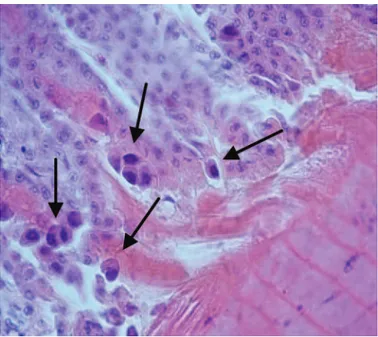
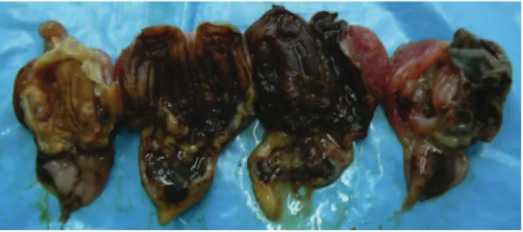
In dead field chickens delivered to the Department of Poultry Diseases, growth retardation, paleness of mus-cles, organs, and skin of legs were observed. The crop, esophagus, proventriculus, gizzard, duodenum, and in some cases rectum were filled with dark-brownish contents. However, the most severe lesions were found in the stomach, where the hemorrhagic changes in the proventriculus, especially at the junction with the giz-zard, were seen and a detachment of the overlaying ke-ratinoid layer and even its partial decay in the gizzard was found. On the mucosal surface numerous erosions were formed. Furthermore, necropsy revealed atrophy of lymphatic organs (thymus, bursa of Fabricius, and spleen), enlarged liver and kidneys, and sometimes numerous whitish necrotic foci in the liver. The his-topathologic examination of tissues of both field and experimental chickens identified the presence of intra-nuclear inclusion bodies in gizzard epithelial cells, but no such structures were present in the liver (Figure 1).
Molecular Methods
The RNA extracted from supernatants of field sam-ples as well as from CEF cultures subjected to RT-PCR produced no amplicons characteristic of infectious bronchitis virus, infectious bursal disease virus, or avian reovirus. On the other hand, the characteristic product of L1 loop of the hexon gene was obtained in the PCR targeting FAdV. The analysis of a 735 NT segment corresponding to 201 to 936 NT in the hexon gene and deduced 245 AA sequence revealed a 99.5% NT homol-ogy (731/735 NT) and 99.2% AA homolhomol-ogy (243/245) with the CELO strain.
Positive PCR amplification of the entire short fi-ber gene (1,233 bp) was obtained for the PL/G060/08 strain. The deduced 384 AA sequence from 13 to 396 AA of the short fiber protein was compared with rel-evant sequences of FAdV-1 strains available in the Gen-Bank (Figure 2). The FAdV PL/G060/08 isolated in Poland showed 98.9% (13 NT differences) and 99.7% (2 differences) NT sequence similarity to CELO and OTE strains, respectively. Six out of 13 NT differences be-tween CELO and PL/G060/08 led to the following AA substitutions: N223→K, I314→T, R328→G, F331→G,
S334→A, and A369→C (arrow indicates that the first
AA was substituted for the second AA). All resulting AA differences were located in the head domain of the short fiber protein and led to the divergence of 1.6% (6/385 AA) between the compared strains. The high-est sequence homology (99.9%) was found between the Polish strain and the FAdV 08-3622 strain isolated in the United Kingdom, in which only 1 NT difference was found (silent mutation).
The 2,291-bp fragment of the long fiber gene was translated into 762 AA (40 to 804 AA, CELO long fi-ber protein numfi-bering) and compared with the respec-tive sequences of other FAdV-1 (Figure 3). Sequences of FAdV-1 strains inducing gizzard erosion were identical. However, 18 NT differences between CELO and patho-genic strains were noticed (NT similarity of 99.2%) but resulted in only 1 AA substitution (T275→A, shaft
do-main of the long fiber protein). The AA divergence in long fiber protein between PL/G060/08 and reference CELO strains reached 0.1% (1/763 AA). Only 6 NT differences between the OTE and PL/G060/08 strains were noticed, and 4 of them altered AA sequences (S99→T, V316→D, A556→T, A620→S). Three of the
de-tected AA differences were located in the shaft domain and 1 was located in the head domain of the long fiber protein. The AA divergence between the PL/G060/08 and OTE strains was 0.5% (4/763 AA).
Results of Experimental Infections
In group 1 of the infected 1-d-old chicks, the first clinical signs of the disease were observed 2 d PI and in-cluded depression, ruffled and dulled feathers, and de-creased feed and water consumption. Mortality started at d 3 PI and by d 6 PI all animals were found dead.
Figure 2. Comparison of the 385 amino acid sequence of short fiber protein of the PL/G060/08 and chicken embryo lethal orphan (CELO) strains of fowl adenovirus serotype 1. UK = United Kingdom. Color code: none = tail of short fiber; light gray = shaft domain; dark gray = head domain; boxed = altered amino acid.
The postmortem examination of dead chickens revealed erosions and detachment of the overlaying keratinoid layer in the gizzard (Figure 4). No clinical signs or le-sions were observed in infected older chickens of group 2.
The DNA of FAdV was detected in the liver, proven-triculus, gizzard, intestines, and lungs of dead chickens inoculated at 1 d of age. In group 2 (inoculated at 21 d of age), the virus was present in the intestines and gizzards only at d 10 PI.
DISCUSSION
The virus that was isolated from the digestive tract of sick broiler chickens in the central part of Poland belonged to FAdV-1. Cases of the disease with similar clinical course were reported in the early 1990s in Asia (Goodwin, 1993; Tanimura et al., 1993). In Poland, the gizzard erosions in defective embryos and newborn chickens were reported in 1993 (Borzemska et al., 1993). The etiological factor was not fully identified but the presence of intracellular basophilic inclusion bodies in the damaged gizzards and 30% FAdV seroprevalence in breeders strongly suggested that adenovirus was the cause of the disease. A few years later, cases of gizzard erosion in 2- to 3-wk-old meat turkeys were also noted and a serological study indicated FAdV as the caus-ative factor of the disease (Minta and Koncicki, 1996). Cases of FAdV infections in chickens associated with pathological lesions in the gizzard have been observed recently in Europe and isolated viral strains were ana-lyzed (Marek et al., 2010). Notwithstanding, most re-ports about adenoviral gizzard erosions originated from Japan from the beginning of 2000 (Ono et al., 2001, 2003a,b).
The epidemiological inquiry conducted in the infect-ed Polish farms indicatinfect-ed that the virus was transmit-ted transovarially. Although there is a lack of direct evidence of the FAdV infection in breeders (no clinical material), the origin of diseased chickens points at one particular breeder flock. The fact that reinforces this hypothesis is that within the same farm no FAdV in-fections were detected in poultry houses with progeny
originating from a breeder flock other than the FAdV-suspected breeder flock. This fact additionally con-firms that proper biosecurity measures are effective in preventing horizontal transmission of FAdV-1 causing gizzard erosion (Ono et al., 2007). The vertical trans-mission of FAdV was discussed elsewhere (McConnell Adair and Fitzgerald, 2008).
Although 21-d-old birds were apparently resistant to experimental infection of SPF chickens using the PL/ G060/08 isolate, the course of the disease was fatal for chicks infected at 1 d of age, thus providing evidence that FAdV can be the sole causative agent capable of inducing clinical disease. The results of the challenge experiment seem significantly different from the study in which the experimental intraocular and intranasal infection of SPF chickens using Japanese 99ZH isolate did not induce clinical signs or mortality and the only lesions occurred in the gizzards of infected chickens, ir-respective of their age (Okuda et al., 2001; Ono et al., 2004). What is more, depression and anorexia were ob-served in a few birds following parenteral inoculation. It might suggest increased pathogenic properties of the FAdV strain isolated in Poland.
The mechanism of FAdV virulence is still not clear (McConnell Adair and Fitzgerald, 2008). The relation-ship between genotype and virulence, but not between serotype and virulence, was reported (Erny et al., 1991). In other studies the role of short and long fibers in infectivity and pathogenicity was indicated (Pallister et al., 1996). These fibers, of 42.5 and 8.5 nm length in the CELO strain, probably bind to different recep-tors on the cell surface, permitting virus attachment and internalization (Hess et al., 1995). Adenoviruses could bind to different molecules as coxsackievirus and adenovirus receptor, integrins, MHC class I particles, proteoglycans, and others undetermined as receptor in the lungs and nervous system (Hong et al., 1997; Nem-erow and Stewart, 1999; Zabner et al., 1999; Dechecchi et al., 2000; Tan et al., 2001). The protein of 200 kDa in the membrane fraction of a cell in a gizzard was identi-fied as the binding target for the FAdV JM 1/1 strain isolated from gizzard erosion in chicken (Taharaguchi et al., 2007).
Polymerase chain reaction RFLP analysis of long fi-ber gene was used for differentiation of FAdV strains inducing gizzard erosions in chicken broilers in Japan (Okuda et al., 2006). However, in the study by Marek et al. (2010) the usefulness of this method for differen-tiation between pathogenic and apathogenic strains of FAdV was questioned because the restriction patterns were the same regardless of the virulence of tested iso-lates. Among 18 NT differences between sequences of PL/G060/08 and CELO strain with respect to the long fiber gene, only 1 was nonsilent and it was located in the shaft region of the protein. On the other hand, there were 6 NT changes that resulted in 4 AA substi-tutions between OTE and pathogenic PL/G060/08 in the long fiber protein. The Japanese pathogenic 99ZH strain and apathogenic OTE strain had 14 NT
ences, which resulted in 8 AA changes in the long fiber protein (Okuda et al., 2006). Regarding the short fi-ber gene, both apathogenic OTE and pathogenic PL/ G060/08 strains had identical sequences and this result was similar to that obtained in the above-mentioned Japanese studies that also revealed that the NT se-quence of the short fiber gene of both pathogenic and apathogenic strains showed a 100% similarity (Okuda et al., 2006). On the other hand, the CELO sequence of the short fiber gene was different (13 NT changes) and 6 of them were nonsilent and all were located in the head domain, which is believed to bind to receptors permitting the virus internalization (Hess et al., 1995).
The short fiber seems to be essential for some stage in virus growth assembly or release. Tan et al. (2001) reported that a mutant CELO strain with disrupted short fiber gene could not form the virus particle but a mutant CELO strain devoid of the long fiber gene gen-erated the virus; however, it replicated poorly and had a reduced cell entering ability. The authors concluded that the long fiber gene is not essential but important for the in vivo biology of the CELO strain. Based on nucleic and AA sequence analysis of long and short fi-ber genes of the virus isolated in Poland, it seems that the short fiber might be the main influence on patho-genicity of FAdV-1 strains contrary to the long fiber, which had been considered a virulence determinant in the Japanese 99ZH strain. Nevertheless, NT mutations are clustered mainly in the short fiber gene of the PL/ G060/08 strain and in the long fiber gene of the 99ZH strain, and both were associated with similar clinical lesions. Considering this, we assume that other regions of the FAdV genome may also be involved in the virus virulence. The interaction of hexon protein and fiber was suspected to be a probable factor influencing the pathogenicity in human adenoviruses causing conjuncti-vitis and keratoconjuncticonjuncti-vitis (Vainio et al., 2001). Neu-tralization of the infectivity of adenoviruses is primar-ily carried out by antibodies against the hexon protein (Adhikary et al., 2004). This protein constitutes a large proportion of the surface of the virion and 4 regions, loops L1 to L4, contain the type-specific neutralizing epitopes (Adám et al., 1998; Hess, 2000). Antigenic de-terminants (epitopes) react with neutralizing antibod-ies, and manifestation of new or changed epitopes in the hexon could change the antigenic specificity of the virus, enabling it to escape type-specific neutralization (Roy et al., 1998). In our study only the L1 loop of the hexon gene was investigated indicating the serotype of virus; however, analysis of additional loops could give some insight into their role in the pathogenicity.
In summary, the virus causing the outbreaks of giz-zard erosions in broiler chickens in Poland belongs to FAdV-1. Epidemiological inquiry suggests that detected FAdV spread across the country vertically. Experiments on SPF chickens proved that FAdV isolated in Poland can solely induce clinical signs and mortality without coexisting factors, but the pathogenicity of the virus is dependent on the age of infected chickens.
Numer-ous changes in sequences of short and long fiber genes of the PL/G060/08 strain inducing gizzard erosion in chickens were found in comparison with the reference CELO strain. However, the identified differences do not seem to be solely involved in virulence determina-tion. Therefore, further analysis of the FAdV genome is needed to assess its effect on virus pathogenicity.
REFERENCES
Adám, E., I. Nasz, F. Hudecz, A. Lengyel, G. Mezo, and O. Dobay. 1998. Characterization of intertype specific epitopes on adenovi-rus hexons. Arch. Virol. 143:1669–1682.
Adhikary, A. K., T. Inada, J. Numaga, E. Suzuki, H. Ushijima, U. Banik, A. Mukouyama, S. Matsuno, and N. Okabe. 2004. Char-acterisation of hexon and fibre genes of a novel strain of adeno-virus involved in epidemic keratoconjunctivitis. J. Clin. Pathol. 57:95–97.
Borzemska, W., W. Piusinski, G. Kosowska, P. Szeleszczuk, and J. Mikita. 1993. Gizzard erosions in embryos and baby chickens. Med. Weter. 49:159–161.
Cavanagh, D., K. Mawditt, B. Welchman Dde, P. Britton, and R. E.
Gough. 2002. Coronaviruses from pheasants (Phasianus
colchi-cus) are genetically closely related to coronaviruses of
domes-tic fowl (infectious bronchitis virus) and turkeys. Avian Pathol. 31:81–93.
Cowen, B., G. B. Mitchell, and B. W. Calnek. 1978. An adenovirus survey of poultry flocks during the growing and laying periods. Avian Dis. 22:115–121.
Dechecchi, M. C., A. Tamanini, A. Bonizzato, and G. Cabrini. 2000. Heparan sulfate glycosaminoglycans are involved in adenovirus type 5 and 2-host cell interactions. Virology 268:382–390. Erny, K. M., D. A. Barr, and K. J. Fahley. 1991. Molecular
charac-terization of highly virulent fowl adenovirus associated with out-breaks of inclusion body hepatitis. Avian Pathol. 20:597–606. Fitzgerald, S. D. 2008. Adenovirus infections. Pages 251–252 in
Dis-eases of Poultry. Y. M. Saif, A. M. Fadly, J. R. Glisson, L. R. McDougald, L. K. Nolan, and D. E. Swayne, ed. Blackwell Pub-lishing Professional, Ames, IA.
Goodwin, M. A. 1993. Adenovirus inclusion body ventriculitis in
chickens and captive bobwhite quail (Colinus virginianus). Avian
Dis. 37:568–571.
Hess, M. 2000. Detection and differentiation of avian adenoviruses: A review. Avian Pathol. 29:195–206.
Hess, M., A. Cuzange, R. W. H. Ruigrok, J. Chroboczek, and B. Jacrot. 1995. The avian adenovirus penton: Two fibres and one base. J. Mol. Biol. 252:379–385.
Hong, S. S., L. Karayan, J. Tournier, D. T. Curiel, and P. A. Bou-langer. 1997. Adenovirus type 5 fiber knob binds to MHC class I alpha 2 domain at the surface of human epithelial and B lympho-blastoid cells. EMBO J. 16:2294–2306.
Liu, H. J., J. J. Giambrone, and B. L. Nielsen. 1997. Molecular char-acterization of avian reoviruses using nested PCR and nucleotide sequence analysis. J. Virol. Methods 65:159–167.
Marek, A., E. Schulz, C. Hess, and M. Hess. 2010. Comparison of the fibers of fowl adenovirus A serotype 1 isolates from chickens with gizzard erosions in Europe and apathogenic reference strains. J. Vet. Diagn. Invest. 22:937–941.
McConnell Adair, B., and S. D. Fitzgerald. 2008. Group I adenovi-rus infection. Pages 252–266 in Diseases of Poultry. Y. M. Saif, A. M. Fadly, J. R. Glisson, L. R. McDougald, L. K. Nolan, and D. E. Swayne, ed. Blackwell Publishing Professional, Ames, IA. Meulemans, G., M. Boschmans, T. P. Berg, and M. Decaesstecker.
2001. Polymerase chain reaction combined with restriction en-zyme analysis for detection and differentiation of fowl adenovi-ruses. Avian Pathol. 30:655–660.
Minta, Z., and A. Koncicki. 1996. Nowe jednostki chorobowe u in-dyków. Pages 5–7 in Proc. III Miedzynarodowe Targi Pro Ani-mali Aktuyalne problemy w patologii ptaków.
Okuda, Y., M. Ono, I. Shibata, and S. Sato. 2004. Pathogenicity of serotype 8 fowl adenovirus isolated from gizzard erosions of slaughtered broiler chickens. J. Vet. Med. Sci. 66:1561–1566. Okuda, Y., M. Ono, I. Shibata, S. Sato, and H. Akashi. 2006.
Com-parison of the polymerase chain reaction-restriction fragment length polymorphism pattern of the fiber gene and pathogenicity of serotype-1 fowl adenovirus isolates from gizzard erosions and from feces of clinically healthy chickens in Japan. J. Vet. Diagn. Invest. 18:162–167.
Okuda, Y., M. Ono, S. Yazawa, I. Shibata, and S. Sato. 2001. Ex-perimental infection of specific-pathogen-free chickens with sero-type-1 fowl adenovirus isolated from a broiler chicken with giz-zard erosions. Avian Dis. 45:19–25.
Ono, M., Y. Okuda, I. Shibata, S. Sato, and K. Okada. 2004. Patho-genicity by parenteral injection of fowl adenovirus isolated from gizzard erosion and resistance to reinfection in adenoviral gizzard erosion in chickens. Vet. Pathol. 41:483–489.
Ono, M., Y. Okuda, I. Shibata, S. Sato, and K. Okada. 2007. Repro-duction of adenoviral gizzard erosion by the horizontal transmis-sion of fowl adenovirus serotype 1. J. Vet. Med. Sci. 69:1005– 1008.
Ono, M., Y. Okuda, S. Yazawa, Y. Imai, I. Shibata, S. Sato, and K. Okada. 2003a. Adenoviral gizzard erosion in commercial broiler chickens. Vet. Pathol. 40:294–303.
Ono, M., Y. Okuda, S. Yazawa, I. Shibata, S. Sato, and K. Okada. 2003b. Outbreaks of adenoviral gizzard erosion in slaughtered broiler chickens in Japan. Vet. Rec. 153:775–779.
Ono, M., Y. Okuda, S. Yazawa, I. Shibata, N. Tanimura, K. Kimura, M. Haritani, M. Mase, and S. Sato. 2001. Epizootic outbreaks of gizzard erosion associated with adenovirus infection in chickens. Avian Dis. 45:268–275.
Pallister, J., P. J. Wright, and M. Sheppard. 1996. A single gene encoding the fiber is responsible for variations in virulence in the fowl adenoviruses. J. Virol. 70:5115–5122.
Reed, A. C., and H. Muench. 1938. A simple method of estimating fifty per cent endpoints. Am. J. Hyg. 27:493–497.
Roy, S., P. S. Shirley, A. McClelland, and M. Kaleko. 1998. Circum-vention of immunity to the adenovirus major coat protein hexon. J. Virol. 72:6875–6879.
Taharaguchi, S., Y. Kono, H. Ohta, and K. Takase. 2007. Putative host cell receptor for fowl adenovirus detected in gizzard. J. Vet. Med. Sci. 69:1203–1205.
Tan, P. K., A. I. Michou, J. M. Bergelson, and M. Cotten. 2001. De-fining CAR as a cellular receptor for the avian adenovirus CELO using a genetic analysis of the two viral fibre proteins. J. Gen. Virol. 82:1465–1472.
Tanimura, N., K. Nakamura, K. Imai, M. Maeda, T. Gobo, S. Nitta, T. Ishihara, and H. Amano. 1993. Necrotizing pancreatitis and gizzard erosion associated with adenovirus infection in chickens. Avian Dis. 37:606–611.
Vainio, K., E. Borch, and A. L. Bruu. 2001. No sequence variation in part of the hexon and the fibre genes of adenovirus 8 isolated from patients with conjunctivitis or epidemic keratoconjuncti-vitis (EKC) in Norway during 1989 to 1996. J. Clin. Pathol. 54:558–561.
Zabner, J., M. Chillon, T. Grunst, T. O. Moninger, B. L. Davidson, R. Gregory, and D. Armentano. 1999. A chimeric type 2 adenovi-rus vector with a type 17 fiber enhances gene transfer to human airway epithelia. J. Virol. 73:8689–8695.