P
Rev Inves Clin. 2018;70:32-39 ORIGINAL ARTICLE
CYTOTOXIC ACTIVITY OF A BLACK BEAN
(PHASEOLUS VULGARIS L.) EXTRACT AND ITS
FLAVONOID FRACTION IN BOTH IN VITRO
AND IN VIVO MODELS OF LYMPHOMA
Ulises Aregueta-Robles
1‡, Oscar R. Fajardo-Ramírez
1‡, Luis Villela
1,2*, Janet A. Gutiérrez-Uribe
3,4,
José Hernández-Hernández
1, Rosa del Carmen López-Sánchez
1, Sean-Patrick Scott
1and
Sergio Serna-Saldívar
31Tecnológico de Monterrey, Escuela de Medicina y Ciencias de la Salud, Monterrey, N.L.; 2Instituto de Seguridad y
Servicios Sociales de los Trabajadores del Estado de Sonora (ISSSTESON), Centro Médico Dr. Ignacio Chávez, Hermosillo, Son.; 3Tecnológico de Monterrey, Centro de Biotecnología-FEMSA, Monterrey, N.L.; 4Tecnológico de Monterrey, Puebla,
Puebla, Mexico
ABSTRACT
Background: Black bean (Phaseolus vulgaris L.) is a very common legume seed in Mexican diet. Flavonoids and crude extracts from different plants have been reported as effective agents for chemoprevention and cytotoxicity in several cancer cell lines. We investigated the effects of black bean hulls extract (BBE) and its flavonoid fraction (FF) on lymphoma cells. Methods: BBE and FF were characterized by high-performance liquid chromatography. Viability and flow cytometry assays were carried out. Finally, a mouse model was generated to test the in vivo effect of both fractions. Results: Both BBE and FF inhibited cell proliferation in a dose-dependent way. In addition,cells underwent apoptosis, and the cellular population at S-phase increased after exposure to these fractions. Furthermore, mice treated with BBE or FF increased the overall survival by 5 or 6 days, respectively, in comparison with a placebo group (p = 0.056). Discussion: BBE and FF had cytotoxic action by driving OCI-Ly7 cells into apoptosis as well as blocking progression to G2/M phase. In addition, BBE and FF treatments were effective in xenograft models.
Key words: Flavonoids. Black bean extract. Lymphoma. Phaseolus vulgaris.
Corresponding author:
*Luis Villela
Escuela de Medicina y Ciencias de la Salud, Tecnológico de Monterrey
Edificio CITES, 3er piso
Av. Morones Prieto No. 3000 Poniente Col. Los Doctores
C.P. 64710, Monterrey, N.L., México E-mail: [email protected]
‡Both authors contributed equally.
Received for publication: 06-09-2017 Approved for publication: 06-12-2017 doi: 10.24875/RIC.17002395
INTRODUCTION
Diffuse large-B-cell lymphoma (DLBCL) is the most common type of non-Hodgkin’s lymphoma (NHL) in
adults, representing 30–40% of all cases1. In Mexico,
lymphoma is the third and fourth cause of death in males and females of economically productive age (25–45 years), respectively2.
With respect to survival rates (SR) for this entity, the initial response to treatment is a key factor. Patients with partial response or refractoriness to cyclophos-phamide, hydroxydaunorubicin, vincristine (onco-vin) and prednisone (CHOP) had a short SR (median: 10 months)3. In patients receiving treatment regimens
with the monoclonal antibody rituximab (R-CHOP), the SR increased by 10%4. Therefore, research of new
therapeutic molecules to improve the initial response and, consequently, the SR is mandatory.
Several drugs obtained from natural sources have been added to chemotherapy schemes of DLBCL. Vincristine, a molecule isolated from an endemic plant of Madagascar named Madagascar periwinkle (Catharanthus roseus), is currently used to treat leu-kemia and Hodgkin’s lymphoma, among other tumors. Another molecule, doxorubicin, was obtained from Streptomyces peucetyus and is part of regimens for breast cancer, leukemia, and lymphomas5. In this
con-text, several reports have suggested that diet-derived phenolic compounds may have potential antineoplas-tic effects by reversing, inhibiting or delaying tumor development6. Black bean (Phaseolus vulgaris L.),
a commonly consumed seed in the Mexican diet, is extremely rich in flavonoids, polyphenols, tannins, saponins, phytosterols, and other antioxidants7. Some
studies have reported that the black bean hulls extract (BBE) is a protective agent against chemically-induced DNA damage in bone marrow and peripheral blood cells in vivo8, as well as providing protection against
colon carcinogenesis in mice9. Its cytotoxic effect has
been tested in several tumor cell lines such as breast (MCF-7), hepatic (HepG2), colon (CaCo2), and cervix (HeLa)7,10.
Several authors have fractioned the BBE to isolate its molecules and characterize its antineoplastic proper-ties. One study showed that the saponin-rich fraction of BBE stimulates the immune system against cancer cells11,12. Black bean tannins inhibited cellular
prolif-eration of colon, prostate, and breast cell lines13,14. In
addition, flavonoid fractions (FF) and flavonoid single molecules inhibited cell growth and triggered apopto-sis in human prostate cancer cells (PC-3)15, as well as
having proapoptotic effects against CCRF-CEM, Daudi, HeLa and lung adenocarcinoma cell lines16-18.
In this study, we investigated the cytotoxic effect of methanolic BBE and its flavonoids fraction against the
aggressive lymphoma cell line. In addition, we evalu-ated their potential apoptotic effect in vitro as well as their capability to disrupt the cell cycle. Then, we tested their effect on a xenograft model and com-pared with placebo and cyclophosphamide (used as reference control) by oral administration.
MATERIALS AND METHODS
Preparation of the BBE and FF
The BBE was prepared as described by Gutiérrez-Uribe et al.10, whereas the FF was obtained by dissolving 2 g
of dried BBE in 20 mL of methanol (DEQ Monterrey, Mexico) followed by sonication (Branson® 8510) for 5 min, then 20 mL of tri-distilled water was added and homogenized by sonication for 5 more min. The blend was centrifuged at 800 g for 5 min (Centrifuge Allegra and Beckman Coulter®) and the supernatant was recovered. The flavonoids were purified using a C18 Solid Phase Extraction cartridge 20 cc/5 g (Waters®, Milford, and MA) followed by a washing step with 10 mL of 25% MeOH, and the flavonoid-rich fraction was recovered by eluting 10 mL of 60% MeOH. The flavonoid solution was then dried by lyophilization (Freezone Benchtop 20.5 L, Labconco®).
High-performance liquid chromatography
(HPLC) identification and quantification
of flavonoids
Both BBE and FF were analyzed according to the pro-cedure described by Guajardo et al.19. Briefly, the
extracts (BBE or FF) were dissolved in 80% methanol to a concentration of 1 mg/mL before their injection into the HPLC system. HPLC analysis was achieved with gradient elution using (A) water adjusted to pH 2 with trifluoroacetic acid (Sigma, St. Louis, MO), and (B) HPLC-grade acetonitrile (Merck, Darmstadt, Germany) as the mobile phase at a flow rate of 0.5 mL/min. The column employed was Zorbax SB-Aq 4.6 × 150 mm and 3.5 µm (Agilent, USA). Fractionation was achieved with gradient conditions at room temperature as fol-lows: 20% B for the first 6 min, increasing the B con-centration to 50% at 12 min and to 100% at 30 min. At the end of the chromatographic separation, initial conditions were reacquired to achieve accurate equili-bration of the column. The injection volume was 5 µL
of either BBE or FF. Each chromatogram was recorded at 360 nm (bandwidth-16) and 280 nm. BBE and FF compounds were analyzed with HPLC LC/MSD-TOF (Agilent Technologies, Waldbrunn, Germany) to con-firm the identity of flavonoids. On both analyses, stan-dards of myricetin, quercetin, and kaempferol (Sigma, St. Louis, MO) were used to quantify the flavonoids present in the BBE and the FF as reported previously10,19.
Cell culture
The DLBCL-derived cell line (OCI-Ly7) was kindly pro-vided by Professor Ricardo Dalla Favera from Columbia University, (New York, NY). NIH-3T3 and VERO cells were obtained from ATCC (CRL-1658 and CCL-81). Cells were cultured in IMDM, (Invitrogen, Carlsbad, CA) supplemented with 10% fetal bovine serum, (Invitrogen, Carlsbad, CA), 1% pen-strep; and main-tained at 37°C in 5% of CO2 atmosphere.
Cytotoxicity assays
OCI-Ly7 cells were cultured in a white opaque 96-well plate (Corning, Pittston, PA), at a density of 2 × 10⁴ cells/well. Cells were exposed to different
concentrations of BBE, FF or CTX (90% pure per USP reference standards, Laboratorios Sanfer S.A. De C.V., Mexico), used as positive control. After 24 h, cellular viability was monitored using CellTiter-Blue reagent (Promega, Madison, WI). To evaluate the effect over normal cells, NIH-3T3 and VERO cell lines were exposed to the extracts under the same conditions.
Apoptosis and cell cycle assay
Cells were plated in a 12-well plate (Costar, Wilkes Barren, PA) at a density of 2 × 10⁵ cells/well and exposed
to either BBE or FF using the IC50 obtained in the cytotox-icity assays. Following an incubation of 24 h, cells were trypsinized and washed with cold phosphate-buffered saline (PBS); the number of apoptotic cells was deter-mined using the annexin V (AV)-FITC/PI apoptosis assay kit (Miltenyi Biotech, Auburn, CA). A minimum of 1 × 10⁴ events was recorded using FACS Canto™ II system
(BD, San Jose, CA). Fluorescence intensities for both AF and PI were obtained and analyzed in FACSDiva software (BD Biosciences). Double positive (AF and PI) or single
positive (AF) were set as apoptotic cells. For the cell cycle analysis, the Cell CycleTEST™ plus DNA reagent kit BD
(San Jose, CA) was used. A total of 1 × 10⁴ events were
captured and histograms of PI fluorescence intensity were used to determine the distribution of cells in three major phases within the cycle (G1 vs. S vs. G2/M). The percentage of cells in each cycle was determined using the ModFit LT 3.2, Verity Software House (Topsham, ME). All experiments were run in triplicate.
Mice strain and housing conditions
The protocol was approved by the Institutional Committee for laboratory animal welfare of the Escuela de Medicina y Ciencias de la Salud at Tecnológico de Monterrey (approval 2010-011, September 07, 2010), and the animals were treated under institutional guide-lines for the care and use of animals. Male C.B-17/IcrHsd-Prkdc SCID mice, 6–8 weeks old, were obtained from Harlan Laboratories Inc., (Indianapolis, IN) and housed at sterile conditions in the clean room conditioned with positive pressure air flow at the vivarium of the Escuela de Medicina y Ciencias de la Salud at Tecnológico de Monterrey. Environmental conditions were: 21°C ± 2°C and 55% ± 15% relative humidity and light/dark cycles of 12 h. Mice were held individually in sterile ventilated cages (Tecniplast, Italy). Before the experiments, mice were kept under standard animal housing conditions for 2 weeks with free access to sterile water and food (Global 19% Protein Extruded Rodent Diet, Teklad Diets, Madison, WI). Sterile water was supplemented with trimethoprim and sulfamethoxazole as a prophy-lactic antibiotic as part of the protocol for avoiding opportunistic infections. At the end of the experimental protocol, animals used as negative controls underwent anesthesia and euthanasia with a mixture of Ketamine (100 mg/mL, Laboratorios Aranda, Qro, Mexico) and Xylacine (20 mg/mL, 25 mL Laboratorios Aranda, Qro, Mexico). The doses were 200 mg of ketamine and 16 mg of xylacine per kg of body weight administered into the anterolateral region of any caudal limb muscle, followed by cervical dislocation once the mice presented absence of motor reflexes and periorbital signs.
Xenografting model and treatment
To generate the animal model, a suspension of lym-phoma cells (1 × 10⁷ cells suspended in 300 µL of
media) was administrated intraperitoneally as previ-ously described by Schimdt et al.20. Cells used for
injec-tion were maintained in log phase with viability around 98%. The animal model for lymphoma was character-ized by histopathological analysis using hematoxylin and eosin staining, along with immunohistochemis-try for CD79a and BCL-6 to confirm the tumor type. Following the characterizations, mice underwent treatment and were divided into four groups: The pla-cebo group which received only PBS, one group receiv-ing the FF (15 mg/mouse/day, usreceiv-ing the rodent pel-lets as the vehicle), another group receiving the BBE (20 mg/mouse/day, using the rodent pellets as the vehicle), and the control group which received CTX (i.p. At 150 mg/kg of body weight); the administration schedule was one injection every 3 days for a total of 7 applications21. Body weight and mice behavior were
monitored during the experiment. After animal death, tumors were extracted and underwent pathological analysis. The survival time was registered since tumor implantation.
Statistical analysis
Matlab® 2010 software was used for the statistical analysis. All in vitro assays were performed in triplicate reporting data as a mean and standard deviation. IC50 was calculated by a variation of the Hill equation22.
For apoptosis and cell cycle, statistical differences were assessed using the two-tailed t-test to test the null hypothesis of no difference between populations. The differences between tumor volumes and weights were evaluated through ANOVA analyses. In the ani-mal model, statistical differences between treatments from Kaplan–Meier curves were evaluated using the log-rank test (SPSS Statistics 19 was used in this test). p ≤ 0.05 was considered significant.
RESULTS
Characterizations of flavonoids content
The chromatographic analysis of FF identified three glycosylated flavonoids: myricetin 3-O-glucoside at a concentration of 11.29 mg/g, while quercetin 3-O galactoside and kaempferol 3-O glucoside were present at concentrations of 97.68 and 1.00 mg/g, respectively19,23.
Cytotoxic properties
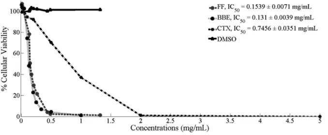
BBE showed a dose-dependent cytotoxic effect on a culture of OCI-Ly7 with a calcu-lated IC50 of 0.131 ± 0.004 mg/mL. A similar response was observed in cells exposed to the FF (IC50: 0.154 ± 0.007 mg/mL), while the IC50 for CTX was 0.745 ± 0.035 mg/mL, showing that both frac-tions (BBE and FF) were more cytotoxic than CTX (p < 0.0005) (Fig. 1).
Specificity for tumor cells is one of the most-wanted characteristics in drugs for cancer therapy; therefore, we exposed non-cancer cells (NIH-3T3 and VERO) to our extracts. For this purpose, we used a high concen-tration of BBE (1 mg/mL), and the percentages of liv-ing cells were 95.31% ± 6.38% and 90.17% ± 7.33% in NIH-3T3 and VERO cells, respectively, both being highly different to the viability found in OCI-Ly7 cells (1.17% ± 0.18). Regarding the FF, with a final concen-tration of 1.32 mg/mL, the viabilities observed were 65.74% ± 10.25% and 80.42% ± 4.36% in NIH-3T3 and VERO cells, respectively. These results showed that the tumor cells tend to be significantly more sen-sitive to the fractions obtained from black bean than non-cancer cells.
Flow cytometry assay
After exposing the tumor cells to BBE using the IC50 obtained in cytotoxicity assays, we observed that 41.6% of cells were positive for both AV, a molecule expressed in cell surface during early stages of apop-tosis, and propidium iodide (PI), a molecule that binds DNA in cells with damaged membranes or late stage of apoptosis (Fig. 2B). While 44.6% were positive for both markers in the cells exposed to FF (Fig. 2C), only 7.4% of cells were AV and PI positive when they were exposed to the vehicle (PBS) (Fig. 2A) (p < 0.0001 and p < 0.001, respectively) almost sim-ilar was observed in cells exposed to cyclophospha-mide (CTX) (Fig. 2D).
Cell cycle analysis
Regarding the cell cycle stages, the normal distribu-tion of log phase OCI-Ly7 cells (treated with vehicle) was 24.51% ± 0.63% in G1, 65.16% ± 0.88% in S,
and 10.33% ± 1.23% in G2/M (Fig. 2E). The addi-tion of BBE to the cell culture changed the distribuaddi-tion to 21.4% ± 0.91% in G1, 77.15% ± 0.93% in S, and 1.53% ± 0.88% in G2/M (Fig. 2F), while in the cells exposed to FF, the distribution was 21.5% ± 0.82% in G1, 77.91% ± 0.87% in S, and 0.59% ± 0.27% in G2/M (Fig. 2G). Interestingly, the treatment with CTX resulted in similar distributions in G1 and S phases (21.69% ± 0.76% and 62.62% ± 0.52%, respectively) compared to control, but more cells (15.4% ± 0.43%) were arrested in G2/M (Fig. 2H).
In vivo analysis of cytotoxicity
All inoculated mice presented characteristics of tumor growth in nodes compared to non-inoculated mice. Histopathological analysis confirmed that neoplastic tissue corresponded to diffuse prolifera-tion of large lymphocytes showing positive nuclear expression of BCL-6 and positive membrane expres-sion for CD79a (Fig. 3A-C). Interestingly, oral admin-istration of either BBE or FF increased the SR com-pared with the group of mice treated with placebo (p=0.056); surprisingly, the response was similar to the group treated with CTX (Fig. 4). The SR for the FF group was 33.3 days (confidence interval [CI]: 95%: 28.1–38.5), while for BBE and CTX were 33.8 (CI 95%: 28.3–39.5) and 33.1 (CI 95%: 28.4–37.8) days, respectively. The SR in the placebo group was 26.7 days (CI 95%: 23.7–29.8).
DISCUSSION
DLBCL represents the most common subtype of NHL, accounting for 30–40% of all newly diagnosed cases. In the United States, 8000 deaths per year are caused by this disease. The current therapeutic schemes for NHL include the administration of doxorubicin and vin-cristine, two molecules obtained from natural sources.
In this respect, 60% of all approved anticancer drugs in the USA during 2010, were obtained from natural sources24. It is well-known that natural dietary
com-pounds have chemoprevention effects by blocking, inhibiting, reversing, or retarding the process of carcino-genesis. In this context, BBE has been widely used for its antineoplastic properties8. The phytochemical content
(tannins, flavonoids, saponins, and phytosterols) of this extract has been linked to the inhibition of proliferation of cancer cells and the scavenging of free radicals7,9.
In this study, chromatographic analysis of the FF obtained from the BBE of P. vulgaris L., identified three molecules (myricetin 3-O glucoside, quercetin 3-O glucoside, and kaempferol 3-O glucoside). In this context, Cheng et al. described an FF from Gynostemma pentaphyllum com-posed of 8 molecules, two of them quercetin derivatives, and showed a cytotoxic effect over prostate cancer cell line (PC-3)15. Furthermore, Jeganathan et al. reported
the same three flavonols contained in Camellia sinen-sis L. (Myricetin 3-O glucoside, Quercetin 3-O gluco-side, and Kaempferol 3-O glucoside), in a widely used
Figure 1. Cell viability assays.OCI-Ly7 cells were exposed to several concentrations of either black beans hulls extract (BBE), its flavonoid fraction (FF), cyclophosphamide (CTX), or Vehicle (DMSO) for 24 h.
tea plant (C. sinensis L.) from Sri Lanka but at different concentrations (0.94 mg/g, 1.5 mg/g, and 1.31 mg/g, respectively)25; this plant has been reported to have
antioxidant, antimicrobial, anticancer, anti-atheroscle-rotic, and anti-proliferative properties.
In this study, we describe the anti-proliferative effect of both BBE and FF over a lymphoma cell line (OCI-Ly7) in a dose-dependent manner. In addition, we found that
both FF and BBE were capable of inducing apoptosis and arresting the cell cycle in S-phase, which also suggests that the glycosylated flavonoids from BBE were responsi-ble for its effect. In terms of selectivity, our experiments demonstrated that the tumor cell line was more sensi-tive to both extracts than non-cancer cells (NIH-3T3 and VERO), which can be explained by the growth rate of tumor cells compared with non-cancer cells.
The results of cytotoxicity assays using the BBE and FF were similar to other studies. For instance, the FF obtained from Rhus verniciflua Stokes induced apop-tosis in B-cell lymphoma (BJAB) and T-cell lymphoma (Jurkat)26. A similar apoptotic effect and arrest of
G2/M phase in human B lymphoma cells (BJAB) were observed by other researchers using the same compo-nent at 100 µg/mL27.
Regarding the cell cycle arrest, our results suggest that both treatments (BBE and FF) blocked the entrance to the G2/M phase since an increase in duration of the S phase was observed. In this context, Cheng et al. pub-lished that both flavonoids and saponins obtained from Gynostemma pentaphyllum were capable of inducing apoptosis and arresting in S and G2/M phases in pros-tate cancer cells (PC-3)15. In addition, they found that
both fractions modulated the expression of cyclin A, a molecule part of the complex needed for this transition
Figure 2. Flow cytometry analysis. Apoptosis results (Annexin-V/PI) of cells exposed to vehicle (A), to black beans hulls extract (BBE) (B), to flavonoid fraction (FF) (C), and cyclophosphamide (CTX). In addition, DNA content was analyzed in cells treated with vehicle (E), BBE (F), FF (G), and CTX (H).
A
E
B
F
C
G
D
H
Figure 3. Characterization of the in vivo model. (A) Hematoxylin and Eosin staining of the tumor excised from mice injected with the OCI-Ly7 cell suspension resulted in a classical aggressive non-Hodgkin’s lymphoma cell population. In addition, both nuclear expression of positive BCL-6 (B) and membrane expression of CD79a (C) were detected.
A B
C
(cyclin-A/cdk2)28 and the expression of caspase-3,
which is an important key in the apoptotic process.
To test the effect of these fractions in vivo, we devel-oped a xenografting model and assessed it using his-tological analysis. The results of biopsies taken from the grafted mice showed positive cells for CD79a and BCL-6 (two commonly used markers for DLBCL). With the animal model well characterized, the group of mice under placebo treatment died around day 26.7, while the group under CTX treatment (150 mg/kg i.p.) died days later (33.1 days), which showed the expected outcome. Surprisingly, the cytotoxic effect observed in vitro was observed in vivo as well, since the groups of mice receiving BBE or FF survived around 33.3 and 33.8 days, respectively.
In addition, oral administration through food is an advan-tage and a straightforward way of treatment using these fractions. A similar observation was reported using either alkaloid or terpenoid fractions adminis-tered in the food of mice grafted with either lymphoma (DAL) or lung cancer cells (L-929); in this study, the fractions increased the SR around 35.64 ± 1.05 days and a reduction in tumor volume were observed29.
Data showed in our study addresses new questions about the components in both BBE and FF, specifically, the effect of the sugar residue on the glycoside forms isolated from this plant, and the outcome of different concentrations and/or ratios among the flavonoids.
Finally, in this study, we found that both BBE and FF from P. vulgaris L. Had cytotoxic properties compara-ble to cyclophosphamide, a widely-used molecule for cancer therapy. In addition, these extracts seemed to be specific for tumor cells, since they were almost innocuous to non-cancer cells (NIH-3T3 and VERO cells). Another important observation was that the administration of the extracts in a xenografting model of lymphoma increased the SR, similar to that was observed with cyclophosphamide treatment. Further studies are needed to identify specific components of the fractions that exhibited the observed cytotoxic properties. For example, Oncamex, the myricetin-de-rived flavonoid, has shown antitumor activity in a pre-clinical model30.
ACKNOWLEDGMENTS
The authors thank Qiong Shen for providing the cells donated by Prof. Dalla Favera, and to María Isabel García Cruz for laboratory administrative and techni-cal assistance. This study was supported by Cátedra de Hematología y Cáncer and Research Chair Funds and NutriOmics Research Group from Tecnológico de Monterrey, and by Consejo Nacional de Ciencia y Tecnología (CONACYT, Mexico) via student scholar-ship no. 39331.
REFERENCES
1. Ahmedin-Jemal DV, Bray F, Center MM, Jacques-Ferlay ME, Ward E, Forman D. Global cancer statistics. CA Cancer J Clin. 2011;61:69-90.
2. Meneses-García A, Ruiz-Godoy L, Beltrán-Ortega A,
Sánchez-Cervantes F, Tapia-Conyer R, Mohar A. Principales neo-plasias malignas en México y su distribución geográfica. Rev Invest Clin. 2012;64:322-9.
3. Villela L, López-Guillermo A, Montoto S, et al. Prognostic fea-tures and outcome in patients with diffuse large B-cell lym-phoma who do not achieve a complete response to first-line regimens. Cancer. 2001;91:1557-62.
4. Coiffier B, Thieblemont C, Van Den Neste E, et al. Long-term outcome of patients in the LNH-98.5 trial, the first randomized study comparing rituximab-CHOP to standard CHOP chemo-therapy in DLBCL patients: a study by the groupe d’etudes des lymphomes de l’adulte. Blood. 2010;116:2040-5.
5. Zhang Z, Yu X, Wang Z, Wu P, Huang J. Anthracyclines poten-tiate anti-tumor immunity: a new opportunity for chemoimmu-notherapy. Cancer Lett. 2015;369:331-5.
6. Kris-Etherton PM, Hecker KD, Bonanome A, et al. Bioactive com-pounds in foods: their role in the prevention of cardiovascular disease and cancer. Am J Med. 2002;113 Suppl 9B:71S-88S. 7. Aparicio-Fernández X, Reynoso-Camacho R, Castaño-Tostado E,
et al. Antiradical capacity and induction of apoptosis on HeLa
cells by a Phaseolus vulgaris extract. Plant Foods Hum Nutr.
2008;63:35-40. Figure 4. Kaplan–Meier lymphoma survival curves. Survival
population was plotted since day 20 after inoculation and 1 day before the first subject decease was reported. Statistical differences between groups were calculated using chi-square test according to the log-rank test. Black beans hulls extract (BBE) group and flavonoid fraction (FF) group were not statistically different from CTX group, with p values of 0.42 and 0.8, respectively. All treated groups, BBE, FF, and CTX, obtained a trend p = 0.056.
8. Azevedo L, Gomes JC, Stringheta PC, et al. Black bean (Phaseolus vulgaris L.) as a protective agent against DNA damage in mice. Food Chem Toxicol. 2003;41:1671-6.
9. Bobe G, Barrett KG, Mentor-Marcel RA, et al. Dietary cooked navy beans and their fractions attenuate colon carcinogenesis in azoxymethane-induced ob/ob mice. Nutr Cancer. 2008;60: 373-81.
10. Gutiérrez-Uribe JA, Serna-Saldívar SR, Moreno-Cuevas JE, Hernández-Brenes C, Guajardo-Touché EM. Cancer Cell Growth
Inhibition by Black Bean (Phaseolus vulgaris L.) Extracts. United
States Patent US No. 2006024394/A1; 2006.
11. Berhow MA, Wagner ED, Vaughn SF, Plewa MJ. Characterization and antimutagenic activity of soybean saponins. Mutat Res. 2000;448:11-22.
12. Shi J, Arunasalam K, Yeung D, et al. Saponins from edible legumes: chemistry, processing, and health benefits. J Med Food. 2004;7:67-78.
13. Bawadi HA, Bansode RR, Trappey A 2nd, Truax RE, Losso JN.
Inhibition of caco-2 colon, MCF-7 and hs578T breast, and DU 145 prostatic cancer cell proliferation by water-soluble black bean condensed tannins. Cancer Lett. 2005;218:153-62. 14. Kandil FE, Smith MA, Rogers RB, et al. Composition of a
chemo-preventive proanthocyanidin-rich fraction from cranberry fruits responsible for the inhibition of 12-O-tetradecanoyl phor-bol-13-acetate (TPA)-induced ornithine decarboxylase (ODC) activity. J Agric Food Chem. 2002;50:1063-9.
15. Cheng TC, Lu JF, Wang JS, et al. Antiproliferation effect and apoptosis mechanism of prostate cancer cell PC-3 by flavonoids
and saponins prepared from Gynostemma pentaphyllum. J Agric
Food Chem. 2011;59:11319-29.
16. Ferguson PJ, Kurowska E, Freeman DJ, Chambers AF, Koropatnick DJ. A flavonoid fraction from cranberry extract inhibits proliferation of human tumor cell lines. J Nutr. 2004;134: 1529-35.
17. Kameswaran TR, Ramanibai R. The antiproliferative activity of flavonoidal fraction of Indigofera tinctoria is through cell cycle arrest and apoptotic pathway in A-549 Cells. J Biol Sci. 2008;8:584-90.
18. Son YO, Lee KY, Lee JC, et al. Selective antiproliferative and
apoptotic effects of flavonoids purified from Rhus verniciflua
stokes on normal versus transformed hepatic cell lines. Toxicol Lett. 2005;155:115-25.
19. Guajardo-Flores D, García-Patiño M, Serna-Guerrero D, Gutiérrez-Uribe JA, Serna-Saldívar SO. Characterization and quantification of saponins and flavonoids in sprouts, seed coats and cotyledons of germinated black beans. Food Chem. 2012;134:1312-9.
20. Schimdt-Wolf IG, Negrin RS, Kiem HP, Blume KG, Weissman IL. Use of a SCID mouse/human lymphoma model to evaluate cytokine-induce killer cells with potent antitumor cell activity. J Exp Med. 1991;174:139-49.
21. Man S, Bocci G, Francia G, et al. Antitumor effects in mice of low-dose (metronomic) cyclophosphamide administered continuously through the drinking water. Cancer Res. 2002;62:2731-5. 22. Goutelle S, Maurin M, Rougier F, et al. The hill equation: a review
of its capabilities in pharmacological modelling. Fundam Clin Pharmacol. 2008;22:633-48.
23. Guajardo-Flores D, Serna-Saldívar SO, Gutiérrez-Uribe JA. Evaluation of the antioxidant and proliferative activities of extracted saponins and flavonols from germinated black beans (Phaseolus vulgaris L.). Food Chem. 2013;141:1497-503. 24. Newman DJ, Cragg GM. Natural products as sources of new
drugs over the 30 years from 1981 to 2010. J Nat Prod. 2012;75:311-35.
25. Jeganathan B, Punyasiri PA, Kottawa-Arachchi JD, et al Genetic variation of flavonols quercetin, myricetin, and kaempferol in the
Sri Lankan tea (Camellia sinensis L.) and their health-promoting
aspects. Int J Food Sci. 2016;2016:6057434.
26. Lee JC, Lee KY, Kim J, et al. Extract from Rhus verniciflua stokes is capable of inhibiting the growth of human lymphoma cells. Food Chem Toxicol. 2004;42:1383-8.
27. Jang HS, Kook SH, Son YO, et al. Flavonoids purified from Rhus
verniciflua stokes actively inhibit cell growth and induce apop-tosis in human osteosarcoma cells. Biochim Biophys Acta. 2005;1726:309-16.
28. Pagano M, Pepperkok R, Verde F, Ansorge W, Draetta G. Cyclin A is required at two points in the human cell cycle. EMBO J. 1992;11:961-71.
29. Chitra V, Sharma S, Kayande N. Evaluation of anticancer activity of Vitex negundo in experimental animals: an in vitro and in vivo
study. Int J Pharm Tech Res. 2009;1:1485-9.
30. Martínez-Pérez C, Ward C, Turnbull AK, et al. Antitumor activity of the novel flavonoid on camex in preclinical breast cancer models. Br J Cancer. 2016;114:905-16.