The usefulness of the buffy coat
smear and panbacterial polymerase chain reaction
in early diagnosis of neonatal sepsis
Guadalupe García-Elorriaga, Nancy Cortés-Torres,* Julio César Ballesteros-del-Olmo,**
Guillermo Del Rey-Pineda,*** César González-Bonilla*
* Unidad de Investigación en Inmunología e Infectología,
** Servicio de Neonatología, Hospital General Dr. Gaudencio González Garza, *** Departamento de Infectología, Hospital Infantil de México Federico Gómez, Banco Central de Sangre, Centro Médico Nacional La Raza,
Instituto Mexicano del Seguro Social. ARTÍCULO ORIGINAL
ABSTRACT
Objective. In this study are evaluated the usefulness of the buffy coat smear and panbacterial polymerase chain reaction (PCR) as diagnostic tests in the early detection of neonatal sepsis. Material and methods. It was studied 49 patients aged up to 28 days who were hospitalized in the Intensive Care Unit (ICUs) of the Neonatology, with a clinical diagnosis of neonatal sepsis and 49 umbilical cord samples of healthy newborns. Blood cultures and 50 µL of plasma were taken for the DNA and performance of the broad-range PCR primer system (panbacterial PCR). Simultaneously, were taken three capillaries with blood for the leukocyte layer (buffy coat) smear, we performed three stains: Gram; Löeffler blue me-thylene (LBM), and acridine orange (AO). Statistical analysis included sensitivity, specificity, positive predictive value (PPV), and negative predictive value (NPV) against the clini-cal diagnosis. Results. With respect to stains of buffy coat smear, they resulted very specific, from 90-97%, with 64-75% sensitivity, 87-94% PPV, and 77-82% NPV. In inverse fas-hion, PCR resulted very sensitive at 96%, with 91% specificity,
92% PPV, and 96% NPV. Conclusions. Buffy coat smear
stains are easy, fast, and specific, while that of PCR was hig-hly sensitive. Thus, both can be utilized as diagnostic tests.
Key words. Bacteriologic stains. Molecular diagnosis. Neo-natal sepsis. Buffy coat.
Utilidad de la tinción de buffy coat y
reacción en cadena de la polimerasa panbacteriana en el diagnóstico temprano de sepsis neonatal
RESUMEN
Introducción. En este estudio se evaluaron la utilidad del frotis de la buffy coat y la reacción en cadena de la polimerasa (PCR) panbacteriana, como pruebas de diagnóstico para la de-tección temprana de la sepsis neonatal. Material y méto-dos. Se estudiaron 49 pacientes de hasta 28 días de edad, hospitalizados en la Unidad de Cuidados Intensivos Neonata-les con diagnóstico clínico de sepsis neonatal, y 49 muestras de cordón umbilical de recién nacidos sanos. Se tomaron he-mocultivos y 50 μL de plasma para la extracción del ADN y un sistema de iniciadores para PCR de amplio rango (PCR panbacteriana). Al mismo tiempo se tomaron tres capilares de sangre para el frotis de la capa de leucocitos (buffy coat), des-pués se realizaron tres tinciones: Gram, azul de metileno de Löeffler y naranja de acridina. El análisis estadístico incluyó la sensibilidad, especificidad, valor predictivo positivo (VPP) y valor predictivo negativo (VPN) en contra del diagnóstico clí-nico. Resultados. Las tinciones de los frotis de buffy coat re-sultaron muy específicas, de 90-97%, con 64-75% de sensibilidad, 87-94% de VPP y 77-82% de VPN. De manera in-versa, PCR resultó muy sensible en 96%, con 91% de especifi-cidad, 92% de VPP y 96% de VPN. Conclusiones. Las tinciones de buffy coat son fáciles, rápidas y específicas, mien-tras que PCR fue sumamente sensible. Por lo tanto, ambas pueden utilizarse como pruebas diagnósticas
INTRODUCTION
Neonatal sepsis is defined as the systemic inflam-matory response syndrome arising from an infectious process that presents during the first 28 days of extrauterine life.1 It is considered as a health
pro-blem worldwide due to its high indices of morbility and mortality.2 In Mexico, there are between 4 and
15 cases per 1,000 live births; in Intensive Care Units (ICUs), this amount is 5-20 times higher.3
To establish an adequate diagnosis of sepsis, we must evaluate clinical and laboratory aspects.4 In a
previous study conducted by our work group,5 we
de-cided to evaluate 11 indices of infection in global fas-hion (high C reactive protein, increased erythrocyte sedimentation rate (ESR), leukopenia, neutropenia, bandemia, increased band/neutrophils, plaquetopenia, anemia, inmadure/neutrophil ratio, fibrinogen and haptoglobin) arbitrarily assigning one point for each test alteration observed. In patients with > 5 positive indices, sensitivity and sensitivity were found near equilibrium (71.4 and 68%, respectively) with 69% efficiency in the clinical test. The blood culture is the microbiological gold standard, but its sensitivity ran-ges between 20 and 80%.6
It has been reported in some articles7-9 that by
means of supernatant study of the centrifuged blood capillary, different stains can be carried out for de-tection of bacteria, and even of fungi and yeasts; this test is called the buffy coat smear.
In addition to traditional methods for identifying microbial presence, it has been demonstrated that re-cognition of DNA bacterial sequences possesses pan-specific advantages, such as those associated with the case of the gene 16S rRNA through the polymerase chain reaction (PCR)10,11 and real-time PCR.12
Diagnosis of sepsis, is clinical and based on cul-ture results. Although blood culcul-ture is the gold stan-dard for the diagnosis, culture reports would be available only 48-72 h, and have low sensitivity. The earliest signs are often subtle and nonspecific, these signs include changes in respiration, apnea and/or tachypnea; in heart rate, including frequent episodes of bradycardia and/or tachycardia; in venti-lation and cardiac rate. In addition, neonates with sepsis commonly exhibit alterations in core body temperature, as manifested by fever and or hypo-thermia. Changes in skin perfusion commonly accompany sepsis, as manifested by mottling, cooling of the extremities, and a general “ill look” to the baby overall. Other subtle findings of sepsis include the development of feeding intolerance, vomiting, or diarrhea.
Accurate and timely diagnosis of early onset neo-natal sepsis remains challenging to the clinician and the laboratory. Many potential markers (acute pha-se reactants, cell surface markers, cytokines) are not routinely available to the laboratory. Most tests are measured on a continuous scale where there is overlap between infected and non-infected infants. Total leukocyte count, immature to total neutrophil ratio, platelet count, and CRP (C reactive protein), have low sensitivity and specificity or varying delayed responses early in the course of infection. Leukocyte indices and CRP are considered to be ‘‘late’’ markers and are not sensitive enough for early diagnosis of neonatal sepsis.
OBJECTIVE
Of the study was to evaluate the usefulness of Gram, acridine orange, and Löeffler blue methylene stainings of the buffy coat smear and of the panbacterial PCR as early diagnostic tests for neo-natal sepsis.
MATERIAL AND METHODS
We studied 49 neonatal patients aged up to 28 days who were hospitalized in the Intensive Care Unit (ICUs) of the Neonatology Service Hospital Ge-neral Dr. Gaudencio González Garza from August 2005 to July 2006 with a clinical diagnosis of neona-tal sepsis without antibiotic treatment only in 4% or with a maximum of a 48-h antibiotic treatment, or even with > 3 days without a clinical response, sin-ce the hospital is a Tertiary medical care. We used various combinations of antibiotics, the scheme used was ampicillin/amikacin, second cefotaxime/van-comycin and to a lesser extent cefotaxime/amikacin. We performed a blinded aleatorized study. As the control group, 49 blood samples in which the absence of pathogens was confirmed by blood culture, were provided by the Umbilical Cord Pluripotent Hemato-poietic Cell Bank that pertains to the Central Blood Bank of the Mexican Institute of Social Security Institute’s (IMSS’s) La Raza National Medical Center (BSC-CMNR). The protocol was accepted by the local Research and Ethics Committee of the Hos-pital, and all parents or guardians signed the letter of informed consent.
Sample calculation
independent samples. Considering P1 (proportion of the population 1) and the detection rate of the “bu-ffy coat” which is 75 % (8) and P2 (the proportion of the population 2) and the detection rate of panbacte-rial PCR in vitreus is 95%,10 with a value of alpha
equal 0.05 for an expected proportion of 80%, giving a total of 49 diseased patients and 49 new born without disease. This program is based on the nor-mal approximation to the binomial distribution.
Clinical diagnosis of neonatal sepsis
Alteration of rates of infection at least 5 (leuko-cytosis or leucopenia, neutrophilia or neutropenia, thrombocytopenia, presence of bands, presence of vacuolated neutrophils or toxic granulations, bands/ total neutrophils ratio, immature/mature ratio alte-red, hemoglobin, ESR, CRP increased and positive blood culture. The diagnosis of neonatal sepsis ba-sed on a number of risk factors and nonspecific cli-nical and laboratory paramentes, there is no validated scale. Aproximately, sepsis screen per se
had a sensitivity and negative predictive value (NPV) of 48.3 and 78.3%.
Inclusion criteria
Newborn patients with changes in respiration, heart rate, ventilation and cardiac rate. Alterations in core body temperature, changes in skin perfu-sion, cooling of the extremities, and a general “ill look” to the baby overall. The development of fee-ding intolerance, vomiting, or diarrhea. In addition, we evaluated 11 indices of infection in global fashion (high C reactive protein, increased ESR y, leukope-nia, neutropeleukope-nia, bandemia, increased band/neutro-phils, plaquetopenia, anemia, inmadure/neutrophil ratio, fibrinogen and haptoglobin) arbitrarily assig-ning one point for each test alteration observed. In patients with > 5 positive indices, sensitivity and sensitivity were found near equilibrium.
We performed the following on patients under aseptic conditions: venous puncture or catheter aspi-rate, obtaining approximately 500 µL of blood for buffy coat smear and PCR. Additionally, the remain-der of the laboratory tests was conducted, taking samples for complete blood work, blood culture (BacT/Alert®), C-reactive protein, lumbar puncture for cytochemical study and culture, uroculture, and umbilical cord culture. Procedures for these tests were carried out in the Hospital’s laboratories by authorized personnel with conventional techniques. The control group was made buffy coat smear and
PCR.The gold standard was the clinical diagnosis, which includes: clinical parameters, laboratory and blood culture.
DNA extraction
Care was always taken during and after obtaining patient samples to prevent extraneous environmen-tal contamination. For example, the skin surface was prepared by swabbing with alcohol prior to ob-taining the sample; samples were immediately placed in sterile microfuge tubes. The DNA was isolated with guanidine isothiocyanate and phenol utilizing 500 µL of TRIzol reagent (Gibco BRL) according to the procedure described by Chomczynski.13 The DNA
was resuspended in 50 µL of distilled water after precipitation with ethanol at 75%. This solution was heated at 55 ºC for 20 min. We determined its absor-bance relationship at 260/280 nm.
Panbacterial PCR
The broad-range PCR primer system has been used previously to screen synovial DNA prepara-tions.10 Briefly, this is a non-nested assay system
that employs two primers:
• 5'-GCGTTAATCGGAATTACTGGGCGTAAG-3', and
• 5'-GGTTGCGCTCGTTGCGGGACTTAACC-3'.
Cycling parameters were 4 min at 95 oC, then 35
cycles for 1 min at 95 oC, I min at 52 oC, 1 min at
72 oC, and finally 10 min at 72 oC. This primer
sys-tem is referred to as panbacterial, because it ampli-fies 16S ribosomal RNA (rRNA) gene sequences from a number of bacterial species. As designed, the system amplifies a DNA fragment of ~577 bp (de-pending on the organism),10 spanning the region
from nucleotides 501-1,077 in the standard (Escheri-chia coli) 16S rRNA gene.
As with other similar systems, the rationale for the design of this broad-range primer system resides in the observation that prokaryotic 16S rRNA gene sequences have been conserved over evolution, and that these sequences have been especially well con-served in particular regions that function importan-tly in the ribosome.14 Design of consensus primers
Borrelia; Campylobacter; Escherichia; Listeria; Neis-seria; Proteus; Rickettsia; Salmonella; Staphylococ-cus; Yersinia; Bordetella; Chlamydia; Klebsiella; Mycoplasma; Pseudomonas; Shigella; Serratia; Streptococcus, and others. Sensitivity generally lies within the detection range of 10-50 bacterial cells.
For each assay run, water controls (i.e., no added template) were interspersed among the samples to be analyzed. Pure DNA from E. coli was utilized as a positive control for the panbacterial screening sys-tem. Amplification products were analyzed on ethi-dium bromide-stained agarose gels. The tests were performed in duplicate.
Buffy coat smear
We mounted three capillaries for each patient and control with blood samples in sterile fashion and fixed them with heat. These were centrifuged at 3,000 rpm for 15 min to obtain three layers in the capillary; the plasma; the leukocyte layer or “buffy coat”, and the erythrocyte layer. We cut the capilla-ry with a diamond-point marker at the height of the erythrocyte layer and also at that of the plasma, slightly above the buffy coat, and emptied the con-tents into three clean, degreased, and sterile contai-ners. The drops of buffy coat were resuspended in PBS, and we proceeded to carry out the smears. The latter were first allow to air dry and then methanol-dried for 2 min.
For each patient and control, we performed three stains: Gram; Löeffler blue methylene (LBM), and acridine orange (AO). Observation by microscope was conducted for a minimum of 15 min per plate prior to being determined as negative. The tests were performed in duplicate.
RESULTS
Within the study group of patients, 68% were males, while 32% were females; on the other hand, in the control group there was gender equity. The percentage of mortality was 18.9% in patients with sepsis.
The risk factors most frequently found in the group of patients with a clinical diagnosis of sepsis were the following: parenteral nutrition, (100%); ca-therer use, (97%); prolonged fasting, (92%); mecha-nical ventilation, (89%); cesarean birth (67%); malnutrition (57%); low birth weight, (38%); pre-mature rupture of membranes, (31%), and surgical procedures (27%). There were no associated risk fac-tors with regard to the control group.
In the hematologic and serum examinations per-formed, we established the following changes in se-veral parameters, (Table 1). In addition, there was development in the cultures performed to identify probable sources of infection: 33% in urocultures; 27% in blood cultures; 15% in cerebrospinal fluid (CSF) culture; 15% in catheters, and 3% in umbili-cal cord secretion. We used various combinations of antibiotics, the scheme used was ampicillin/amika-cin, second cefotaxime/vancomycin and to a lesser extent cefotaxime/amikacin. There were no positive cultures in the control group.
We found 33 cases (67%) (Table 2) in the distinct cultures carried out that resulted positive, but the actual number of patients whit multiple infections demonstrated by culture test was 29 (59%).
We also analyzed all of the cultures together to determine which microorganisms predominated as causes of infection. Among the results, it was no-teworthy that CNS(coagulase-negative Staphylococ-cus) was the cause of 44.8% of the infections, followed by E. coli, the cause of 31%. We also found
Candida sp. and Klebsiella in 10.8% each, Pseudo-monas sp. and Enterobacter sp. in 5.4% each, and
Enterobacter cloacae in 2.7%.
With respect to bacteria found in the buffy coat smear, we found bacterial forms in 39 patients (80%), in which extracellular Gram-positive cocci predominated in 15 of the patients; of these, three died. We also observed extra- and intracellular Gram-positive cocci (phagocyted by polymorpho-nuclears) in seven patients; of these, three died. Similarly, we found 14 cases with Gram-negative
a
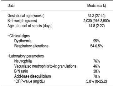
Table 1. Neonatal data, clinical signs and laboratory parameters.
Data Media (rank)
Gestational age (weeks) 34.2 (27-40)
Birthweigth (grams) 2,030 (915-3,500)
Age at onset of sepsis (days) 14.8 (2-27)
• Clinical signs
Dysthermia 95%
Respiratory alterations 54-0.5%
• Laboratory parameters
Neutrophilia 76%
Vacuolated neutrophils/toxic granulations 46%
B/N ratio 38%
Acid-base disequilibrium 70%
*CRP-value (mg/dL) 5.8% (0-25.2)
bacilli and three cases with Gram-positive bacilli (Table 3).
In the smears carried out in control-group pa-tients without sepsis, we found bacteria in four ca-ses (8%), observing in two caca-ses extracellular Gram-positive cocci and in the remaining two, Gram-positive bacilli.
With regard to the number of patients with a cli-nical diagnosis of sepsis, performed by physician and detected by panbacterial PCR, we found that in the
group of patients, we were able to appreciate a 577-bp band in 47 of cases (96%), while in the control group, we only observed this in four cases (8%).
In table 4, we grouped patients taking into ac-count the microbial group identified and compared the latter with the results obtained from the stains and the panbacterial PCR, because these are interes-ting for understanding the importance of the appli-cation of our methodologies. In the remaining positive cultures, we did not observe agreement with our data.
With all of these results, we calculated percenta-ges of sensitivity, specificity, Positive predictive va-lue (PPV), and NPV for each of the stains and for PCR with traditional formulae according to 2 x 2 contingency tables.15 We considered that the gold
standard for each of these was the clinical diagnosis (> 5 changed parameters), in this respect, we deter-mined the usefulness with regard to confirmation of the clinical suspicion, diagnostic support, treatment restoration in the case of a positive test, and compa-tibility with the remainder of laboratory and medical office examinations (Table 5).
DISCUSSION
We observed that in the group of patients, there was a predominance of more than double of masculine over feminine gender (68 and 32%, respectively). This tendency is cited by other authors.3,16
As in the majority of cases reported, the clinical signs and symptoms observed were characterized by their variety and unspecificity. The main clinical manifestations found were dysthermia in 95% of ca-ses, coinciding with the findings of other authors,17
and acid-base disequilibrium in 70%.
As we mentioned previously, to carry out a correct early diagnosis, we require the support of several laboratory data; a sole datum does not support the diagnosis. In this study, the changed pa-rameters that stood out were, in first place, C-reactive
2 .
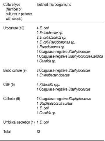
Table 2. Isolated microorganisms in cultures of the study group.
Culture type Isolated microorganisms (Number of
cultures in patients with sepsis)
Uroculture (13) 4 E. coli 2 Enterobacter sp. 2 E. coli/Candida sp. 1 E. coli/Pseudomonas sp. 1 Pseudomonas sp.
1 Coagulase-negative Staphylococcus 1 Coagulase-negative Staphylococcus/Candida 1 Candida sp.
Blood culture (9) 8 Coagulase-negative Staphylococcus 1 Enterobacter cloacae
CSF (5) 4 Klebsiella spp.
1 Coagulase-negative Staphylococcus
Catheter (5) 2 Coagulase-negative Staphylococcus 1 Staphylococcus aureus
1 E. coli 1 Candida sp.
Umbilical secretion (1) 1 E. coli
Total 33
E. coli: Escherechia coli. CSF: cerebrospinal fluid.
3 .
Table 3. Microorganisms observed in buffy coat smear stains.
Bacteria type Patients Controls
Positive cases Mortality Positive cases Mortality (n = 39) in positive cases (n = 4) in positive cases
Extracellular Gram-positive cocci 15 3 2 0
Extra- and intracellular Gram- positive cocci 7 3 0 0
Gram-negative bacilli 14 0 0 0
García-Elorriaga G, et al.
Early diagnosis of neonatal sepsis.
i
Rev Invest Clin
2012; 64 (3): 275-283
280
b e T 4
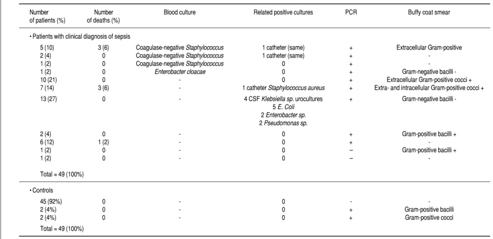
Table 4. Comparison of polymerase chain reaction (PCR) and buffy coat smear results with different positive cultures.
Number Number Blood culture Related positive cultures PCR Buffy coat smear
of patients (%) of deaths (%)
• Patients with clinical diagnosis of sepsis
5 (10) 3 (6) Coagulase-negative Staphylococcus 1 catheter (same) + Extracellular Gram-positive
2 (4) 0 Coagulase-negative Staphylococcus 1 catheter (same) +
-1 (2) 0 Coagulase-negative Staphylococcus 0 +
-1 (2) 0 Enterobacter cloacae 0 + Gramnegative bacilli
-10 (21) 0 - 0 + Extracellular Gram-positive cocci +
7 (14) 3 (6) - 1 catheter Staphylococcus aureus + Extra- and intracellular Gram-positive cocci +
13 (27) 0 - 4 CSF Klebsiella sp. urocultures + Gramnegative bacilli
-5 E. Coli 2 Enterobacter sp. 2 Pseudomonas sp.
2 (4) 0 - 0 + Gram-positive bacilli +
6 (12) 1 (2) - 0 +
-1 (2) 0 - 0 – Gram-positive bacilli +
1 (2) 0 - 0 –
-Total = 49 (100%)
• Controls
45 (92%) 0 - 0 -
-2 (4%) 0 - 0 + Gram-positive bacilli
2 (4%) 0 - 0 + Gram-positive cocci
Total = 49 (100%)
CSF: cerebrospinal fluid. E. Coli: Escherichia coli.
b e T 5
Table 5. Statistical values obtained from buffy coat smear stains and from panbacterial polymerase chain reaction (PCR) compared with the clinical diagnosis.
Clinical diagnosis
Panbacterial PCR (%) Gram stain (%) Staining with AO (%) Staining with LBM (%)
Sensitivity 96 72 75 64
Specificity 91 95 90 97
protein, found altered in 84 %, on which there are data that this is a highly sensitive, but not a highly specific, marker,5 in that it is changed in many
in-flammatory processes, thus causing false-positives for sepsis. In second place, we found total amount of neutrophils, altered in 76%, according to Manroe’s reference ranges for neutrophils indices.18
In the blood cultures, there was low recuperation of microorganisms (only 18%), which was worrisome. One of the causes for this may be high dilution in the blood culture due to the small volumes of blood. Normally, a relationship of blood volume:mean of 1:10 is recommended;19 also recommended is that
these tests be carried out at a minimum of twice to increase possibility of detection.20 This was very
di-fficult to perform with the majority of patients, due to their weight, associated pathologies, etc. Other causes can be the transitory or intermittent nature of the bacteremia, the possible low density of the bacteria present, or even an inhibitory effect of the antibiotic therapy intra- or extrapartum.21
The main microorganism isolated was coagulase-negative Staphylococcus, 89% of positive blood cul-tures and 35% of the remaining culcul-tures performed. The finding of CNS as the outstanding agent in blood cultures coincides especially with that publis-hed in previous studies conducted by our work group2,5 and in Mexico in general,3,21 where it is
ob-served that these are isolated from approximately 46-57% of positive blood cultures. It is noteworthy that in all patients with blood culture positive to coagulase-negative Staphylococcus, the disease was detected after the third day of life, that is, the for-mer can be considered as cases of late-origin or no-socomial sepsis, while in the case of the patient with a blood culture positive for E. cloacae, this was de-termined on the first day of life. Thus, we can think that this was early sepsis. As can be observed in ta-ble 2, another of the principal microorganisms isola-ted from the distinct cultures was E. coli
(approximately 30% of the isolations). This coinci-des with that reported by other authors in Mexico.16
About the matter to the diagnosis of neonatal sep-sis, our work team previously reported that, the smear of the buffy coat stained with Gram had a good specificity, taking as gold standard the blood culture.22 We decided to investigate what type of
stain was the most appropriate for detecting bacte-ria in blood. The Gram stain was not ideal for stai-ning blood; however, several authors utilize this stain because it is essential for classifying bacteria such as Gram-positive or -negative and to renovate the antibiotic picture. In addition, blue methylene
and acridine orange stains have been employed with excellent results observed. We found that Gram-posi-tive cocci predominated over Gram-negaGram-posi-tive bacilli, as can be observed in table 3. This is to a certain de-gree contrary to what we expected, because it was ci-ted that neonatal sepsis is caused mainly by Gram-negative bacilli, especially enterobacterias (between 40 and 60%); among these, the mortality rate ranges between 30 and 90%.3,23 However, this
did not occur in this study. The highest percentage of death was related with the finding of Gram-posi-tive cocci, (14%) and none with Gram-negaGram-posi-tive baci-lli. We also found Gram-positive bacilli in the study group; it is probable that this was Listeria mono-cytogenes, in that the latter is the most commonly found cause of sepsis in neonates.17
It is important to note that in control group, we found four cases of samples with bacteria, two with Gram-positive cocci and two with Gram-negative ba-cilli, which can be due to various causes: auto-con-tamination from the sample-taking stain, to inadequate asepsis at site of the puncture or mate-rial contamination, false-positive on observing it, or transitory bacteremia, because the blood culture was negative. Among the findings of Gram-positive cocci in patients, we discovered two variants: extra-cellular Gram-positive cocci in 15 patients, and intracellular Gram-positive cocci in seven (phagocyted in polymorphonuclears). Of these, we establish the distinction that despite that there is the same amount of deaths in both cases, we noted that there was a tendency toward greater mortality in the case of intracellular cocci. Contrary to what would be thought if bacteria were observed outside of the leukocytes, which could reflect a poor response or deficient immune mechanisms (poor opsonization),8
what is certain is that leukocytes infected in peri-pheral blood usually indicate a disease with a fatal result.24 Thus, it has been considered as
entertai-ning a poor prognosis. It is thought that the genus Staphylococcus especially can utilize the phagocyte as a “Trojan horse” and be transported at a distance, which sometimes can be the origin of new septic foci.25 Studies on bacteremia in rabbits suggest that
phagocyted Staphylococcus pathogens are free from damage at the intracellular location, because they derive from a microambiance that is protected from destruction by the immune system of the host, and in this manner they multiply.7,25
sepsis. Both tests have the advantage that seem to be good and early predictors of the beginning of sep-sis, and also they seem to have a prognostic value for the severity, but they are expensive and inacces-sible.26 Also, it was described a novel manual
me-thod that included use of a serum separator tube to early diagnostic of sepsis, and it is cost-effective.27 It
is important to observe the low recuperation of mi-croorganisms in blood cultures on comparing these with the positive results of PCR as well as of the bu-ffy coat smear, thus exhibiting the importance of their application. Also of great value was positive PCR by itself due to its high sensitivity, in that among six cases in which one patient died, the blood culture, other cultures, and the buffy coatsmear re-sulted negative. One aspect that commands our at-tention was the case of the patient with a diagnosis of sepsis, but with negative cultures, PCR, and buffy coatsmear. Perhaps this was a microorganism of di-fficult detection, for which a search is not common-ly in the laboratory, for example, Ureaplasma sp.
Panbacterial PCR was able to discard for us a great percentage of patients those who were and who were not infected. This type of PCR only detects bacterial-origin sepsis; therefore, it would be recommended to identify the class and morphology of the bacteria with Gram staining of the buffy coat smear.
The five cases with three deaths attract our attention; in these, we found CNS in the blood culture,
in agreement with the PCR and the buffy coatsmear staining; in addition, there was a catheter culture that coincided with the blood culture. After this we had two patients with CNS-positive blood cultures, positive PCR, but negative buffy coatsmear, compri-sing two examples of false-negatives for the buffy coatsmear; perhaps the amount of bacteria in the blood was not sufficient for detection on the slide. There were also seven patients, of whom three died. In the seven, we detected extra- and intracellular Gram-positive cocci, but the blood culture did not develop; this can be due to that the bacteria are within the leukocytes, diminishing in this manner the number of circulating bacteria and with this, the possibility of recuperating these in the culture.
In synthesis, in order to confirm or discard the clinical suspicion of infection, buffy coat smear stai-ning is cheap, it can be applied to routine labora-tory, is very specific, but its sensitivity is low, the panbacterial PCR, is expensive, but its sensitivity is high, both methods were useful, although the per-formance of the blood culture is necessary for anti-microbial sensitivity. The staining has the limiting
that depends on the expertise of the analyst and the laboratories implement it must have good systems of control and quality assurance including the analysis of panels submitted by an outside institution. According to the clinical data, sepsis arising from nosocomial-source bacteria predominated.
It is important to possess knowledge on the mi-croorganisms present in a hospital service or insti-tution, as well as the sensitivity in vitro of the antibiotics employed empirically, because from these we are able to establish adequate programs of pre-vention and to select early antimicrobial schemes that can improve survival in neonates who acquire an intrahospital infection.
ACKNOWLEDGEMENTS
This study was financially supported by Immuno-logy and InfectoImmuno-logy Research Unit, CMNR, IMSS, Mexico City.
ABBREVIATIONS
• CRP: C reactive protein. • B/N: band/neutrophil. • E. coli:Escherechia coli.
• CSF: cerebrospinal fluid. • AO: acridine orange.
• BM: Löeffler blue methylene.
REFERENCES
1. Goldstein B, Giroir B, Randolph A. International pediatric sep-sis consensus conference: definitions for sepsep-sis and organ dys-function in pediatrics. Pediatric Crit Care Med 2005; 6: 1-8. 2. Ballesteros-del-Olmo JC, Merás-Montoya AI. Infecciones en
Neonatología en un tercer nivel de atención. Boletín de la Aca-demia Mexicana de Pediatría 2006. Disponible en: http:// www.academiamexicanadepediatria.com.mx/publicaciones/aca-demicos opinan/otras.php
3. Rodríguez-Weber MA, López-Candiani C, Arredondo-García JL, Gutiérrez-Castrellón P, Sánchez-Arriaga F. Morbilidad y mortalidad por sepsis neonatal en un hospital de tercer nivel de atención. Sal Pub Mex 2003; 45: 90-5.
4. Sundaram V, Kumar P, Dutta S, Mukhopadhyay K, Ray P, Gautam V, et al. Blood culture confirmed bacterial sepsis in neonates in a North Indian tertiary care center: changes over the last decade. Jpn J Infect Dis 2009; 60: 46-50.
5. Ballesteros JC, Rodríguez C, Morales M, E G, García-RP, Vega-G J, et al. Indicadores de infección temprana en sep-ticemia neonatal. Rev Mex Pediatr 1996;63: 17-24.
6. Kumarcs V, Neelagaud YF. Incubation period for culture posi-tivity to detect septicemia in neonates. Indian J Med Microbiol 2005; 23: 270-1.
7. McCabe W, LaPorte J. Intracellular bacteria in the peripheral blood in Staphylococcal bacteremia. Ann Intern Med 1962; 57: 141-3. 8. Faden H. Early diagnosis of neonatal bacteremia by buffy coat
9. Lemus-Varela ML, Alberto VS, Arriaga-Dávila JJ. Clinical and laboratory parameters in neonatal nosocomial sepsis. Gac Med Mex 2008; 144: 409-11.
10. Gérard HC, Wang Z, Wang GF, El-Gabalawy H, Goldbach-Mansky R, Li Y, et al. Chromosomal DNA from a variety of bacterial species is present in synovial tissue from patients with various forms of arthritis. Arthritis Rheum 2001; 44: 1689-97. 11. Dutta S, Narang A, Chakraborty A, Ray P. Diagnosis of
neona-tal sepsis using universal primer polymerase chain reaction be-fore and after starting antibiotic therapy. Arch Pediatr Adolesc Med 2009; 163: 6-11.
12. Ohlin A, Bäckman A, Björkqvist M, Mölling P, Jurstrand M, Schollin J. Real-time PCR of the 16S-rRNA gene in the diagno-sis of neonatal bacteraemia. Acta Paediatr 2008; 97: 1376-80. 13. Chomczynski P. A reagent for the single-step simultaneous
iso-lation of RNA, DNA and proteins from cell and tissue samples. Biotechniques 1993; 15: 532-7.
14. Gutell RR, Larsen N, Woese CR. Lesson from an evolving rRNA: 16S and 23S structures from a comparative perspective. Microbiol Rev 1994; 58: 10-26.
15. Ponce-de-León S. Como leer revistas médicas para aprender so-bre una prueba diagnóstica. Rev Invest Clin 1988; 40: 73-83. 16. Gutiérrez-Muñoz UH, Gutiérrez-Muñoz J, Rosas-Barrientos V.
Factores de riesgo en sepsis neonatal en un hospital de tercer nivel en la Ciudad de México. Revista de Especialidades Médi-co-Quirúrgicas, ISSSTE 2005; 10: 21-4.
1 7 . Murgía M, Mancilla J. Programa de actualización continúa en neonatología. Libro 7. México: Intersistemas Editores; 2 0 0 4 .
18. Manroe BL, Weinberg AG, Rosenfeld CR, Browne R. The neo-natal blood count in health and disease. I. Reference values for neutrophilic cells. J Pediatr 1979; 95: 89-98.
19. Isaacman DJ, Karasic RB, Reynolds EA, Krost SI. Effect of number of blood cultures and volume of blood and detection of bacteremia in children. J Pediatr 1996; 128: 190-5. 20. Mangurten HH, Le Beau LJ. Diagnosis of neonatal bacteremia
by a microblood culture technique. J Pediatr 1997;90: 990. 2 1 . Domínguez-Sosa J, Vila-Ruiz F, Setién-Castillo IA.
Prevalen-cia y resistenPrevalen-cia bacteriana en una unidad de cuidados
intensi-vos neonatales. Enfermedades Infecciosas y Microbiología 2005; 25(3).
22. Ballesteros-del-Olmo JC, Tena-Reyes D, García-Elorriaga G, Vega-García A, Ramírez-Ortiz M, Sosa-Maldonado J, et al. Sensibilidad y especificidad del frotis de buffy coat teñido con Gram en el diagnóstico de sepsis neonatal. Rev Mex Pediatr 2008; 75: 97-102.
23. Zamora-Castorena S, Murgía-de Sierra MF. Cinco años de ex-periencia con sepsis neonatal en un centro pediátrico. Rev In-vest Clin 1998; 50: 463-70.
24. Kostiala A, Jormalainen S, Kosunen T. Detection of experimen-tal bacteremia and fungemia by examination of buffy coat pre-pared by a micromethod. Am J Clin Pathol 1979; 72: 437-43. 25. Melly MA, Thomison JB, Rogers DE. Fate of staphylococci
within human leukocytes. J Exp Med 1960; 112: 1121-30. 26. Figueras-Aloy J, Gómez-López L, Rodríguez-Miguélez JM,
Salvia-Roiges MD, Jordán-García I, Ferrer-Codina I, et al. Serum soluble ICAM-1, VCAM-1, L-selectin, and P-selectin le-vels as markers of infection and their relation to clinical severi-ty in neonatal sepsis. Am J Perinatol 2007; 24: 331-8.
27. Barman P, Sengupta S, Singh S. Study of a novel method to assist in early reporting of sepsis from the microbiology labo-ratory. J Infect Dev Ctries 2010; 4(12): 822-7.
Reimpresos:
Guadalupe García-Elorriaga, Ph.D. Unidad de investigación en
Inmunología e Infectología
Centro Médico Nacional La Raza, IMSS Av. Jacarandas y Seris s/n
02990, México, D.F.
Ph.: (+52) (55) 5724-5900, Ext. 24321 Fax: (+52) (55) 5353-0989
E mail: [email protected]