Otras secciones de este sitio:
☞ ☞ ☞ ☞
☞ Índice de este número
☞ ☞ ☞ ☞
☞ Más revistas
☞ ☞ ☞ ☞
☞ Búsqueda
Others sections in this web site:
☞ ☞ ☞ ☞
☞ Contents of this number ☞
☞ ☞ ☞
☞ More journals ☞
☞ ☞ ☞ ☞ Search Artículo:
Cytotoxin production in phytopathogenic and entomopathogenic Serratia
marcescens
Derechos reservados, Copyright © 2001: Asociación Mexicana de Microbiología, AC Revista Latinoamericana de Microbiología
Número
Number 4
Octubre-Diciembre
October-December 2001
Volumen
Volume43
Cytotoxin production in phytopathogenic and
entomopathogenic
Serratia marcescens
M.M. Escobar,* G.V. Carbonell,* L.O.S. Beriam,** W.J. Siqueira*** and T. Yano*
Rev
ista
Lat
inoamer
Vol. 43, No. 4 October - December. 2001 pp. 165 - 170
* Departamento de Microbiología e Imunologia, Universidade Estadual de Cam-pinas, SP, Brasil.
** Laboratório de Bacteriologia Vegetal, Centro Experimental, Instituto Biológico de São Paulo, Campinas, SP, Brasil.
*** Laboratório de Cultura de Tecidos, Centro de Genética, Biologia Molecular e Fitoquímica do Instituto Agronômico de Campinas, Campinas, SP, Brasil.
INTRODUCTION
Serratia marcescens is a Gram-negative bacillus
classi-fied as a member of the family Enterobacteriaceae
com-monly found in soil, water, plants and animals.7 This
bacte-rium has been recognized as an important nosocomial
pa-thogen, causing urinary and respiratory tract infections,11,14
bacteremias,17 meningitis,2 and septic arthritis.13
In plants, S. marcescens can be found on healthy plants or as a phytopathogen, such as in onion (Allium cepa L.), causing
de-cay of stores bulbs.1 The disease caused on vegetables like soft
rot of potato, tomato, etc., is very similar to those caused by
Erwinia strains.8
S. marcescens produces several exoenzymes such as
gela-tinase, lecithinase, proteinase and chigela-tinase,9 that may be
har-mful to insects, and the pathogenicity depends on stress
condi-tions affecting the host insect.10 A detailed study on the
spe-cies and biotypes of S. marcescens associated with insects
ABSTRACT. In this work, culture filtrates of entomopathogenic and
phytopathogenic Serratia marcescens strains induced cytotoxic effects on CHO, Vero and HEp-2 cell lines. Morphological changes on sensitive cells were characterized by cell rounding and detachment as soon as 30 min of incubation, culminating in cell death after 24 h. The cytotoxic effect was completely neutralized by specific antiserum indicating that occur antigenic similarity among cytotoxins produced by these strains. The toxicity assays on plants showed that the culture supernatants did not provoke any visible morphological change and did not affect their growth. By contrast, the plants treated with bacte-rial suspension showed disease symptom, such as shriveling and de-cay of stores bulbus in onion and lettuce plantlets. In conclusion, this study show that phytopathogenic and entomopathogenic S.
marces-cens may produce a cytototoxin similar to that produced by clinical
isolates and it is toxic to different mammalian cell lines. These results are especially important for studies involving this bacterium as biolo-gical control agent.
Key words: Serratia marcescens, cell culture, cytotoxins, virulence
factors.
RESUMEN. En este trabajo, filtrados del cultivo de cepas de Serratia
marcescens entomopatógenas y fitopatógenas indujeron efectos
cito-tóxicos sobre las líneas celulares CHO, Vero y Hep-2. Los cambios morfológicos sobre las células sensibles se caracterizaron por el redon-deamiento de las células y el despegado de las placas, a partir de los 30 min de incubación y terminando a las 24 h, con la muerte celular. El efecto citotóxico fue neutralizado completamente por un antisuero es-pecífico, lo que sugiere que existe similitud antigénica entre las cito-toxinas producidas por estas cepas. Los ensayos de toxicidad sobre plantas, mostraron que los sobrenadantes del cultivo no provocaron ningún cambio morfológico visible y no afectaron su crecimiento. Por el contrario, las plantas tratadas con la suspensión bacteriana mostraron síntomas de la enfermedad, tales como en plantas de cebolla y lechuga. En conclusión, este estudio muestra que S. marcescens entomopatógena y fitopatógena puede producir una citotoxina similar a la producida por aislados clínicos y que es tóxica a diferentes líneas celulares de mamí-feros. Estos resultados son especialmente importantes para estudios que involucran esta bacteria como un agente de control biológico.
Palabras clave: Serratia marcescens, citotoxinas, factores de
viru-lencia.
showed that the major group was the pigmented S.
marces-cens biotype A2a.7 Pigmented biotypes of S. marcescens are
more frequently recovered from natural environments and are
rarely responsible for an outbreak of nosocomial infection.2
Recently, it was demonstrated that clinical isolates of S.
marcescens can produce a toxin active on epitelial cells
grown in vitro.3 This cytotoxin is extracellular, heat-labile
and culture conditions identified as optimal were
incu-bation at temperatures ranging from 30 to 37oC for 24h,
under shaking, in medium adjusted to pH 8.5.5
In the present study we investigated the ability of S.
marcescens isolated from plants and insects to produce
cytotoxins active on mammalian cell lines and to compare it with the cytotoxin produced by clinical isolates of S.
marcescens. The effect of the toxin in plants and the
sero-logical relationship with cytotoxin produced by clinical S.
marcescens were also evaluated.
MATERIAL AND METHODS
Bacterial strains. The phytopathogenic Serratia
mar-cescens strains isolated from lettuce (numbers 5/6 and 6/5)
Expe-Escobar et al. Cytotoxin production in phytopathogenic and entomopathogenic Serratia marcescens
Rev Latinoam Microbiol 2001; 43 (4): 165-170
166
rimental do Instituto Biológico de São Paulo, Campinas, Brasil. The previously characterized cytotoxin-producing
S. marcescens (number 458) isolated from urinary
infec-tion3 was used as a reference strain.
Preparation of bacterial culture filtrates. S.
marces-cens strains were cultured in 10 ml of Trypticase Soy Broth
(TSB, Difco Lab., Detroit, MI) at 37oC for 18 h, with
sha-king at 110 rpm. Bacterial cultures were centrifuged (10,000
x g for 10 min at 4oC) and the culture supernatants were
fil-trated through 0.2 µm filters. Alternatively, after
centrifuga-tion of bacterial culture, the supernatant was 10-fold fractio-nated with ammonium sulfate at 80% saturation and the pe-llet was extensively dialyzed with Tris-HCl buffer (pH 7.0).
Cytotoxicity assays. The cell lines HeLa (human cervix),
Vero (African Green Monkey kidney), HEp-2 (human la-rynx) and CHO (Chinese hamster ovary) cells, obtained from the American Type Culture Collection (Rockville, MD) were tested for the cytotoxicity as previously
descri-bed.3 Briefly, the cells were grown in tissue culture flasks in
Eagle medium supplemented with 10% fetal calf serum. Ce-lls were detached from the flasks with trypsin-EDTA, resus-pended to approximately 104 cells/ml in Eagle medium and 0.1 ml samples were pippeted into each well of a 96-well microtiter plate. Confluent monolayers were inoculated with bacterial culture filtrates and the plates incubated at 37oC in
5% CO2 for 24 h. As negative controls some wells received
only TSB medium or E. coli K12/711 supernatant filtrates, and as positive control, filtrates of E. coli H30 (producing verocytotoxin) and S. marcescens 458 were used. Cell mo-nolayer morphology was observed under an inverted mi-croscope and checked for cytotoxic effect daily for 3 days.
Determination of cell viability. Cellular viability was
determined for assays with mammalian cells, as described
previously.5 After cytotoxic assay (incubation with filtrates
for 24 h), the media containing the bacterial filtrates were re-moved and cultures were washed with sterile phosphate-bu-ffered saline (PBS), 0.025 M, pH 7.4. To each well, 0.2 ml
Eagle medium containing 50 µg/ml neutral red was added
and the plate was incubated for 3 h at 37oC. The media
con-taining the dye were removed and each well was washed for 2-3 min with formol-calcium solution (40% formaldehyde, 10% anhydrous calcium chloride) to remove unincorporated neutral red. Finally, 0.2 ml of an acetic acid-ethanol mixture (1.0 ml glacial acetic acid in 100 ml 50% ethanol) was added to each well and the plate was kept for 15 min at room tem-perature in order to remove the dye from the viable cells. Plates were transferred to a spectrophotometer (Titertek Multiskan model 340) and read with a 540 nm filter. Control cell cultures received medium without bacterial filtrates. Cell viability was determined by comparison to the absor-bance values obtained for control wells (without toxin), whi-ch were taken as 100% cell viability.
Toxicity assays on lettuce tissue culture. Seeds of lettuce
were surface-sterilized by immersion in 70% ethanol for 1 min, followed by 2.5% sodium hypochloride for 60 min and thoroughly rinsed in sterile distilled water. They were transfe-rred to Petri dishes containing medium for germination
(water-agar) and maintained in culture room at 25 ± 3oC.
Af-ter 7 days, cotyledons of plantlets growth were removed, cut and inoculated on plates containing Murashige & Skoog (MS)
medium12 in the presence of culture filtrate of
phytopathoge-nic, entomopathogenic and clinical S. marcescens two-fold serially diluted. Ten explants for each plate were inoculated and plates were incubated as described above, for observation of the callus formation. Evaluation was carried out after 15 days of growth, using as parameters: callus absence; small ca-llus on some points of explants; small caca-llus on all periphery of explants; callus more developed and involving all explants.
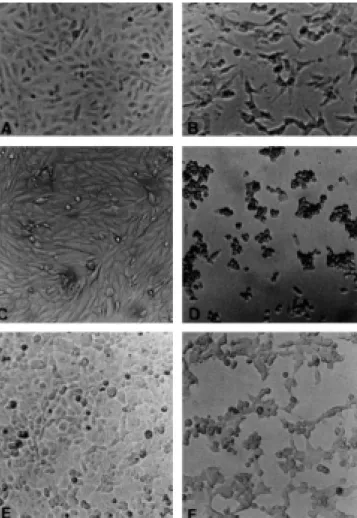
Figure 1. Effect of environmental S. marcescens supernatants number
edigraphic.com
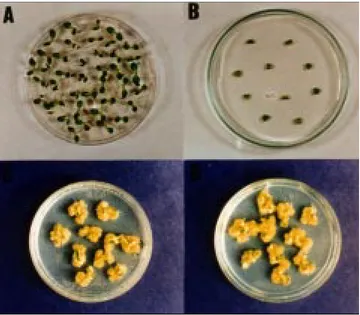
Phytotoxicity on onion bulbs. The assay was carried out
for all S. marcescens strains by injecting 1 ml of culture fil-trates or 1 ml of the same filfil-trates concentrated 10-fold with ammonium sulfate (80%) in onion bulbs. The bulbs were maintained at room temperature during 5 days with the root immersed in water. As negative control were used bulbs treated with sterile distilled water and as positive control
suspension of S. marcescens (1 x 10⁸ CFU/ml). After 5 days,
the bulbs were sliced by half and observed for decay.
Phytotoxicity on lettuce plantlets. Seeds of lettuce were
sterilized and put into 24 wells microplate, containing water-agar medium and placed in the culture room at 25 ± 3°C un-til development of plantlets. After this period, to each well was applied 0.5 ml of culture filtrates of S. marcescens
stra-S. marcescens 6/5
0 20 40 60 80 100 120
0 h 0,5 h 1 h 2 h 3 h 4 h 24 h Time (h)
Hep-2 Vero CHO
S. marcescens 980
20 40 60 80 100 120
0 h 0,5 h 1 h 2 h 3 h 4 h 24 h Time (h)
Hep-2
S. marcescens 981
0 20 40 60 80 100 120
0 h 0,5 h 1 h 2 h 3 h 4 h 24 h Time (h)
Hep-2 Vero CHO
S. marcescens 5/6
0 20 40 60 80 100 120
0 h 0,5 h 1 h 2 h 3 h 4 h 24 h Time (h)
Hep-2 Vero
Vero
Cellular viability (%)
Cellular viability (%)
Cellular viability (%)
Cellular viability (%)
0
Escobar et al. Cytotoxin production in phytopathogenic and entomopathogenic Serratia marcescens
Rev Latinoam Microbiol 2001; 43 (4): 165-170
168
ins 10-fold concentrated with ammonium sulfate (80%). Ne-gative controls included seeds treated with TSB medium and as positive control, the seeds that received suspension of
phytopathogenic, entomopathogenic and clinical S.
marces-cens. After 7 days of incubation in culture room, the
plant-lets were analyzed with a magnifying glass lens.
Preparation of antiserum. Rabbits were injected
intra-muscularly with cytotoxin produced by the clinical strain.
Samples of 50 µl of purified cytotoxin (75µg/ml) were
emulsified in an equal volume of complete Freund adju-vant, with subsequent injections in incomplete adjuvant. Animals were bled at 2-week intervals and checked for neutralizing titres. Antiserum obtained was inactivated at 56°C for 30 min.
Seroneutralization assay. The seroneutralization assay
was carried out as described previously,15 with antiserum
produced in rabbits against purified cytotoxin produced by the clinical S. marcescens strain 458. Anti-cytotoxin sera were serially diluted in Eagle medium and mixed with fil-trates of phytopathogenic and entomopathogenic S.
mar-cescens. Negative controls with preimmune serum were
also included. The mixtures were incubated for 1 h at 37°C prior to inoculation in CHO cells.
Figure 5. Figure 3.
edigraphic.com
RESULTS
Cytotoxicity assays on mammalian cells. Cytopathic
effects were observed in CHO, Vero and HEp-2 cells trea-ted with culture supernatant from phytopathogenic and entomopathogenic S. marcescens. After 24 h of incubation, there was a change from spindle-shaped cells characteristic of normal cells to round and shrivelled cells, and these changes were followed by gradual destruction of the mono-layer (Fig. 1). The HeLa cells were unresponsive and did not exhibit cytopathic effects under conditions in which other cell lines did.
Determination of cellular viability. Cellular viability of
the Vero, CHO and HEp-2 cells was determined for all S.
marcescens strains and the results were closely similar. Figure
2 shows that cell death occured rapidly, as early as 0.5 h after incubation with supernatant of 6/5, 5/6, 981 and 980 environ-mental strains. Toxicity assays on plant tissue culture. As shown in Figure 3, the culture filtrates of phytopathogenic, entomopathogenic and clinical S. marcescens were not toxic to lettuce seedling in vitro. All the explants inoculated in MS medium supplemented with several dilutions of supernatants filtrates showed callus developmented, involving all explants.
Phytotoxicity on onion bulbs assay. Figure 4 shows
the results obtained with culture filtrates of phytopathoge-nic, entomopathogenic and clinical S. marcescens inocula-ted in onion bulbs. It can be observed that apparently did not cause any tecidual alteration, in contrast with bulbs ino-culated with bacterial suspension, that shows decay .
Phytotoxicity on lettuce plantlets assay. Culture filtrates
of phytopathogenic, entomopathogenic and clinical S.
mar-cescens did not cause morphological changes on analyzed
let-tuce plantlets, even when concentrated 10 times with 80% ammonium sulfate. With the inoculation of bacterial suspen-sion, it can be observed the appearance of symptoms such as shriveling and necrotic stains on plantlets cotyledons (Fig. 5).
Seroneutralization assays. The cytotoxic activity from
phytopathogenic and entomopathogenic S. marcescens on CHO cells was completely neutralized by antiserum obtai-ned against cytotoxin of the clinical S. marcescens, with ti-tre of 1/128.
DISCUSSION
Cytotoxins have been considered as important virulence
factors in several species of bacteria6 and are detected on the
basis of the degree of the damage imposed to several
mam-malian cell lines, evidenced by morphological changes.6
Serratia marcescens isolated from clinical specimens can
produce an extracellular cytotoxin active on epithelial cell
li-nes in culture.5 However, as far as we know there are no data
on cytotoxin production by environmental S. marcescens.
In this work, supernatants of the S. marcescens isolated from plants and insects were capable of damaging mammalian cell lines, in vitro. Morphological changes on the sensitive cell lines were characterized by cell rounding and detachment (Fig. 1), culminating in cell death. The cytopathic effect ob-served resemble those described in cells inoculated with
cyto-toxin produced by clinical isolates.3
The sensitivity of several cell lines to cytotoxin produ-ced by entomopathogenic and phytopathogenic was com-pared in this study. Figure 2 shown that CHO, Vero and HEp-2 lines were similarly sensitive for all strains tested, with cytopathic effects observed as soon as 30 min of incu-bation with culture filtrates and it was confirmed by mea-sured of the cellular viability ranging 40%. Moreover, the HeLa cells were resistant to all culture filtrates, as shown in cellular viability assays.
The cytotoxic activity of the supernatants obtained from culture of phytopathogenic and entomopathogenic S.
mar-cescens on CHO cells was completely neutralized by
speci-fic polyclonal antiserum anti-cytotoxin produced by clini-cal S. marcescens. It indicates that these strains produces toxins antigenically related. Moreover, the cytotoxin shows similar biological activity on CHO cells with the cytotoxin produced by clinical S. marcescens.
The toxicity assays on plants shows that the culture su-pernatants not provoked any visible morphological chan-ges and did not affect their growth. By contrast, the plants treated with bacterial suspension shown disease symptom, such as shriveling and decay of stores bulbus in onion (Fig. 4) and lettuce plantlets (Fig. 5). On the basis of the-se results, it appears that the extracellular factor capable of damaging cultured cells was not responsible by disease in plants.
In conclusion, this study shows that phytopathogenic and entomopathogenic S. marcescens may produces a cytoto-toxin similar to produced by clinical isolates and it is toxic to different mammalian cell lines. These results are especially important for studies involving this bacterium as biological control agent. At present, more detailed study of biological activity of this cytotoxin is in progress in our laboratory.
ACKNOWLEDGMENTS
We are grateful to Dr. Rauly Maximo Rabelo Moreti (Laboratório de Cultura de Células, Centro de Genética, Biologia Molecular e Fitoquímica do Instituto Agronômico de Campinas, Brasil), for excellent technical support.
REFERENCES
1. Beriam, L.O.S., Siigaglia, C., and Rodrigues Neto, J. 1990.
Serra-tia marcescens associada a podridão de cebola armazenada.
Escobar et al. Cytotoxin production in phytopathogenic and entomopathogenic Serratia marcescens
Rev Latinoam Microbiol 2001; 43 (4): 165-170
170
2. Campbell, J.R., Diacovo, T., and Baker, C.J. 1992. Serratia
marce-scens meningitis in neonates. Ped. Infect. Dis. J. 11:881-886.
3. Carbonell, G.V., Alfieri,. A.F., Alfieri, A.A., Vidotto, M.C., Levy, C.E., Darini, A.L.C., and Yanaguita, R.M. 1997. Detection of cy-totoxic activity on Vero cells in clinical isolates of Serratia
marce-scens. Braz. J. Med. Biol. Res. 30:1291-1298.
4. Carbonell, G.V., Della Colleta, H.H.M., Yano, T., Darini, A.L.C., Levy, C.E., and Fonseca, B.A.L. 2000. Clinical relevance and viru-lence factors of pigmented S. marcescens. FEMS Immun. Med. Mi-crobiol. 28:143-149.
5. Carbonell, G.V., Fonseca, B.A.L., Figueiredo, L.T.M., Darini, A.L.C., and Yanaguita, R.M. 1996. Culture conditions affect cyto-toxin production by Serratia marcescens. FEMS Immun. Med. Mi-crobiol. 16:299-307.
6. Finlay, B.B., and Falkow, S. 1989. Common themes in microbial pathogenicity. Microbiol. Rev. 53:210-230.
7. Grimont, P.A.D., Grimont, F., and Lysenko, O. 1979. Species and biotype identification of Serratia strains associated with insects. Curr. Microbiol. 2:139-142.
8. Ito, M.F., Paradela Filho, O., Rodrigues Neto, J., Beriam, L.O.S., Longo, R.S., and Santos, J.M. 1996. Ocorrência de Serratia
marce-scens Bizio sobre lagartas de Heliothis viremarce-scens (Fabr.). Bragantia,
55(2):289-292.
9. Kaška, M., Lysenko, O., and Chaloupka, J. 1976. Exocellular pro-teases of Serratia marcescens and their toxicity to larvae of
Galle-ria mellonella. Folia Microbiol, 21:465-473.
10. Lysenko, O. 1976. Chitinase of Serratia marcescens and its toxici-ty to insects. J. Invertebr. Pathol. 27:385-386.
11. Meltz, D.J., and Grieco, M.H. 1973. Characteristics of Serratia
marcescens pneumonia. Arch. Int. Med. 132:359-364.
12. Murashige, T. and Skoog, F. 1962. A revised medium for rapid growth and bioassays with tobacco tissue culture. Physiol. Plant. 5:473-497.
13. Nakashima, A.K., McCarthy, M.A., Martone, W.J., and Anderson, R.L. 1987. Epidemic arthritis caused by Serratia marcescens and associated with a benzalkonium chloride antiseptic. J. Clin. Micro-biol. 25:1014.
14. Okuda, T., Endo, N., and Osada, Y. 1981. Outbreak of nosocomial urinary tract infections caused by Serratia marcescens. J. Clin. Mi-crobiol. 20:691-695.
15. Parreira, V.R., and Yano, T. 1998. Cytotoxin produced by
Escheri-chia coli isolated from chickens with swollen head syndrome
(SHS). Vet. Microbiol. 62:111-119.
16. Thelestam, M., and Florin, I. 1994. Assay of cytopathogenic toxins in cultured cells. Methods Enzymol 235:679-690.
17. Volkow-Fernández, P., Léon-Rosales, S.P., Sifuentes-Osornio, J., Calva-Mercado, J.J., Ruiz-Palacios, G.M., and Ma. Cérbon. 1993. Epidemia de bacteremias primarias por una cepa endémica de
Ser-ratia marcescens en una unidad de terapia intensiva. Salud Pública
de México 35:440-7.
Correspondence to:
Gleize Villela Carbonell
Departamento de Microbiologia e Imunologia, IB, Universidade Estadual de Campinas
13081-970 Campinas, SP, Brasil.