Psychology & Neuroscience
The Role of Wernicke’s Area in Language Comprehension
Alfredo Ardila, Byron Bernal, and Monica RosselliOnline First Publication, August 8, 2016. http://dx.doi.org/10.1037/pne0000060
CITATION
Ardila, A., Bernal, B., & Rosselli, M. (2016, August 8). The Role of Wernicke’s Area in Language Comprehension. Psychology & Neuroscience. Advance online publication. http://
The Role of Wernicke’s Area in Language Comprehension
Alfredo Ardila
Florida International University
Byron Bernal
Nicklaus Children’s Hospital, Miami, Florida
Monica Rosselli
Florida Atlantic University, Davie
The aphasia literature frequently states that Wernicke’s area is responsible for language understanding. The aim of this study was to pinpoint the core function of Wernicke’s area. Neuroimaging and clinical data indicate that Wernicke’s area participates in phonological and lexical recognition. A much larger brain network is involved in semantic understanding and language comprehension beyond words. We concluded that the “classic” Wernicke’s area is the core area that modulates the recognition of individual words.
Keywords: Wernicke’s area, aphasia, language understanding, word recognition, temporal lobe
The aphasia literature frequently states that Wernicke’s area is responsible for language un-derstanding. For example, Wood (1971) defined Wernicke’s area as “a region in the superior convolution of the temporal lobe of the cere-brum identified as the center for understanding speech heard” (p. 23). Nicolosi, Harryman, and Kresheck (2004) referred to Wernicke’s area as the “region in the superior convolution of the temporal lobe of the cerebrum which is identi-fied as the center for understanding oral lan-guage; corresponds approximately to Brodmann areas 22, 39, and 40” (p. 343). Even general information sources assign the function of speech comprehension to Wernicke’s area. The Encyclopedia Britannica, for example, defines Wernicke’s area as the following:
[the] region of the brain that contains motor neurons involved in the comprehension of speech. This area
was first described in 1874 by German neurologist Carl Wernicke. The Wernicke area is located in the poste-rior third of the upper temporal convolution of the left hemisphere of the brain. Thus, it lies close to the auditory cortex. This area appears to be uniquely important for the comprehension of speech sounds and is considered to be the receptive language, or language comprehension, centre. (http://www.britannica.com/science/Wernicke-area)
Many other authors and information sources consider Wernicke’s area to be involved in lan-guage understanding. This interpretation of the function of Wernicke’s area, however, is not entirely correct. Wernicke’s area probably has a more limited function—namely, recognizing phonemes and words (vocabulary). For exam-ple, Démonet et al. (1992) used positron emis-sion tomography to analyze brain activation during phonological and lexical semantic pro-cessing. The authors found that phonological processing was associated with greater activa-tion of the left superior temporal gyrus, whereas lexical semantic processing was associated with greater activity of the left middle and inferior temporal gyri. These findings are congruent with the aphasia literature, in which pathology of the left superior temporal gyrus (Brodmann area [BA] 22) causes defects in phoneme dis-crimination, whereas lexical impairments are found in cases of middle temporal gyrus (BA21) damage. Semantic deficits are frequently ob-served in cases of posterior inferior temporal– occipital (BA37) pathology (e.g., Benson &
Ar-Alfredo Ardila, Department of Communication Sciences and Disorders, Florida International University; Byron Ber-nal, Radiology Department and Miami Children’s Research Institute, Nicklaus Children’s Hospital, Miami, Florida; Monica Rosselli, Department of Psychology, Florida Atlan-tic University, Davie.
Correspondence concerning this article should be ad-dressed to Alfredo Ardila, Department of Communication Sciences and Disorders, Florida International University, 11200 Southwest 8th Street, AHC3-431B, Miami, FL 33199. E-mail: [email protected]
This
document
is
copyrighted
by
the
American
Psychological
Association
or
one
of
its
allied
publishers.
This
article
is
intended
solely
for
the
personal
use
of
the
individual
user
and
is
not
to
be
disseminated
broadly.
dila, 1996; Luria, 1976). These observations suggest that Wernicke’s area participates in two basic language recognition functions: phoneme discrimination and lexical knowledge. BA37 is not usually included in the classic Wernicke’s area. Pathology in this temporal– occipital area results in the highest number of semantic para-phasias that are found across different aphasia syndromes (Ardila & Rosselli, 1993), suggest-ing significant semantic disturbances.
Mesulam (2001) believed that Wernicke’s area is located at the left temporoparietal junc-tion. He further stated:
Wernicke’s area can be said to provide a critical gate-way for linking the sensory patterns of words to the distributed associations that encode their meaning. Its dysfunction interferes with the comprehension of words and with the translation of thoughts into words. (p. 426)
In a more recent paper (Mesulam, 2013), he suggested that the anterior temporal lobe of the left hemisphere should also be included in the language network as a third major hub that plays a critical role in language comprehension, particularly the comprehension of words that denote concrete entities. A similar perspective was proposed by Ardila, Bernal, and Rosselli (2016a).
Language understanding unquestionably re-quires the participation of more extended brain areas. As an illustration, Ferstl, Neumann, Bo-gler, and von Cramon (2008) performed a meta-analysis of 23 neuroimaging studies on text comprehension that sought to pinpoint the ex-tension of the brain network that is involved in processing language in context. The authors found that, independent of the baseline, the an-terior temporal lobes were bilaterally active. The processing of coherent compared with in-coherent text also engaged the left dorsomedial prefrontal cortex and posterior cingulate cortex. Right hemisphere activation was seen most no-tably in the analysis of contrasts that tested specific subprocesses, such as metaphor com-prehension. The results generally suggested that when language understanding is processed within a specific context, it is associated with an extensive brain activation circuit that involves not only the left but also the right hemisphere. Evidently, language comprehension within a particular context implies diverse abilities (e.g., abstraction, metaphor understanding, etc.)
be-yond the purely auditory language understand-ing of individual words.
Some authors have clearly emphasized this point. For example, Tanner (2007) explicitly emphasized that Wernicke’s area is not the cen-ter for oral language understanding—it is only an important step in language comprehension. He further stated:
Auditory comprehension is an ongoing process of re-ceiving environmental information and continuously adjusting the parameters of what is to be perceived and associated. An individual’s unique learning and mem-ory experiences play important roles in this process. This process can virtually engage the brain as a whole and the totality of a person’s mind. (Tanner, 2007, p. 66)
Wernicke (1970) himself pointed out that the left temporal lobe is involved in recognizing words:
The whole area of the convolution encircling the Syl-via fissure, in association with the cortex of the insula, serves as [the] speech center. The first frontal gyrus, being motor, is the center for representation of move-ments, and the first temporal gyrus, being sensory, is the center for word-images. (p. 280)
Binder (2015) stated that although Wer-nicke’s area has been traditionally presumed to be involved in language comprehension, mod-ern imaging and neuropsychological studies in-dicate that this region plays a much larger role in speech production. Indeed, the posterior peri-sylvian region, usually referred to as Wer-nicke’s area, does not support the main function that is traditionally ascribed to it (i.e., speech comprehension). According to Binder, language comprehension requires a widely distributed se-mantic network. This point of view is congruent with the proposals of Ferstl et al. (2008) and Tanner (2007).
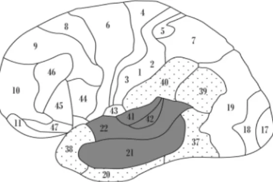
Ardila, Bernal, and Rosselli (2016b) sug-gested that there is a core Wernicke’s area that includes not only BA22 and BA21 (as usually suggested) but also BA41 and BA42. This core Wernicke’s area participates in the phonologi-cal and lexiphonologi-cal recognition of words. There is also a fringe or peripheral zone around this core Wernicke’s area that is involved in language associations. The fringe or peripheral Wer-nicke’s area corresponds to BA20, BA37, BA38, BA39, and BA40 (i.e., the “extended Wernicke’s area”). The classic Wernicke’s area (i.e., the “core Wernicke’s area”) is only in-volved in the phonological and lexical
recogni-2 ARDILA, BERNAL, AND ROSSELLI
This
document
is
copyrighted
by
the
American
Psychological
Association
or
one
of
its
allied
publishers.
This
article
is
intended
solely
for
the
personal
use
of
the
individual
user
and
is
not
to
be
disseminated
tion of words (see Figure 1)—that is, in discrim-inating the phonemes that are included in words and recognizing the sequence of phonemes cor-responding to a word. Semantic recognition re-quires a more extended network, including at least the adjacent inferior temporal– occipital area, BA37.
DeWitt and Rauschecker (2013) proposed that Wernicke’s area may be better construed as two cortical modules (i.e., an auditory word-form area in the auditory ventral stream and an “inner speech area” in the auditory dorsal stream), thus emphasizing the heterogeneity of this language processing area. Dronkers, Red-fern, and Knight (2000) proposed that tradi-tional language areas, such as Wernicke’s area, may serve somewhat different functions than originally described. They suggested that the analysis of more specific deficits and their ana-tomical correlates can lead to improvements in the mapping of language functions in the brain. The transhemispheric contribution of the right hemisphere to language comprehension should also be noted. Prosody importantly con-tributes to the meaning that is conveyed by the same text. This can vary between languages because some languages more critically depend on this feature, such as tonal languages (e.g., Mandarin, Cantonese, and Vietnamese). In non-tonal languages, intonation adds emotional color and meaning to text. With intonation, we can turn a declarative statement into an inter-rogative statement, convey resentment with
sar-casm, or even mean the opposite (i.e., irony). Prosody plays an important role in adding an affective component to sentence comprehen-sion. Interestingly, the same classic dissociation between expressive and receptive functions that exists in the left hemisphere for declarative lan-guage has been proposed to exist in the right hemisphere for prosody (Dahan, 2015; Wein-traub, Mesulam, & Kramer, 1981). However, a recent study reported rather bilateral hemi-spheric involvement. Emotional speech com-prehension, for example, has been found to in-volve areas that are adjacent to the superior temporal sulcus bilaterally (Hervé, Razafi-mandimby, Jobard, & Tzourio-Mazoyer, 2013), although this finding could be partially ex-plained by other variables. Thus, at least one study on prosodic decoding lateralization found that left hemisphere involvement was more pre-cisely associated with the verbal complexity of the prosodic emotional stimuli than with pros-ody itself (Mitchell & Ross, 2008). Nonethe-less, an undeniable fact is the substantive con-tribution of the right hemisphere to sentence comprehension through prosodic decoding.
Therefore, it is crucial to emphasize that the “classic” Wernicke’s area is the core area in the recognition of individual words. Ultimately, a much larger brain network is involved in lan-guage understanding.
References
Ardila, A., Bernal, B., & Rosselli, M. (2016a). How extended is Wernicke’s area? Meta-analytic con-nectivity study of BA20 and integrative proposal.
Neuroscience Journal, 2016,Article ID 4962562. http://dx.doi.org/10.1155/2016/4962562
Ardila, A., Bernal, B., & Rosselli, M. (2016b). How localized are language brain areas? A review of Brodmann areas involvement in oral language. Ar-chives of Clinical Neuropsychology, 31,112–122. http://dx.doi.org/10.1093/arclin/acv081
Ardila, A., & Rosselli, M. (1993). Language devia-tions in aphasia: A frequency analysis.Brain and Language, 44,165–180. http://dx.doi.org/10.1006/ brln.1993.1011
Benson, D. F., & Ardila, A. (1996). Aphasia: A clinical perspective. New York, NY: Oxford Uni-versity Press.
Binder, J. R. (2015). The Wernicke area: Modern evidence and a reinterpretation. Neurology, 85,
2170 –2175. http://dx.doi.org/10.1212/WNL .0000000000002219
Figure 1. A “core Wernicke’s area” is involved in pho-neme and word recognition, roughly corresponding to BA41, BA42, BA21, and BA22. An “extended Wernicke’s area” or “Wernicke’s system” also includes BA20, BA37, BA38, BA39, and BA40; these Brodmann areas (BAs) participate in word associations (i.e., associating words with other information).
This
document
is
copyrighted
by
the
American
Psychological
Association
or
one
of
its
allied
publishers.
This
article
is
intended
solely
for
the
personal
use
of
the
individual
user
and
is
not
to
be
disseminated
Dahan, D. (2015). Prosody and language comprehen-sion. Wiley Interdisciplinary Reviews: Cognitive Science, 6, 441– 452. http://dx.doi.org/10.1002/ wcs.1355
Démonet, J. F., Chollet, F., Ramsay, S., Cardebat, D., Nespoulous, J. L., Wise, R., . . . Frackowiak, R. (1992). The anatomy of phonological and semantic processing in normal subjects.Brain: A Journal of Neurology, 115, 1753–1768. http://dx.doi.org/10 .1093/brain/115.6.1753
DeWitt, I., & Rauschecker, J. P. (2013). Wernicke’s area revisited: Parallel streams and word process-ing.Brain and Language, 127,181–191. http://dx .doi.org/10.1016/j.bandl.2013.09.014
Dronkers, N. F., Redfern, B., & Knight, R. (2000). The neural architecture of language disorders. In M. S. Gazzaniga (Ed.),The new cognitive neuro-sciences (pp. 949 –958). Cambridge, MA: MIT Press.
Ferstl, E. C., Neumann, J., Bogler, C., & von Cra-mon, D. Y. (2008). The extended language net-work: A meta-analysis of neuroimaging studies on
text comprehension. Human Brain Mapping, 29,
581–593. http://dx.doi.org/10.1002/hbm.20422 Hervé, P. Y., Razafimandimby, A., Jobard, G., &
Tzourio-Mazoyer, N. (2013). A shared neural sub-strate for mentalizing and the affective component
of sentence comprehension. PloS ONE, 8(1),
e54400.
Luria, A. R. (1976).Basic problems of neurolinguis-tics. New York, NY: De Gruyter Mouton. http:// dx.doi.org/10.1515/9783110800159
Mesulam, M. M. (2001). Primary progressive apha-sia. Annals of Neurology, 49, 425– 432. http://dx .doi.org/10.1002/ana.91
Mesulam, M. (2013). Primary progressive aphasia: A dementia of the language network. Dementia & Neuropsychologia, 7, 2–9. http://dx.doi.org/10 .1590/S1980-57642013DN70100002
Mitchell, R. L., & Ross, E. D. (2008). fMRI evidence for the effect of verbal complexity on lateralisation of the neural response associated with decoding
prosodic emotion. Neuropsychologia, 46, 2880 –
2887. http://dx.doi.org/10.1016/j.neuropsycholo-gia.2008.05.024
Nicolosi, L., Harryman, E., & Kresheck, J. (2004).
Terminology of communication disorders: Speech–language– hearing. Philadelphia, PA: Lip-pincott Williams & Wilkins.
Tanner, D. C. (2007). A redefining Wernicke’s area: Receptive language and discourse semantics. Jour-nal of Allied Health, 36,63– 66.
Weintraub, S., Mesulam, M. M., & Kramer, L. (1981). Disturbances in prosody: A right-hemi-sphere contribution to language.Archives of Neu-rology, 38, 742–744. http://dx.doi.org/10.1001/ archneur.1981.00510120042004
Wernicke, C. (1970). The aphasic symptom-complex: A psychological study on an anatomical basis.Archives of Neurology, 22,280 –282. http:// dx.doi.org/10.1001/archneur.1970.004802100 90013
Wood, K. (1971). Terminology and nomenclature. In L. E. Travis (Ed.),Handbook for speech pathology and audiology (pp. 3–26). Englewood Cliffs, NJ: Prentice Hall.
Received February 18, 2016 Revision received June 15, 2016
Accepted June 15, 2016 䡲
4 ARDILA, BERNAL, AND ROSSELLI
This
document
is
copyrighted
by
the
American
Psychological
Association
or
one
of
its
allied
publishers.
This
article
is
intended
solely
for
the
personal
use
of
the
individual
user
and
is
not
to
be
disseminated