PROGRAMA DE DOCTORADO EN CIENCIAS DE LA VISIÓN
TESIS DOCTORAL:
DESARROLLO DE UNA GUÍA CLÍNICA BASADA
EN LA EVIDENCIA PARA LA ADAPTACIÓN DE
LENTES DE CONTACTO PERMEABLES
AL GAS DE DISEÑO CORNEAL
EN QUERATOCONO
DEVELOPMENT OF AN EVIDENCE-BASED CLINICAL
PRACTICE GUIDELINE TO FIT CORNEAL GAS
PERMEABLE CONTACT LENSES IN KERATOCONUS
Presentada por Dña. Sara Ortiz Toquero para optar al
grado de Doctora por la Universidad de Valladolid
Dirigida por:
AUTORIZACIÓN DEL DIRECTOR DE TESIS
(Art. 7.2 de la Normativa para la presentación y defensa de la Tesis Doctoral en la UVa)
Impreso 1T
D. RAÚL MARTÍN HERRANZ, con D.N.I. 50.447.910-D, Investigador principal del Grupo de
Investigación en Optometría del Instituto Universitario de Oftalmobiología Aplicada de la
Universidad de Valladolid (IOBA) y Profesor Contratado Doctor del Departamento de Física
Teórica, Atómica y Óptica de la Facultad de Ciencias de la Universidad de Valladolid, con
dirección a efecto de notificaciones “Campus Miguel Delibes. Paseo de Belén 7, 47011,
Valladolid” y con e-mail “[email protected]”, como Director de la Tesis Doctoral titulada
“Desarrollo de una guía clínica basada en la evidencia para la adaptación de lentes de contacto
permeables al gas de diseño corneal en queratocono” realizada por Dª Sara Ortiz Toquero,
alumna del Programa de Doctorado en Ciencias de la Visión impartido por el IOBA,
AUTORIZA su presentación, considerando que es APTA para su defensa.
Valladolid, 14 de marzo de 2017
El Director de la Tesis,
Fdo.: Dr. Raúl Martín Herranz
SR/SRA. PRESIDENTE/A DE LA COMISIÓN DE DOCTORADO
Fd
d
Fd
Fd
Fd
Fd
d
d
Fd
d
d
d
Fd
d
d
d
Fd
d
d
d
d
d
d
d
d
d
d
d
d
Fd
d
Fd
d
d
F
d
d
d
d
F
F
d
d
F
o.
o
o.
.
.
.
.
.
.
.
.
:
:
:
:
:
:
:
:
:
:
:
:
:
:
:
:
:
D
D
D
D
D
D
D
D
D
D
D
D
D
D
D
D
Dr
D
D
D
D
D
D
D
D
D
. Ra
a
a
a
a
a
a
a
a
a
a
a
a
a
ú
ú
ú
ú
ú
ú
ú
ú
ú
ú
ú
ú
ú
ú
ú
ú
ú
ú
ú
ú
ú
úl
úl
ú
ú
ú
ú
ú
M
M
M
M
M
M
M
M
M
M
M
M
M
M
M
M
M
M
M
M
M
M
M
M
M
M
M
a
a
a
a
a
a
a
a
a
a
a
a
a
a
a
a
ar
tí
í
í
í
í
í
í
í
í
í
í
í
í
í
í
í
í
í
í
í
í
í
í
í
n Herr
r
r
rr
rr
rr
r
r
r
rr
rr
r
r
rr
rr
rr
rr
rr
rr
r
r
r
r
anz
Firmado digitalmente por NOMBRE MARTIN HERRANZ RAUL - NIF 50447910D
A mi abuela Vicenta
Ahora ya sí que me puedes llamar “doctora”
“Las personas tranquilas y silenciosas,
son las que tienen las mentes más fuertes y ruidosas”
Stephen Hawking
“Todo parece imposible hasta que se hace”
Nelson Mandela
“Nuestra recompensa se encuentra en el esfuerzo y no
en el resultado, un esfuerzo total es una victoria completa”
Mahatma Gandhi
“Your work is going to fill a large part of your life, and the
only way to be truly satisfied is to do what you believe
is great work. And the only way to do great work is to
love what you do. If you haven't found it yet, keep looking.
Don't settle”
IX
XIII
XV
XVII
XXIII
Capítulo
Índice
Organización
de la tesis
Abreviaturas
Capítulo 0: ÍNDICE
3
Capítulo 0: ÍNDICE
Capítulo 0: ORGANIZACIÓN DE LA TESIS
7
Capítulo 0: ORGANIZACIÓN DE LA TESIS
Capítulo 0: ORGANIZACIÓN DE LA TESIS
Capítulo 0: ORGANIZATION OF THE DOCTORAL THESIS
13
Capítulo 0: ORGANIZATION OF THE DOCTORAL THESIS
Capítulo 0: ORGANIZATION OF THE DOCTORAL THESIS
17
Capítulo 0: ABREVIATURAS
19
Capítulo
Síntesis
general
Capítulo 1: SÍNTESIS GENERAL - Estado actual del tema
23
Capítulo 1: SÍNTESIS GENERAL - Estado actual del tema
Capítulo 1: SÍNTESIS GENERAL - Estado actual del tema
Capítulo 1: SÍNTESIS GENERAL - Estado actual del tema
Capítulo 1: SÍNTESIS GENERAL - Estado actual del tema
Capítulo 1: SÍNTESIS GENERAL - Estado actual del tema
Capítulo 1: SÍNTESIS GENERAL - Estado actual del tema
Capítulo 1: SÍNTESIS GENERAL - Estado actual del tema
Capítulo 1: SÍNTESIS GENERAL - Estado actual del tema
Capítulo 1: SÍNTESIS GENERAL - Estado actual del tema
Capítulo 1: SÍNTESIS GENERAL - Estado actual del tema
Capítulo 1: SÍNTESIS GENERAL - Justificación
45
Capítulo 1: SÍNTESIS GENERAL - Hipótesis
47
Capítulo 1: SÍNTESIS GENERAL - Objetivos
49
Capítulo 1: SÍNTESIS GENERAL – Material y métodos
51
Capítulo 1: SÍNTESIS GENERAL – Material y métodos
-Capítulo 1: SÍNTESIS GENERAL – Material y métodos
55
-Capítulo 1: SÍNTESIS GENERAL – Material y métodos
57
-Capítulo 1: SÍNTESIS GENERAL – Material y métodos
Capítulo 1: SÍNTESIS GENERAL – Resultados
61
Capítulo 1: SÍNTESIS GENERAL – Resultados
Capítulo 1: SÍNTESIS GENERAL – Resultados
Capítulo 1: SÍNTESIS GENERAL – Resultados
Capítulo 1: SÍNTESIS GENERAL – Resultados
Capítulo 1: SÍNTESIS GENERAL – Resultados
Capítulo 1: SÍNTESIS GENERAL - Discusión
73
Capítulo 1: SÍNTESIS GENERAL - Discusión
Capítulo 1: SÍNTESIS GENERAL - Discusión
Capítulo 1: SÍNTESIS GENERAL – Conclusiones
79
Capítulo
Summary of the
Doctoral Thesis
Capítulo 2: SUMMARY OF THE DOCTORAL THESIS – State of the art
83
Capítulo 2: SUMMARY OF THE DOCTORAL THESIS – State of the art
Capítulo 2: SUMMARY OF THE DOCTORAL THESIS – State of the art
Capítulo 2: SUMMARY OF THE DOCTORAL THESIS – State of the art
Capítulo 2: SUMMARY OF THE DOCTORAL THESIS – State of the art
Capítulo 2: SUMMARY OF THE DOCTORAL THESIS – State of the art
Capítulo 2: SUMMARY OF THE DOCTORAL THESIS – State of the art
Capítulo 2: SUMMARY OF THE DOCTORAL THESIS – State of the art
Capítulo 2: SUMMARY OF THE DOCTORAL THESIS – Motivation
101
Capítulo 2: SUMMARY OF THE DOCTORAL THESIS – Hypothesis
103
Capítulo 2: SUMMARY OF THE DOCTORAL THESIS – Objectives
105
Capítulo 2: SUMMARY OF THE DOCTORAL THESIS – Methods
107
Capítulo 2: SUMMARY OF THE DOCTORAL THESIS – Methods
-Capítulo 2: SUMMARY OF THE DOCTORAL THESIS – Methods
111
-Capítulo 2: SUMMARY OF THE DOCTORAL THESIS – Methods
Capítulo 2: SUMMARY OF THE DOCTORAL THESIS – Results
115
Capítulo 2: SUMMARY OF THE DOCTORAL THESIS – Results
Capítulo 2: SUMMARY OF THE DOCTORAL THESIS – Results
Capítulo 2: SUMMARY OF THE DOCTORAL THESIS – Results
Capítulo 2: SUMMARY OF THE DOCTORAL THESIS – Results
Capítulo 2: SUMMARY OF THE DOCTORAL THESIS – Discussion
125
Capítulo 2: SUMMARY OF THE DOCTORAL THESIS – Discussion
Capítulo 2: SUMMARY OF THE DOCTORAL THESIS – Conclusions
129
Capítulo
Impacto de la
corrección con LC
RPG en la calidad de
vida de los pacientes
con queratocono
CHAPTER 3: The influence of the refractive correction
on the quality of life in keratoconus patients
CAPÍTULO 3: Impacto de la corrección con LC RPG en la calidad de vida de los pacientes con queratocono
133
3. Impacto de la
corrección con LC RPG
en la calidad de vida de
The influence of the refractive correction on the vision-related
quality of life in keratoconus patients
Sara Ortiz-Toquero1,2,3•Sofia Perez3•Guadalupe Rodriguez1,2,3•Victoria de Juan1,2,3•
Agustin Mayo-Iscar4•Raul Martin1,2,3,4
Accepted: 24 August 2015 / Published online: 3 September 2015 ÓSpringer International Publishing Switzerland 2015
Abstract
Purpose The aim of this study was to assess the impact of
refractive correction [spectacles vs rigid gas-permeable contact lenses (RGP CLs)] on the vision-related quality of life (VR-QoL) obtained with the standardized question-naire, NEI-VFQ-25, in keratoconus patients compared with healthy myopic subjects.
Methods The Spanish version of NEI-VFQ-25 was
administered two consecutive times to 25 keratoconus patients (RGP CL wearers) and 25 healthy myopic subjects (RGP and soft CL wearers). The first time was to assess the VR-QoL for spectacle wearing, such as those for refractive correction, and the second time was for CL wearing.
Results Keratoconus patients showed a lower VR-QoL
impairment (P\0.01) than healthy subjects in the total and all subscale score of NEI-VFQ-25 related to wearing spectacles. With CL correction, keratoconus patients showed a VR-QoL improvement with statistically signifi-cant differences (P\0.04) in only four subscales,
including distance activities, mental health, color vision and peripheral vision, compared with healthy subjects. In the keratoconus group, compared to spectacle use, CL wear improved the VR-QoL score (P=0.01) and all subscales except for ocular pain (P\0.01) and mental health (P=0.25).
Conclusions The use of the NEI-VFQ-25 to explore the
difference in the VR-QoL between healthy subjects and patients with keratoconus provides further evidence of improved VR-QoL with RGP CL wear compared with spectacles in keratoconus patients. RGP CL management in keratoconus patients could minimize the impact of the disease on the patient’s well-being.
Keywords KeratoconusQuality of LifeRefractive correctionRigid gas-permeable contact lens management
Introduction
Keratoconus is a bilateral, asymmetric and progressive corneal disorder, resulting in myopia, irregular astigmatism and reduced vision related to central and paracentral cor-neal thinning, steepening and scarring [1–3]. This ectatic condition affects between 50 and 230 individuals per 100,000 people [3] and commonly appears during the second decade of life and puberty, progressing until the fourth decade of life, when it usually stabilizes [1–3]. In the early stages, keratoconus can be managed with spectacles or soft contact lenses, but as keratoconus progresses, the irregular astigmatism often requires rigid gas-permeable (RGP) contact lenses (CL) that can improve the best-cor-rected visual acuity (BCVA) [3].
The National Eye Institute-Vision Function Question-naire (NEI-VFQ-25) developed by the National Eye & Raul Martin
1 Departamento de Fı´sica Teo´rica, Ato´mica y O´ ptica,
Universidad de Valladolid, Paseo de Bele´n, 7 - Campus Miguel Delibes, 47011 Valladolid, Spain
2 Instituto Universitario de Oftalmobiologı´a Aplicada (IOBA),
Universidad de Valladolid, Paseo de Bele´n, 17 - Campus Miguel Delibes, 47011 Valladolid, Spain
3 Optometry Research Group, IOBA Eye Institute, School of
Optometry, University of Valladolid, Valladolid, Spain
4 Departamento de Estadı´stica e Investigacio´n Operativa e
IMUVA, Universidad de Valladolid, Paseo de Bele´n, 7 - Campus Miguel Delibes, 47011 Valladolid, Spain
123
Qual Life Res (2016) 25:1043–1051uveitis [7], post-retinal detachment surgery [8], dry eyes [9] and others in patients’ VR-QoL.
Using the NEI-VFQ-25, patients with keratoconus show a significantly disproportional impaired VR-QoL [4, 10,
11] that worsens with time [12]. Moreover, these patients show similar results in the NEI-VFQ-25 to those reported for patients with advanced (categories 3 and 4) age-related macular degeneration [13].
The strongest associations with a lower VR-QoL with low visual acuity (worse than 20/40) and steeper corneal curvature ([52 D) have been described to greatly influence the patient’s attitude toward his/her disease and perception of its impact on visual function when there is an increase in the 3 D corneal curvature or a visual acuity decrease of higher than ten letters occur over time [12].
However, all studies on the VR-QoL in keratoconus patients consider the BCVA obtained indiscriminately with spectacles or CL [10, 11, 14–18]. To the best of our knowledge, no studies have previously examined the effect of keratoconus correction (spectacles or RGP CL) over the VR-QoL score.
As keratoconus patients show different visual acuity corrected with spectacles or with RGP CL wear, the aim of this study was to assess the impact of refractive correction (spectacles vs RGP CL) on the VR-QoL obtained with the standardized questionnaire, NEI-VFQ-25, in keratoconus patients. As a second objective, we compared the VR-QoL score obtained with spectacles and CL in healthy myopic (non-keratoconus) subjects to assess whether refractive correction shows different impact in keratoconus patients. These data would be crucial in clinical trials that evaluate the impact of new treatment modalities for keratoconus, such as cross-linking, intracorneal rings and corneal grafts, on the VR-QoL or in future design and validation of patient-reported outcomes (PROs) to assess the VR-QoL in keratoconus patients.
Methods
This is an observational and non-randomized study. Informed consent was obtained from each subject after
corneas and other eye disorders.
The keratoconus group included patients who were successfully fitted with RGP CLs (KAKC RGP design, Conoptica-Hecht Contactlinsen GmbH, Baden-Wu¨rttem-bert, Germany) in at least 1 year of comfortable use. The healthy group was recruited in 1 week and included non-symptomatic myopic contact lens wearers (RGP or soft lenses) with BCVAC 20/20 (Snellen chart) with myopia and astigmatism lower than 3.00 D. All subjects in both groups were aged 18 years or older, free of cognitive impairment, living independently and Spanish speaking.
Patients with any active ocular surface disease (e.g., significant dry eye symptoms or keratitis), corneal opaci-ties, pellucid marginal corneal degeneration, glaucoma, use of medication that could affect ocular physiology or a history of any type of ocular surgery were excluded.
Independent corneal specialists confirmed the diagnoses of keratoconus after a completed eye examination, which included Scheimpflug topographical analysis (Galilei, Ziemer, Port, Switzerland) and biomicroscopy examination (with a demonstration of at least one biomicroscopic sign, including Vogt´s striae, Fleischer´s ring, corneal thinning or scarring). The keratoconus stage has been identified using the Amsler-Krumeich classification [19]. The keratoconus eyes were designated as better and worse eyes based on the BCVA, simulated keratometry and stage of Amsler-Krumeich classification.
verified with the Kolmogorov–Smirnov test (P\0.05 indicated that the data were nonparametric-distributed).
The total and subsection scores, ranging from 0 (worst) to 100 (best), were calculated for the questionnaire VFQ-25 as directed by the National Health Institute (following the standard method recommended by the developers) [20] for determining the mean score (±standard deviation) for healthy and keratoconus patients wearing spectacles or CL. Additionally, a Rasch analysis with the algorithm proposed by Massof [20] to be used with small sample sizes that would not be able to obtain reliable estimates with standard Rasch analysis software [21] was conducted. The correla-tion between the standard method recommended by the developers of the NEI-VFQ-25 [22] and the Massof’s algorithm [20] for approximating Rasch analysis was cal-culated with Spearman coefficient. The total standard and the Massof’s algorithm score obtained with spectacles and CL in each study group was compared with the Wilcoxon test (P\0.05 considered statistically significant) and between healthy and keratoconus groups with the Mann– Whitney U test (P\0.05 considered statistically signifi-cant). Finally, the difference between CL and spectacle wear total score was calculated with the standard method and with Massof’s algorithm for each study group and compared with Wilcoxon test (P\0.05 considered statis-tically significant).
The visual acuity and the subsection standard scores obtained with spectacles and CL in each study group were compared with the Wilcoxon test (P\0.05 considered statistically significant). Visual acuity between healthy and keratoconus groups was also compared with the Mann– Whitney U test (P\0.05 considered statistically signifi-cant). Subsection standard scores in healthy and kerato-conus groups were compared with the Mann–Whitney U test (P\0.05 considered statistically significant). Finally, differences between the degree of keratoconus (total standard score) was assessed with a nonparametric Kruskal–Wallis ANOVA (P\0.05 considered statistically significant).
Results
Subjects
Fifty patients (23 women and 27 men) were included in the study. The mean age of the total sample was 33.7±11.2 years (range 18–58 years).
Twenty-five subjects (17 women and 8 men) comprised the healthy group with a mean age of 30.3±11.3 years (range 18–55 years) and a mean spherical equivalent refractive error of-3.63±1.69 D (range from-1.50 D to -8.00 D). The mean of simulated keratometry reading was
7.83±0.40 mm. The BCVA with spectacles was 1.00±0.00, and CL was 1.05±0.08 (Snellen chart) (P=0.01). Healthy group showed better BCVA with spectacles and contact lenses than obtained by the kerato-conus patients (P\0.01). Twenty-four subjects were soft CL wearers, and one subject was an RGP CL wearer. The mean number of daily hours of CL use was 8.72±2.93 (range 3–15 h) and 5.44±2.16 days per week (range 1–7 days).
Twenty-five subjects (six women and 19 men) com-prised the keratoconus group with a mean age of 37.1±10.1 years (range 22–58 years) and a mean spher-ical equivalent refractive error of-4.56±3.68 D (range from-0.25 to-11.50 D), and the BCVAs with spectacles and RGP CLs were 0.60±0.30 and 0.93 ±0.17 (Snellen chart), respectively (P\0.01). According to the Amsler-Krumeich classification, there were 13 eyes in the stage 1; 17 eyes in the stage 2; 15 eyes stage 3; and only four eyes in the stage 4.
All subjects were RGP CL wearers. In the better eye, the mean of the simulated keratometry reading was 7.35±0.49 mm and the BCVAs with spectacles and RGP CLs were 0.74±0.26 and 0.96±0.18 (P\0.01) (Snellen visual chart), respectively. In the worse eye, the mean of the simulated keratometry reading was 6.99±0.52 mm and the BCVAs with spectacles and RGP CLs were 0.45±0.26 and 0.90±0.16 (P\0.01) (Snellen visual chart), respectively. The mean number of daily hours of CL wear was 9.36±4.73 (range 1–17 h) 6.08±1.53 days per week (range 2–7 days).
VR-QoL: keratoconus versus healthy subjects
Total score of NEI-VFQ-25 and all subscales scores were lower in the keratoconus group than in the healthy group with both refractive corrections (spectacles and CLs) (Table1), except the general vision subscale with CLs when keratoconus patients showed a slightly better score than healthy subjects (nonsignificant,P=0.38 Table1).
Healthy subjects showed a high score (P\0.01) in the VR-QoL than in keratoconus patients in the total and all subscales for spectacle wear. Nevertheless, these differ-ences reduced with the use of CL, and there were statisti-cally significant differences (P\0.04) in the VR-QoL in only the following four subscales: distance activities, mental health, color vision and peripheral vision.
VR-QoL: spectacles versus CL wear
In the keratoconus group, the total and all subscales scores for CLs were higher than for spectacles, except in ocular pain and mental health (Table1). The ocular pain subscale with spectacles showed a better score than with CLs (P\0.01) in both study groups, but the peripheral vision
Qual Life Res (2016) 25:1043–1051 1045
Spectacles 76.00±20.00 40–100 42.50±21.52 20–100 \0.01
CL 68.00±16.33 40–100 72.00±22.36 20–100 0.38
P* 0.02 \0.01
Ocular pain
Spectacles 92.50±11.97 50–100 75.00±21.80 37.50–100 \0.01
CL 71.00±18.65 37.50–100 63.50±27.46 12.50–100 0.46
P* \0.01 \0.01
Near activities
Spectacles 94.00±14.13 41.67–100 57.67±27.94 0–100 \0.01
CL 91.00±11.51 58.33–100 81.33±20.73 33.33–100 0.13
P* 0.34 \0.01
Distance activities
Spectacles 93.33±9.32 66.77–100 51.66±30.90 0–100 \0.01
CL 91.67±8.33 75–100 80.00±18.79 41.67–100 0.04
P* 0.57 \0.01
Social functioning
Spectacles 96.50±7.67 75–100 71.88±29.32 0–100 \0.01
CL 96.50±7.67 75–100 89.00±17.79 37.50–100 0.13
P* 1.00 \0.01
Mental health
Spectacles 73.90±16.85 25–90 54.33±25.91 7.5–87.50 \0.01
CL 73.35±14.46 25–87.50 50.80±19.40 10–75 \0.01
P* 0.53 0.25
Role difficulties
Spectacles 89.50±12.85 62.50–100 53.00±38.74 0–100 \0.01
CL 90.00±13.50 50–100 75.00±29.97 0–100 0.12
P* 0.65 \0.01
Dependency
Spectacles 96.87±7.27 75–100 68.40±39.70 0–100 \0.01
CL 98.00±5.52 75–100 87.00±25.00 0–100 0.07
P* 0.32 \0.01
Driving
Spectacles 85.78±23.71 0–100 48.75±31.68 0–100 \0.01
CL 82.58±20.77 75–100 79.76±20.34 33.33–100 0.71
P* 0.16 \0.01
Color vision
Spectacles 98.00±10.00 50–100 83.33±26.24 0–100 \0.01
CL 100±0.00 100–100 95.00±10.21 75–100 0.02
P* 0.32 0.01
Peripheral vision
score was better with CLs (P\0.01) than with spectacles in healthy and keratoconus subjects. The mental health subscale showed non-statistically significant differences (P=0.25) between spectacles and CLs in both groups.
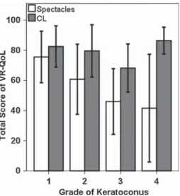
An additional analysis in each keratoconus group showed a lower score in the spectacles VR-QoL than that obtained with CLs (Fig.1). The spectacles score was significantly worse for a high keratoconus degree (P\0.01 Kruskal–Wallis ANOVA). However, the CL score was nonsignificantly different (P=0.06 Kruskal– Wallis ANOVA) between the keratoconus groups.
Standard and Massof’s algorithm (Rasch analysis) scoring comparison
High correlation between the Massof’s algorithm and the NEI-VFQ-25 standard score in contact lens (0.93 Spear-man correlation coefficient) and spectacle wear (0.97 Spearman correlation coefficient) was found. The differ-ences between CL and spectacles wear in healthy and keratoconus patients show the same trend with the standard method (P=0.41 in healthy andP\0.01 in keratoconus
group) or with the Massof’s algorithm (P=0.17 in healthy and P\0.01 in keratoconus group). Both study groups showed statistically significant differences (standard method P\0.01 and Massof’s algorithm P\0.01) in spectacles wear score; however, CL wear score showed non-statistically significant differences (standard method P=0.07 and Massof algorithmP=0.14).
Finally, the difference between CL and spectacle wear total score showed statistically significant differences between healthy and keratoconus patients with the standard method (P\0.01) and with Massof’s algorithm (P\0.01).
Discussion
In the early stages of keratoconus, the refractive error can be corrected with spectacles, but, as it progresses, corneal irregularities induce higher-order aberrations that cannot be corrected with traditional ophthalmic lenses. To compen-sate for these corneal irregularities, it is necessary to pre-scribe RGP CLs, which reduces visual distortion due to the tear that remains between the CL and the anterior surface of the cornea, correcting most of the corneal higher-order aberration-induced keratoconus and providing a generally higher BCVA than that obtained with spectacles [2, 3]. Therefore, it is reasonable to think that the VR-QoL of these patients may be affected by the method of refractive error correction using (spectacles or CL), but to the best of our knowledge, there are no previous reports of the effect of refractive correction in VR-QoL in keratoconus patients. For this reason, our aim in this study was to explore the influence of the refractive correction (spectacles or CL) on the VR-QoL in keratoconus patients assessed with the NEI-VFQ-25. We compared the VR-QoL in healthy myopic subjects to evaluate the effect of refractive correction in non-keratoconus patients. In taking this approach, we may have detected a significantly understated VR-QoL with the use of spectacles in the keratoconus group.
There are several reports regarding the impact of kera-toconus in VR-QoL using NEI-VFQ-25 [10–12, 15, 16,
18]. The National Eye Institute developed the NEI-VFQ to assess health-related quality of life of patients with visual impairments [23] showing off a high correlation between Table 1continued
Healthy (n=25) Keratoconus (n=25) P**
Mean±SD Min–max Mean±SD Min–max
P* 0.41 \0.01
P* Wilcoxon test (P\0.05 considered statistically significant).P** Mann–WhitneyUtest (P\0.05 considered statistically significant)
Fig. 1 NEI-VFQ-25 total score in the different grades of keratoconus with spectacles and CL correction. The CL score was not significantly different (P=0.06 Kruskal–Wallis ANOVA) for different kerato-conus degrees; however, the spectacles score decreased significantly (P\.01 Kruskal–Wallis ANOVA) with the stage of keratoconus
Qual Life Res (2016) 25:1043–1051 1047
preferable [26–28], a Rasch analysis using the Massof approximation [20] was conducted because the small sample size would not be able to obtain reliable estimates with standard Rasch analysis software [21]. We found a great correlation between the standard method recom-mended by the developers of the NEI-VFQ-25 [22] and the Massof’s algorithm [20,21,28] but the small sample size included in our study requires that these results could be interpreted with caution and further studies with high sample sizes could be necessary.
The use of the NEI-VFQ-25 has some limitations. This questionnaire has a dysfunctional rating scale to the response options, using too many categories, long descriptors for categories, using neutral and conceptually overlapping categories and a branch questions design in some items, avoiding the evidence-based guidelines for rating scales design [29]. Some reports, in AMD patients, found a weak validation that is not included modern psy-chometric methods such as item response theory models or Rasch analysis [30]. Others in patients undergoing cataract surgery [29] suggest that the NEI-VFQ-25 is more com-plicated than others (such as VF-14 or CatScale). However, there are not previous reports that compare the respon-siveness of the questionnaire in terms of effect size or difficulty with other questionnaires in keratoconus patients as have been described in cataract surgery patients [31]. These limitations could influence in the item calibration and provoke some loss of measurement quality, affecting to the quality of clinical studies and their results. But because it is difficult to directly compare research findings using different PROs [32], we used the NEI-VFQ-25 to assess VR-QoL in keratoconus patients, and our results agree with previous reports and the NEI-VFQ-25 could be useful to assess VR-QoL in keratoconus patients.
Aydin Kurna et al. [10] studied the VR-QoL in 30 patients with keratoconus (total score 75.2 ±17.2) and 30 healthy subjects (total score 93.2±5.6) and reported findings that are in agreement with the present study [CL score of 77.64 ±16.51 and 87.38±6.62, respectively, (Table 1)]. However, Aydin Kurna et al. included 20 RGP CL wearers and 10 spectacles wearers in the keratoconus group, and the different visual acuity obtained by these two
atsu-Ogawa et al. [16] evaluated the VR-QoL in 45 kera-toconus patients divided into three study groups according to the BCVA and observed a similar impact of the kera-toconus on the VR-QoL that we found. Nevertheless, they included patients with RGP, soft CLs and spectacles in the same group, which can affect the BCVA obtained with each refractive correction method. Moreover, Kymes et al. [11] studied the VR-QoL with NEI-VFQ-25 in a large sample of keratoconus patients, including 75 % who wear CLs in both eyes, 6 % who wear a CL in one eye only and 19 % who wear spectacles. Jones-Jordan et al. [15] examined the relationship of keratoconus asymmetry between both eyes over time on the VR-QoL, including 85 % CL wearers, but this author also does not take into account the method of refractive correction in their results. In summary, there are not previous reports of the impact of refractive method (spectacles vs RGP contact lenses) in VR-QoL of keratoconus patients.
analysis validation in these group of patients [35, 36]. Other questionnaires, such as the Quality of Life Impact of Refractive Correction (QIRC) or The Contact Lens Impact on Quality of Life (CLIQ) could be proposed [37]. But it would be necessary a validation and reliability testing with a large collection of calibrated items that measure a defined latent trait (specific item bank) in keratoconus patients to develop better and higher-quality studies of VR-QoL in these patients, as occur in other eye diseases such as glaucoma [38].
Our study suggests that the BCVA obtained with spectacles is an important factor contributing to kerato-conus patient’s VR-QoL impairment. This is not surpris-ing, as keratoconus patients prefer RGP CLs due to the better vision provided. However, the impact of CL use has a pronounced effect on their overall well-being. All the previous reports of VR-QoL in keratoconus patients use the questionnaires without consideration of the spectacles or contact lens wear, and our results suggest that this could be an important flaw in quality-of-life studies in these patients. The optical correction—spectacles or contact lenses—shows a relevant impact in keratoconus patient’s answer in the NEI-VFQ-25, so this variable may be included in the methodology and statistical analysis of future studies of VR-QoL in keratoconus patients. These findings could have considerable implications for treat-ment decisions, as clinicians may need to consider referring patients to a CL practitioner [14]. In the man-agement of keratoconus, it is essential to avoid significant vision loss, which substantially impacts the VR-QoL [14]. Our data suggest that RGP CLs fitting reduces the impact of the disease on the patients’ VR-QoL. Moreover, there were nonsignificant differences in the total VR-QoL score (related to contact lenses wear) between keratoconus and healthy subjects, but this result will be confirmed in future studies.
The current data are also now extremely relevant to the field of research on keratoconus management, where new treatments are available. de Paranhos et al. [39] evaluated the impact of intracorneal ring segment (ICRS) implanta-tion on the VR-QoL in 42 keratoconus patients with CL intolerance, using the NEI-RQL instrument. They found an improvement of the VR-QoL postoperatively but did not describe whether any of the patients were fit with CLs after the surgery because CLs or spectacles are needed after ICRS surgery to improve the patient’s visual acuity [40,
41]. Future studies of the efficacy of these therapies will need to include the same refractive correction before and after the treatment to avoid the negative bias of the effect in the VR-QoL of spectacles correction.
The main strength of the present study is its use of a control group comprising non-keratoconus healthy subjects in which the VR-QoL is therefore unaffected by refractive
correction (spectacles or CL) to compare with the kerato-conus patients. So, this approach has permitted to compare the effect of refractive correction in healthy and kerato-conus subjects; to the best of our knowledge, these results have not been previously described. Moreover, we found significant differences between spectacle and CL use in the healthy myopic group for general vision, ocular pain and peripheral vision, which is quite comprehensive, as wear-ing CLs provides more discomfort than wearwear-ing spectacles [42] and CLs are known to improve the field of view [43] compared to spectacles [44]. Differences in general vision between spectacles and CL wear in the healthy group could be the subject of further research. Potential limitations include the small sample size of the study groups; however, the results maintained significant differences between the healthy and keratoconus groups (previously described) [12,
14, 16, 18]. Other study approach with a cross-sectional design involving two keratoconus groups (one spectacle wearers and other RGP wearers) could be of interest to clarify the role of the refraction correction in VR-QoL in these patients. However, both groups may be similar in terms of vision, corneal curvature, age, socioeconomic status and gender, and these requirements could be very difficult or impossible to find because the primary indica-tion for spectacle wearing in moderate and advance kera-toconus stage patients is the intolerance to CL wear. For this reason, our study design permits a comparison between both refractive correction options in the same subjects, avoiding intrapersonal differences. This is a novel use of the NEI-VFQ-25, so these results could be interpreted with caution, and developing and validating (with adequate Rasch analysis) new PROs that follow the recommenda-tions to be a psychometrically robust tool to assess VR-QoL in keratoconus patients [29–32] could be necessary. Future studies with larger cohorts of keratoconus patients and longitudinal examinations of the changes in the VR-QoL are needed to confirm the effect of the method for correcting the refraction on the quality of life in kerato-conus and healthy subjects.
Conclusions
The results of this study could support the use of the NEI-VFQ-25 to explore the difference in the VR-QoL between healthy subjects and patients with keratoconus as well as provide further evidence that a higher VR-QoL is obtained with RGP CLs than with spectacles in these patients.
The results underscore the importance of adopting a comprehensive approach to the examination and care of patients with keratoconus, highlighting the need for RGP CL fitting in the management of keratoconus. Nevertheless, this study emphasizes the need to consider the optical
Qual Life Res (2016) 25:1043–1051 1049
that aims to collect more reliable, comparable and objec-tive information about keratoconus patients.
Acknowledgments The authors would like to thank Angel Vin˜as (MSc) for his help in recruiting the healthy study group.
Compliance with ethical standards
Funding S. Ortiz-Toquero was supported by Junta Castilla y Leo´n (Consejeria de Educacio´n), Program: Estrategia Regional de Inves-tigacio´n Cientı´fica, Desarrollo Tecnolo´gico e Innovacio´n 2007–2013, co-funding by Social European Fund. None of the authors has a financial or proprietary interest in any material or method mentioned. A. Mayo has been partially supported by the Ministerio de Ciencia e Innovacio´n (Spanish Government), Grant MTM2011-28657-C02-01, and by Consejerı´a de Educacio´n de la Junta de Castilla y Leo´n, Grant VA212U13.
Conflict of interest All authors declare that they have no conflict of interest.
Ethical approval All procedures performed in studies involving human participants were in accordance with the ethical standards of the institutional and/or national research committee and with the 1964 Helsinki Declaration and its later amendments or comparable ethical standards.
Informed consent Informed consent was obtained from all indi-vidual participants included in the study.
References
1. Rabinowitz, Y. S. (1998). Keratoconus.Survey of Ophthalmol-ogy, 42(4), 297–319.
2. Romero-Jime´nez, M., Santodomingo-Rubido, J., & Wolffsohn, J. S. (2010). Keratoconus: A review. Contact Lens and Anterior Eye, 33(4), 157–166.
3. Barnett, M., & Mannis, M. J. (2011). Contact lenses in the management of keratoconus.Cornea, 30(12), 1510–1516. 4. Mangione, C. M., Lee, P. P., Gutierrez, P. R., Spritzer, K., Berry,
S., Hays, R. D., & National Eye Institute Visual Function Questionnaire Field Test Investigators. (2001). Development of the 25-item National Eye Institute Visual Function Questionnaire. Archives of Ophthalmology, 119(7), 1050–1058.
5. Chatziralli, I. P., Sergentanis, T. N., Peponis, V. G., Papazisis, L. E., & Moschos, M. M. (2013). Risk factors for poor vision-related quality of life among cataract patients. Evaluation of baseline data. Graefes Archive for Clinical and Experimental Ophthal-mology, 251(3), 783–789.
6. Orr, P., Rentz, A. M., Margolis, M. K., Revicki, D. A., Dolan, C.
9. Paulsen, A. J., Cruickshanks, K. J., Fischer, M. E., Huang, G. H., Klein, B. E., Klein, R., et al. (2014). Dry eye in the beaver dam offspring study: prevalence, risk factors, and health-related quality of life. American Journal of Ophthalmology, 157(4), 799–806.
10. Aydin Kurna, S., Altun, A., Gencaga, T., Akkaya, S., & Sengor, T. (2014). Vision related quality of life in patients with kerato-conus. Journal of Ophthalmology, 2014, 694542. doi:10.1155/ 2014/694542.
11. Kymes, S. M., Walline, J. J., Zadnik, K., Gordon, M. O., & Collaborative Longitudinal Evaluation of Keratoconus study group. (2004). Quality of life in keratoconus.American Journal of Ophthalmology, 138(4), 527–535.
12. Kymes, S. M., Walline, J. J., Zadnik, K., Sterling, J., & Gordon, M. O. (2008). Changes in the quality-of-life of people with keratoconus.American Journal of Ophthalmology, 145(4), 611–617.
13. Age-Related Eye Disease Study Research Group. (1999). The Age-Related Eye Disease Study (AREDS): Design implications. AREDS Report No. 1. Controlled Clinical Trials, 20(6), 573–600.
14. Sahebjada, S., Fenwick, E. K., Xie, J., Snibson, G. R., Daniell, M. D., & Baird, P. N. (2014). Impact of keratoconus in the better eye and the worse eye on vision-related quality of life.Investigative Ophthalmology & Vision Science, 55(1), 412–416.
15. Jones-Jordan, L. A., Walline, J. J., Sinnott, L. T., Kymes, S. M., & Zadnik, K. (2013). Asymmetry in keratoconus and vision-re-lated quality of life.Cornea, 32(3), 267–272.
16. Tatematsu-Ogawa, Y., Yamada, M., Kawashima, M., Yamazaki, Y., Bryce, T., & Tsubota, K. (2008). The disease burden of keratoconus in patients’ lives: Comparisons to a Japanese nor-mative sample.Eye & Contact Lens, 34(1), 13–16.
17. Gothwal, V. K., Reddy, S. P., Fathima, A., Bharani, S., Sumalini, R., Bagga, D. K., et al. (2013). Assessment of the impact of keratoconus on vision-related quality of life.Investigative Oph-thalmology & Vision Science, 54(4), 2902–2910.
18. Labiris, G., Giarmoukakis, A., Sideroudi, H., Gkika, M., Fanariotis, M., & Kozobolis, V. (2012). Impact of keratoconus, cross-linking and cross-linking combined with photorefractive keratectomy on self-reported quality of life. Cornea, 31(9), 734–739.
19. Choi, J. A., & Kim, M. S. (2012). Progression of keratoconus by longitudinal assessment with corneal topography. Investigative Ophthalmology & Vision Science, 53(2), 927–935.
20. Massof, R. W. (2007). An interval-scaled scoring algorithm for visual function questionnaires. Optometry and Vision Science, 84(8), 689–704.
21. Dougherty, B. E., & Bullimore, M. A. (2010). Comparison of scoring approaches for the NEI VFQ-25 in low vision.Optometry and Vision Science, 87(8), 543–548.
25. Massof, R. W., & Rubin, G. S. (2001). Visual function assess-ment questionnaires.Survey of Ophthalmology, 45(6), 531–548. 26. Pesudovs, K. (2010). Item banking: A generational change in
patient-reported outcome measurement. Optometry and Vision Science, 87(4), 285–293.
27. Massof, R. W. (2005). Application of stochastic measurement models to visual function rating scale questionnaires.Ophthalmic Epidemiology, 12(2), 103–124.
28. de Boer, M. R., Moll, A. C., de Vet, H. C., Terwee, C. B., Vo¨lker-Dieben, H. J., & van Rens, G. H. (2004). Psychometric properties of vision-related quality of life questionnaires: A systematic review.Ophthalmic and Physiological Optics, 24(4), 257–273. 29. Khadka, J., Gothwal, V. K., McAlinden, C., Lamoureux, E. L., &
Pesudovs, K. (2012). The importance of rating scales in mea-suring patient-reported outcomes. Health and Quality of Life Outcomes, 10, 80.
30. Khadka, J., McAlinden, C., & Pesudovs, K. (2012). Validation of the National Eye Institute Visual Function Questionnaire-25 (NEI VFQ-25) in age-related macular degeneration. Investigative Ophthalmology & Vision Science, 53(3), 1276.
31. McAlinden, C., Gothwal, V. K., Khadka, J., Wright, T. A., Lamoureux, E. L., & Pesudovs, K. (2011). A head-to-head comparison of 16 cataract surgery outcome questionnaires. Ophthalmology, 118(12), 2374–2381.
32. Khadka, J., McAlinden, C., Gothwal, V. K., Lamoureux, E. L., & Pesudovs, K. (2012). The importance of rating scale design in the measurement of patient-reported outcomes using questionnaires or item banks. Investigative Ophthalmology & Vision Science, 53(7), 4042–4054.
33. McAlinden, C., Khadka, J., de Paranhos, J. F. S., Schor, P., & Pesudovs, K. (2012). Psychometric Properties of the NEI-RQL-42 Questionnaire in Keratoconus.Investigative Ophthalmology & Vision Science, 53(11), 7370–7374.
34. McAlinden, C., Skiadaresi, E., Moore, J., & Pesudovs, K. (2011). Subscale assessment of the NEI-RQL-42 questionnaire with Rasch analysis. Investigative Ophthalmology & Vision Science, 52(8), 5685–5694.
35. McAlinden, C., Pesudovs, K., & Moore, J. E. (2010). The development of an instrument to measure quality of vision: the Quality of Vision (QoV) questionnaire. Investigative Ophthal-mology & Vision Science, 51(11), 5537–5545.
36. McAlinden, C., Skiadaresi, E., Gatinel, D., Cabot, F., Huang, J., & Pesudovs, K. (2013). The Quality of Vision questionnaire: Subscale interchangeability. Optometry and Vision Science, 90(8), 760–764.
37. Khadka, J., McAlinden, C., & Pesudovs, K. (2013). Quality assessment of ophthalmic questionnaires: Review and recom-mendations.Optometry and Vision Science, 90(8), 720–744. 38. Khadka, J., McAlinden, C., Craig, J. E., Fenwick, E. K.,
Lamoureux, E. L., & Pesudovs, K. (2015). Identifying content for the glaucoma-specific item bank to measure quality-of-life parameters.Journal of Glaucoma, 24(1), 12–19.
39. de Paranhos, J., Avila, M. P., Paranhos, A, Jr, & Schor, P. (2010). Evaluation of the impact of intracorneal ring segments implan-tation on the quality of life of patients with keratoconus using the NEI-RQL (National Eye Institute Refractive Error Quality of life) instrument.British Journal of Ophthalmology, 94(1), 101–105. 40. Gore, D. M., Shortt, A. J., & Allan, B. D. (2013). New clinical
pathways for keratoconus.Eye, 27(3), 329–339.
41. Moreira, L. B., Bardal, R. A., & Crisigiovanni, L. R. (2013). Contact lenses fitting after intracorneal ring segments implanta-tion in keratoconus.Arquivos Brasileiros de Oftalmologia, 76(4), 215–217.
42. Martin, R., Sanchez, I., de la Rosa, C., de Juan, V., Rodriguez, G., de Paz, I., et al. (2010). Differences in the daily symptoms associated with the silicone hydrogel contact lens wear.Eye & Contact Lens, 36(1), 49–53.
43. Benjamin, W. J. (2005). Visual optics of contact lens wear. In E. S. Bennett & B. A. Weissman (Eds.), Clinical contact lens practice(pp. 1–42). USA: Philadelphia JB Lippincott.
44. Koller, G., Haas, A., Zulauf, M., Koerner, F., & Mojon, D. (2001). Influence of refractive correction on peripheral visual field in static perimetry. Graefes Archive for Clinical and Experimental Ophthalmology, 239(10), 759–762.
Qual Life Res (2016) 25:1043–1051 1051
Capítulo
Estado actual
del proceso de
adaptación de LC RPG
en pacientes con
queratocono
CHAPTER 4: Current status of GP CL fitting
process in keratoconus
CAPÍTULO 4: Estado actual del proceso de adaptación de LC RPG en pacientes con queratocono
147
4.1. Éxito de
adaptación a
A
RTICLESuccess of Rigid Gas Permeable Contact Lens Fitting
Sara Ortiz-Toquero,
M.Sc., Mario Martin,
O.D., Guadalupe Rodriguez,
M.Sc., Victoria de Juan,
Ph.D.,
and Raul Martin,
Ph.D.Objectives:To assess the percentage of successful rigid gas permeable
(GP) contact lenses (CLs)fit for both refractive and therapeutic reasons.
Methods:New CLs (soft or GP)fittings were retrospectively analyzed and
divided into refractive and therapeutic prescriptions. A standardizedfitting
protocol that included complete CLs information after a first eye
examination, a diagnosticfitting visit, a dispensing visit, and a prescribing
visit was used in allfittings. A GPfitting was defined as successful if
full-time wear and optimal ocular surface physiology were both achieved at the review assessment 2 to 3 weeks after lens dispensing.
Results:Of 232 new CLsfittings analyzed, 166 were refractivefittings
(71.6%) and 66 were therapeutic (28.4%). Of the refractivefittings, 88
subjects (53%) were initiallyfitted with GP CLs and 61 (69.3%) of these
met the criteria for successful GPfitting. Within this group, a different
percentage of successfulfits were found for neophyte (72%), previous soft
lens wearers (62%), and previous GP wearers (92.3%). Of the therapeutic
fittings, 61 subjects (92.4%) were initially fitted with GP CLs and 59
(96.7%) of these met the criteria for successful GPfitting.
Conclusions:Following a standardized CLsfitting protocol, a relatively
high percentage of successful GPfits was achieved for refractive (7/10
subjects) and therapeutic (9/10 subjects) prescriptions. These results will improve the information available to patients and aid in their CL choices by providing them with a realistic attitude. It will also help eye care practitioners in their clinical activities by providing evidence-based information.
Key Words:Gas permeable—Contact lenses—Success—Fitting.
(Eye & Contact Lens2016;0: 1–6)
R
igid contact lenses (CLs) have been used to correct refractive errors since 1888. The lenses were initially designed as large diameter scleral lenses that used oxygen-impermeable materials (glass and later, polymethylmethacrylate). Close to a century later, in the 1970s, small diameter corneal lenses with gas permeable (GP)materials1
were introduced. These provided rigid GP lenses that improved patient tolerance and reduced CL-related complications.2 Later advances in manufacturing technology permitted the devel-opment of high oxygen permeability materials that were approved for continuous GP wear.
At present, more than 125 million people are estimated to wear CLs worldwide.1
The GP fitting rate around the world has been reported to be less than 11% over the past decade.1,3Nevertheless, GP lenses present major advantages over soft lenses, such as greater tolerance in patients with dry eye or giant papillary con-junctivitis, in addition to more tear turnover, which provides a bet-ter physiological inbet-teraction between the lens and the ocular surface, and high oxygen transmissibility.4–6
The lenses generally provide patients with excellent vision and more effectively correct high astigmatism.4,5
Gas permeable lens wearers experience a lower number of CL-related complications than soft CLs wearers, and they have a lower incidence of serious complications such as microbial keratitis.7The proportion of GP CLsfittings is clearly low compared with soft lens prescriptions. The low prescription rate for GP lenses suggests that these lenses are not thefirst choice option whenfitting CLs for refractive reasons in healthy eyes (e.g., for myopia, hyperopia, and regular astigmatism correction).3
It has been suggested that a clini-cian’s goal should be“to prescribe a CL from a physiologically adequate material that will have minimal mechanical impact on the corneal surface while providing the required optical correction.”8If practitioners followed this recommendation, a large number of GP lens prescriptions would be expected, but GP lenses represent less than 11% of the patients who wear this type of lens, demonstrating a poor acceptance by practitioners and a substantial failure of pa-tients to accept this type of lenses, which may be related to initial discomfort or other problems adapting to the lens.4,9,10 Several factors can influence a GP CLsfitting, including initial discomfort with the lenses and the additional time required to successfullyfit and manage a patient, particularly for novice practitioners. Addi-tionally, large investments on promoting and developing new soft lens designs and materials9,10
likely affect practitioner’s recommen-dations toward soft lenses.
However, many eye care practitioners propose GP CLs in specialty cases and for challenging patients, such as those with keratoconus or pellucid marginal degeneration, for corneal distor-tion or irregularity after refractive surgery, or in orthokeratology treatments,1,3,11
especially for the control of myopia.12
The aim of this study was, therefore, to assess the percentage of successful GP CLsfits in healthy subjects (refractive prescriptions fitted for only refractive reasons) and in special subjects (fitted with a therapeutic objective) to provide evidence for the current percentage of successful GP lensfits, to aid eye care practitioners
From the Departamento de Física Teórica (S.O.-T., G.R., V.d.J., R.M.), Atómica y Óptica, Universidad de Valladolid, Valladolid, España; Universi-dad de Valladolid (S.O.-T., G.R., V.d.J., R.M.), Instituto Universitario de Oftalmobiología Aplicada (IOBA), Valladolid, España; and Optometry Research Group (S.O.-T., M.M., G.R., V.d.J., R.M.), IOBA Eye Institute, School of Optometry, University of Valladolid, Valladolid, Spain.
S. Ortiz-Toquero was supported by Junta Castilla y León (Consejeria de
Educación), Program: Estrategia Regional de Investigación Científica,
De-sarrollo Tecnológico e Innovación 2007 to 2013, co-funding by Social
European Fund. The remaining authors have no funding or conflicts of
interest to disclose.
Address correspondence to Raul Martin, Ph.D., IOBA Eye Institute, University of Valladolid, Paseo de Belen, 17, 47011 Valladolid, Spain; e-mail: [email protected]
Accepted January 4, 2016.
DOI: 10.1097/ICL.0000000000000254
Eye & Contact Lens Volume 0, Number 0, Month 2016 1
MATERIALS AND METHODS
A retrospective analysis was conducted that included new subjects who were evaluated for thefirst time andfitted with any type of CL at the Optometry Group of the IOBA Eye Institute (University of Valladolid, Spain), which is a tertiary referral clinic that treats patients with irregular corneas and other eye disorders, during the period from January 2010 through December 2014. The study was approved by the Human Sciences Ethics Committee of the University of Valladolid. Informed consent was obtained from each subject, and all subjects were treated in accordance with the Declaration of Helsinki.
Subjects who received CLsfittings were divided into two major study groups: those with a refractive factors, including healthy subjectsfitted just to correct their ametropia (myopia, hyperopia, or regular astigmatism correction), and those where CLs fitting involve therapeutic factors, including subjects who presented some type of ocular pathologic condition (e.g., keratoconus, pellucid marginal degeneration, trauma, or aphakia), secondary irregular cornea (e.g., after refractive surgery or eye trauma), pediatric subjects, cosmetic, or prostheticfitting and subjects who received treatment to manage myopia (orthokeratology treatment).
The following data were collected for all subjects included in the study: age, gender, medical history, previous CLs experience, refraction, best-corrected visual acuity (BCVA) with spectacles and CLs, manual keratometry readings (OM-4 keratometer; Topcon Corp., Tokyo, Japan), type of thefirst diagnostic lensfitted (GP, hydrogel, or silicone hydrogel), type of thefinal lensfitted (GP, hydrogel, or silicone hydrogel), and maximum number of hours of wearing time for the GPfitting.
The success of the GP fittings was defined as adaptation to regular, full-time GP wear (at least 6–8 hours of comfortable wear-ing time) and an optimal physiology of the ocular surface without CL-related complications (grade .2; Efron Grading Scale6). Meanwhile, failed GP fittings were determined as unsuccessful wearers who were unable to reach either a regular daily wear schedule or who presented subjective discomfort and/or any CL-related complication.
GP Fitting Procedure
All the GP CLsfit at this clinic were produced in Spain by one of the three companies: Conoptica-Hecht Contactlinsen, Lenticon, and Menicon. All lensesfit were an aspheric design and made of a medium to high Dk material. Thefitting procedure for the GP lenses involved a standardized protocol to achieve a determination of successful lens parameters and having the subject wear the lenses (Fig. 1). At the initial visit, demographic information and the subject history were collected, and a complete eye examination was
practitioner proposed the most adequate lens design and/or material to meet the needs of the subject. When GP lenses were chosen, the parameters of thefirst diagnostic lenses were selected from a trial set. Thefirst GP diagnostic lens that was calculated was inserted into the subject’s eye. After an adaptation period of approximately 30 min, the GP lens fitting assessment was evaluated (static and dynamicfit), and sodiumfluorescein was instilled. An acceptable fit was achieved when a well-centered lens allowed adequate blink-ing and when a correctfluorescein pattern was obtained, according to the ISO 11980.2 (Ophthalmic optics, CLs and CL care products, Guidance for clinical investigation).13
If any parameter of the diag-nostic lens evaluation was inadequate, the GP was changed, and a second diagnostic lens was selected. Thefitting assessment was repeated until a correct lens placement was achieved. Once the parameters for the lens were determined (back optic zone radius [BOZR] and total diameter), overrefraction was performed to deter-mine the power of the GP lens and the BCVA, and the GP lens was ordered from the manufacturer.