Abstract
Multiple myeloma is a neoplastic monoclonal gammopathy. It is characterized by malignant clonal proliferation of plasma cells in the bone marrow microenvironment and monoclonal protein in urine or blood. Non-secretory myeloma (NSMM) is a rare presentation of multiple myeloma, which occurs in 1% of patients. There is limited data on the clinical course, therapeutic response and outcome of these patients. This paper reports the case of a 64-year-old Hispanic female who had a rapid progression of the disease as well as massive osteolytic activity. The physical examination was un-remarkable, and all vital signs were within normal limits. Histopathologic findings at the time of diagnosis showed hypercellular bone marrow and neoplastic plasmatic cells suggestive of multiple myeloma. The flow-cytometric analysis of the bone marrow cells disclosed an abnormal monoclonal population of plasma cells representing 25% of the nucleated cells in the bone marrow. A bone marrow aspirate revealed hipercellular-ity plasmocytic infiltration (45.6%). Bone marrow trephine biopsy showed interstitial pattern in immunohistochemistry test was positive for CD38 in 25% plasmatic cells. Polyclonal light chains were positive in less than 5% plasma cells and myeloperoxidase was positive in myeloid series. Non-secretory multiple myeloma is considered to be treated as secretory multiple myeloma. There are unspecific protocol treatments for this disease due to the lack of evidence and guidelines.
KEYWORDS: Non-secretory multiple myeloma; Multiple myeloma; Monoclonal; Gammopathy.
Resumen
El mieloma múltiple es una gammapatía monoclonal maligna. Se distingue por pro-liferación clonal de células plasmáticas en la médula ósea y proteína monoclonal en orina y sangre. El mieloma múltiple no secretor es una variante rara de mieloma que ocurre en 1% de los pacientes. Hay datos muy limitados del curso clínico, la respuesta terapéutica y el progreso de los pacientes. Se comunica el caso de una paciente de 64 años de edad, hispana, con progresión rápida de la enfermedad y actividad osteolítica generalizada. La paciente se encontraba sin signos de enfermedad a la exploración física y los signos vitales eran normales. Los hallazgos histopato-lógicos al momento del diagnóstico mostraron médula ósea hipercelular y células plasmáticas neoplásicas sugerentes de mieloma múltiple. El análisis por citometría de flujo de la médula ósea detectó una población anormal de células plasmáticas de tipo monoclonal que representaban 25% de las células nucleadas. El aspirado de médula ósea reveló hipercelular con infiltración por células plasmáticas (45.6%). El estudio de la biopsia de hueso por inmunohistoquímica fue positivo para CD38 en 25% de las células plasmáticas. Las cadenas ligeras policlonales kappa y lambda fueron positivas en menos de 5% de las células plasmáticas y la mieloperoxidasa fue positiva en la serie mieloide. El mieloma múltiple no secretor debe tratarse como uno secretor. No hay protocolos de tratamiento específicos contra esta enfermedad debido a la falta de evidencia y guías de práctica clínica.
PALABRAS CLAVE: Mieloma múltiple no secretor; mieloma múltiple; monoclonal; gammapatía.
True non-secretory multiple myeloma: An
infrequent variant.
Ana Karen Núñez-Cortés,1,3,4 Juan Carlos Olivares-Gazca,1,2,4Yahveth Cantero-Fortiz,1,4,5María Fernanda
Vallejo-Villalobos,4Guillermo J Ruiz-Delgado,1,2,4Sergio Sánchez-Sosa,5,6Guillermo J Ruiz-Argüelles1,2,4
Correspondence Guillermo J Ruiz Argüelles [email protected]
This article must be quoted
Núñez-Cortés AK, Olivares-Gazca JC, Cantero-Fortiz Y, Vallejo-Villalobos MF, et al. True non-secretory multiple mye-loma: An infrequent variant. Hematol Méx. 2019 January-March;20(1):54-58.
1Laboratorios Clínicos de Puebla,
Pue-bla, México.
2 Universidad Popular Autónoma del
Estado de Puebla, Puebla, México.
3 Benemérita Universidad Autónoma de
Puebla, Puebla, México.
4 Centro de Hematología y Medicina
Interna de Puebla, Puebla, México.
5 Universidad de las Américas Puebla,
Puebla, México.
6 Hospital Ángeles de Puebla, Puebla,
México.
Received: August 3rd, 2018 Accepted: October 10th, 2018
BACKGROUND
Multiple myeloma (MM) is a neoplastic monoclo-nal gammopathy. It is characterized by malignant clonal proliferation of plasma cells in the bone marrow microenvironment and monoclonal protein in urine or blood; its manifestations are mediated by tumoral proliferation of immu-noglobulin-secreting plasma cells. This entity may cause multiple complications like anemia, hypercalcemia, renal insufficiency, infections, osteolytic bone lesions and is associated with
organ dysfunction.1-3
Non-secretory myeloma (NSMM) is a rare pre-sentation of multiple myeloma, which occurs in 1% of patients. It is characterized by absence of monoclonal immunoglobulins in serum or urine in those who have the typical manifesta-tions of MM. According to the altered process, the disease can be non-producer when malig-nant plasma cells are incapable to synthesize immunoglobulins or non-secretor when the components synthesized in the cell loss their ability to be secreted or transported through the
cell.4,5 There is no evidence of this phenomenon;
however, it was recently reported that NSM indi-viduals can have a somatic mutation (frameshift) in the gene encoding the constant region of the
immunoglobulin molecule.4
There is limited data on the clinical course, thera-peutic response and outcome of these patients. Our patient had a rapid progression of the disease as well as massive osteolytic activity.
CLINICAL CASE
64-year-old Hispanic female. The patient symp-toms started with left gluteal pain, she sought a private medical center and was diagnosed with cyatic nerve compression. The patient was suggested to change life style activities, she continued without improvement so she sought
attention with a neurosurgeon. She started treat-ments with ozone sessions, without an adequate response. At this moment, she asked for care in the Centro de Hematología y Medicina Interna de Puebla (CHMI) with same symptomatology. Previous medications included ketorolac 10 mg. Without important family and social history data. Physical examination was unremarkable and all vital signs were within normal limits. The com-plete blood cell counts disclosed anemia 12.3 g/ dL as well as slight hypercalcemia 11 g/dL.
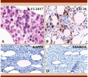
A bone marrow aspirate revealed hipercellularity with plasmocytic infiltration (45.6%). Bone mar-row biopsy showed interstitial involvement with mature and immature plasma cells. No increase of reticulin fibers or collagen fibrosis was iden-tified. Immunohistochemistry was positive for CD38 in 25% plasma cells. Neoplastic plasma cells showed clonal light chains and only a few reactive mature plasma cells showed polyclonal staining pattern (< 5%). Figure 1
Figure 1. Bone marrow biopsy. A. There is interstitial involvement with myeloma. The neoplastic plasma cells displayed features from mature to plasmablastic type (HE. 40 X). B. Immunohistochemistry CD38 stain showed 25% neoplastic plasma cells. C-D. Immuno-histochemistry light chains only show a few mature positive plasma cells (< 5%).
A
C
B
D
B-15-2417 CD-38
Histopathological findings at the time of diag-nosis showed hypercellular bone marrow and neoplastic plasmatic cells suggestive of multiple myeloma. The flow-cytometric analysis of the bone marrow cells disclosed an abnormal mono-clonal population of plasma cells representing 25% of the nucleated cells in the bone marrow. Citogenetic test was unremarkable, without 13q14.3, p53 or ATM deletion.
Serum and urine immunofixation did not show monoclonal immunoglobulin chains whereas serum free light Kappa and Lambda chains and beta 2 immunoglobulin were within nor-mal ranges. High sensitivity reactive protein C 44.0 mg/L, and total proteins in serum were 6.9 g/dL with no monoclonal spike.
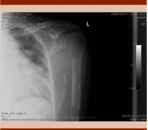
Imaging studies revealed osteolytic cranial, rib cage, pelvis and bilateral femoral bone lesions (Figures 2-3). Computed tomography with mul-tiple lesions of neoplastic character that involve the cranium irregularly and vertebral bodies of the cervical and dorsal sections.
PET-CT revealed lytic lesions as well as hyper metabolic in the axial and appendicular skeleton.
Solid and hypermetabolic lesions in kidneys and adrenal glands were suggestive of secon-dary deposits. According to Durie and Salmon criteria for MM we classified our patient as stage IIIA due to advanced lytic bone lesions at
diag-nosis.6 Treatment was started with bortezomib,
thalidomide and dexamethasone (VTD) and pain control measures, with no improvement in the symptoms. She continued with severe lumbar pain even though she was treated with opioids. Bisphosphonates were not included as supportive therapy due to renal failure, changing
her current classification to a Stage IIIB.6 As a
result of the prescription of opioids for pain control the patient had a bronchial aspiration episode and died.
DISCUSSION
Multiple myeloma is characterized by IgG and IgA secretion as monoclonal components and in some rare cases IgD, IgM or IgE are found in the
serum.5 Some patients with multiple myeloma
(1-5%) are characterized by the absence of detectable monoclonal proteins in serum and urine describing it a as non-secretory multiple myeloma (NSMM) and in 85% of these patients the presence of cytoplasmic M-proteins within plasma cells have been demonstrated indicating
immunoglobulin synthesis.5,7
Figure 3. Osteolytic lesions in iliac bones, left ischia and in the superior third of both femurs.
Figure 2. Osteolytic lesions in left clavicle, rib cage
Manifestations of NSMM are very similar to multiple myeloma except for renal insuf-ficiency; the primary manifestations include lytic bone lesions (as seen in multiple myeloma and usually takes place in skull, sternum, ribs, humerus and pelvis), anemia and
hypercalce-mia.2,8,9 Our patient presented intense back
pain irradiated to the ribs caused by bone destruction or lytic bone lesions with a low response to nonsteroidal anti-inflammatory drugs, calcium value was slightly higher than the normal range (8.5-10.2 mg/dL) suggest-ing hypercalcemia as a consequence of bone
demineralization.9 According to the literature
the development of anemia under these condi-tions is common and is related to bone marrow infiltration, usually is identified as anemia of chronic disease in which cytokines production
decrease red cells production.2,10,11
The diagnosis of NSMM in comparison with mul-tiple myeloma (in which the diagnosis is based on 10% of clonal bone marrow plasma cells and monoclonal proteins in serum and urine) is based on the presence of 30% of monoclonal bone marrow plasma cells or presence of plas-mocytoma proven by biopsy due to the clinical features (lytic bone lesions, hyperkalemia, ane-mia and serum and urine protein electrophoresis
within normal range);2,8 this patient showed upon
diagnosis a biopsy of the bone marrow that re-vealed 45% of plasma cells.
Cytogenetic analysis for t(4;14) resulted nega-tive. High risk disease and poor prognosis are defined by the presence of hypodiploidy, t(4;14), or deletion 17p13; high levels of serum
B2-microglobulin or lactate dehydrogenase.2 In
this case we had none of these markers positive.
The loss of protein production with subsequent disease progression is typically seen in patients with high-risk myeloma and is often associated with a “de-differentiated” pathological specimen
that may or may not express CD138 or CD38,
and that often expresses CD20.12
PET imaging has been used as a potential tool to assess bone disease for active and quiescent bone lesions as well to identify extra medullary manifestations of the disease. Bone marrow plasmacytosis along with PET/CT is used to assess the response in non-secretory disease; this dual evaluation is the best since the two technologies
complement.12
Since non secretory myeloma typically presents with cytopenias and bone demineralization, measures of disease response include imaging
studies and bone marrow evaluation.13
Optimal induction includes the use of a three-drug combination that involves either proteasome inhibitors together with immunomodulatory agents. Bortezomib‐based triplet combinations are among the established standards of care as induction therapy for previously untreated pa-tients with multiple myeloma who are eligible for high‐dose therapy with autologous stem cell
transplantation.14 The patient was considered to
be suitable for the triple drug therapy, dexame-thasone, thalidomide and bortezomib at a dose of 40 mg QW, 100 mg QD and 1.75 mg QW respectively. Prednisone was tapered throughout the treatment.
Thalidomide plus dexamethasone is an effective savage treatment that does not induce cytopenia and appears to be a valuable option in advanced stages of disease or in frail patients when
hema-tologic toxic effects are a concern.12
CONCLUSION
guide-lines. Specific criteria to classify non-secretory multiple myeloma are not defined. We can say it is challenging to evaluate the treatment progress since there is no quantitative way to measure it.
REFERENCES
1. Cantero-Fortiz Y, León-Peña AA, León-González M, Ruiz-Delgado GJ. Central Nervous System Involvement in multiple myeloma. Rev Hematol Mex 2016;17(1):63-6. 2. Palumbo A, Anderson K. Multiple myeloma. N Engl J Med
2011;364(11):1046-60.
3. Kyle RA, Rajkumar SV. Multiple myeloma. N Engl J Med 2004;351:1860-74.
4. Coriu D, Weaver K, Schell M, Eulitz M, Murphy CL, Weiss DT, Solomon A. A molecular basis for nonsecretory myeloma. Blood 2004;104(3):829-31.
5. Avet-loiseau H, Garand R, Lodé L, Harousseau J, Bataille R, Dc W, Garand R, Lode L. nonsecretory multiple myeloma variants brief report translocation t (11;14)(q13;q32) is the hallmark of IgM, IgE, and nonsecretory multiple myeloma variants. Blood 2013;101(4):1570-1.
6. Durie BGM, Salmon SE. A clinical staging system for multiple myeloma. Cancer 1975;36:842-54.
7. Drayson M, Tang LX, Drew R, Mead GP, Carr-smith H, Bradwell AR. Serum free light-chain measurements
for identifying and monitoring patients with nonsecre-tory multiple myeloma. Brief report serum free light-chain measurements for identifying and monitoring patients with nonsecretory multiple myeloma. Blood 2011;97(9):2900-2.
8. Taroeno-Hariadi KW, Purwanto I, Kurnianda J, Mangun-sudirjo S, Harijadi A. Non secretory multiple myeloma – a case report. Med J Indones 2007;16(4):257-61. 9. Euch ME, Ismail FBF, Rezgui A, Karmani M, Derbali F,
Amri R. Non-secretory multiple myeloma with lytic bone lesions about a new observation. Open J Intern Med 2012;2012:179-82.
10. Birgegård G. Managing anemia in lymphoma and multiple myeloma. Ther Clin Risk Manag 2008;4(2):527-39. 11. Birgegård G, Gascón P, Ludwig H. Evaluation of anaemia in
patients with multiple myeloma and lymphoma: Findings of the European Cancer Anaemia Survey. Eur J Haematol 2006;77(5):378-86.
12. Lonial S, Kaufman JL. Non-secretory myeloma: a clinician’s guide. Oncology 2013;27:924-8930.
13. International Myeloma Working Group. Criteria for the classification of monoclonal gammopathies, mul-tiple myeloma and related disorders. Br J Haematol 2003;121:749-57.