Otras secciones de este sitio:
☞ ☞ ☞ ☞
☞ Índice de este número ☞
☞ ☞ ☞
☞ Más revistas ☞
☞ ☞ ☞
☞ Búsqueda
Others sections in this web site: ☞
☞ ☞ ☞
☞ Contents of this number ☞
☞ ☞ ☞
☞ More journals ☞
☞ ☞ ☞ ☞ Search Artículo:
Is there an indication for palliative surgical procedure of shoulder and upper limb malignancies?
Derechos reservados, Copyright © 2003: Academia Mexicana de Cirugía
Cirugía y Cirujanos
Número
Number 2
Abril-Junio
April-June 2003
Volumen
edigraphic.com
Is there an indication for palliative surgical procedure of
shoulder and upper limb malignancies?
Kuauhyama Luna-Ortiz MD, Eduardo Maafs-Molina MD, Angel Herrera-Gómez MD and Jose Luis Barrera-Franco MD Academician
* Department of Surgical Oncology Instituto Nacional de Cancerología, San Fernando # 22, Tlalpan, 14000 México D.F., México
Corresponding author: Kuauhyama Luna Ortiz, MD Department of Surgical Oncology Instituto Nacional de Cancerología Av. San Fernando # 22, Tlalpan, 14000 México, D.F., México E-mail: [email protected]
Recibido para publicación: 29-08-2002 . Aceptado para publicación: 11-02-2003.
Summary
Aims: Major amputation is not a frequent procedure at present, however, there is a group of patients still considered for these treatments. Our objective was to evaluate the results of this surgical intervention as well as to evaluate morbidity, local con-trol, free disease, and palliation.
Methods: The study included 57 patients on whom interscapu-lothoracic disarticulation (STDI) was carried out at the Insti-tuto Nacional de Cancerología were (Mexico City) from 1974 to 1993. Patients with oncologic pathology were operated with a palliative aims. Twenty eight male and twenty nine female patients were found with average age of 44 years (range: 13 to 79 years). Clinical situation was characterized by tumor (100%), pain (46%), functional incapacity (39%), ulceration (32%), and bleeding (9%), Karnofsky 80% (range, 50 to 100). Time of evolution was 25 months (range: 1-121 months) and size was > 10 cm in 86% of cases.
Results: Postoperative complications were present in ten, followed by patients follow infection in five cases, hematoma in the upper lung lobe in two and femoral thrombosis, skin wound necrosis, and bleeding in one case each. Palliation was evaluated to eliminate tumoral load, ulceration, bleed-ing, functional incapacity, and pre- and postoperative pain. Global survival was 12 months (range, 1-139 months). Conclusions: Interscapulothoracic disarticulation is an infre-quently performed surgery with low morbidity and mortality, low recurrence, no effect on survival, and excellent palliative results.
Key words: Interscapulothoracic disarticulation, Forequarter amputation.
Resumen
Objetivo: las amputaciones mayores no son procedimientos frecuentes en la actualidad, sin embargo, hay un grupo de pacientes aún considerados para estos tratamientos. El ob-jetivo fue evaluar el resultado con esta intervención quirúrgi-ca, así como evaluar la morbilidad, control local, periodo li-bre de enfermedad y paliación.
Métodos: el estudio incluyó 57 pacientes en quienes se rea-lizó desarticulación interescapulotorácica (DIET) en un pe-riodo de 1974 a 1993. Los pacientes con patología oncológi-ca quienes fueron operados con objetivo paliativo. Veintiocho hombres y 29 mujeres, con edad promedio de 44 años (mar-gen 13 a 79 años). La presentación clínica fue caracterizada por tumor (100%), dolor (46%), incapacidad funcional (39%), ulceración (32%) y hemorragia (9%). Promedio de Karnofsky 80% (margen 50 a 100%). El tiempo de evolución fue de 25 meses (margen 1 a 121 meses), y la medida del tumor ma-yor de 10 cm en 86% de los casos.
Resultados: Las complicaciones postoperatorias fueron en 10 casos con infección en cinco casos, hematoma en el ló-bulo superior del pulmón en dos casos, trombosis femoral, necrosis de la piel y hemorragia en un caso respectivamen-te. La paliación fue evaluada en términos de eliminar carga tumoral, ulceración, hemorragia, incapacidad funcional, do-lor pre y postoperatorio. La supervivencia global fue de 12 meses (margen 1-139 meses).
La desarticulación interescapulotorácica es una cirugía que se realiza con poca frecuencia con baja morbilidad y morta-lidad, baja recurrencia, sin efecto en la supervivencia y con resultados de paliación excelente.
Palabras clave: desarticulación interescapulotorácica, am-putación.
Introduction
Interscapulothoracic disarticulation (STDI) was first
car-ried out in 1808 by Cuming(1) in a non-oncologic patient.
Crosby(2) in 1836 was the first to carry out STDI in the U.S.
for treatment of cancer. Berger described the technique in 1887, Buchanan presented 141 cases before 1890, and
George T. Pack(3) reported 180 cases in the literature from
1900 to 1942 and 31 cases from his own experience. At that time, major amputation was considered the
Interscapulothoracic disarticulation
edigraphic.com
diagnosis and advent of treatments with chemotherapy andradiotherapy (RT) diminished this type of radical surgery, replaced in great measure by more conservative limb
sur-gery such as the Tikhoff-Linberg(5-7), with better quality of
life and same local control. Nevertheless, for some patients STDI is the only treatment possible in terms of curative or palliative intent when conservative measures such as che-motherapy and radiation are usually attempted for local con-trol and pain relief. If these fail, the disease progresses. Re-lentless growth may lead to tumor fungation, sepsis, and hem-orrhage. This may be due to great tumor load as well as to the fact that some patients may develop sarcomas in the soft tissues or osteosarcomas after treatment with RT, 5% and
0.05 to 0.23%, respectively8-10.
STDI may be combined with thoracic wall resection when in addition to involving shoulder articulation and with pleu-rectomy or neumonectomy, these structures are implicated
by contiguity(11,12). In a few cases, STDI is indicated in
ill-nesses such as lymphedema of thoracic limb after radical dissection of axial with or without lymphomatous sarcoma (Stewart-Treeves syndrome), acute trauma management, and neurofibromas of brachial plexus, the latter associated to Von
Recklinghausen(13-15) at 10 to 15%.
This study is a retrospective analysis of patients who un-derwent palliative STDI for tumors involving shoulder gir-dle region. The objective of this report was to analyze STDI in terms of morbidity, local control, disease-free intervals, and palliation.
Material and methods
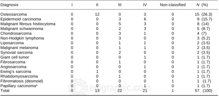
From 1974 to 1993, 57 patients were treated surgically with STDI (28 males and 29 females with mean age of 43 years, range, 13 to 79 years) at the Skin and Soft Tissues Tumors Surgical Service of the Instituto Nacional de Can-cerología in Mexico City. There were 20 soft-tissue sarco-mas, 21 bone tumors, 11 skin cancers, three lymphosarco-mas, and two cases of other tumors (Table I). Staging was per-formed according to the American Joint Committee on
Can-cer (AJCC)16 for each histology type. Forty four (77%)
pa-tients were in advanced clinical stage, as shown in Table I. The clinical picture was as follows: tumoral mass in all 57 (100%) patients; mean time of evolution of symptoms was 25 months (range, 1 to 121 months) prior to first visit to the hospital, and Karnofsky performance status (KPS) mean was 80% (range, 50 to 100%). Diameter of the tumor in 24 patients ranged from 10 to 20 cm; in 12 patients, diameter was between 21 and 30 cm, in 10 it was 31 to 40 cm, and in three patients, diameter ranged from 41 to 50 cm tumor was not measured in eight patients. These patients had one or more of the following symptoms: 26 (46%), pain; 22 (39%), had functional incapacity; 18 (32%), ulceration; five (9%)
patients had bleeding; two (4%) had weight loss, and one (2%) had miasis.
Preoperative evaluation was carried out to discard me-tastasis. Information was obtained based on X-ray and mographies performed during the first years; computed to-mography (CT) was employed during subsequent periods. Twenty eight (49%) patients presented metastatic disease at moment of diagnosis (Table II). Fourteen (25%) patients received neoadjuvant, adjuvant, or palliative treatment based on chemotherapy and 20 (35%) received radiotherapy. The surgical objective was palliative in all cases due to large size of tumors. Follow-up of these patients was every 3 months during the first year and every 4 months thereafter.
Indication for STDI was for great tumoral extension or invasion of neurovascular bundle as well as the impossibil-ity of carrying out conservative limb surgery, severe intrac-table pain with loss of limb function, and one or more of the following local tumor-related complications: tumor funga-tion; hemorrhage; sepsis, and ulceration. Surgery was done
according to Berger method as modified by Pack(1). In all
cases, surgical margin of 5 cm was afforded; when this was impossible, extensive surgery was effected, including tho-racic wall resection with reconstruction of same, employing Marlex mesh to maintain thoracic stability, and/or flap rota-tion when it was impossible to cover mesh by of direct cuta-neous closure.
Results
In 52 patients only, STDI was carried out, while com-bined STDI with resection of thoracic wall was performed in four cases; an additional patient had STDI plus total thy-roidectomy because of shoulder metastatic disease from pap-illary thryoid carcinoma.
Mean surgical time was 157.2 min (range: 1 to 5 h). Mean bleeding during surgery in 51 patients was 806 cc (range, 30 to 3,000 cc) and porto vac drainage ranged from 2 to 20 days (mean: 6.9 days).
Postoperative complications occurred in 10 patients (18%). Infection was present in five (9%), hematoma in up-per lung lobule in two (4%), and femoral thrombosis, skin wound necrosis, and bleeding in one patient each (2%).
The study group consisted of twenty six (46%) patients who experienced preoperative pain; among these, 14 (53%) manifested postoperative phantom limb and four (15%) ex-perienced the pain component. Thirty one (54%) patients did not present preoperative pain; however, 15 (48%) had postoperative phantom limb with pain component in eight (26%) patients.
edigraphic.com
patients died within the first month, nine (16%) patientswith-in the second and third months, 13 (23%) patients between the third and sixth months, 11 (19%) patients between the sixth and twelfth months, and 10 (18%) of patients died af-ter 12 months.
Discussion
STDI is one of the most mutilating surgical procedures. Its use has been reduced over past decades mainly due to treatments with neoadjuvant chemotherapy and/or radiother-apy. With these treatments, better rates of local recurrence were obtained, which allowed to increase use of conserva-tive procedures in extremities. However, amputation surgery is not an abandoned idea at present.
This is a retrospective analysis of 20-year series that con-tained a heterogeneous group of tumors in which 25% of patients received different schemes of chemotherapy and in which chemotherapy treatments evolved during the period. For this reason, it was difficult to obtain conclusions based on chemotherapy. KPS in our series was not low (mean, 80%); however, low KPS is not a contraindication for STDI
to improve the patient quality of life(17).
Among prognostic factors we were able to obtain were time of disease, evolution, which was 25 months on aver-age (range, 1-121 months) prior to admission of the pa-tient to our institution. Likewise, tumor size was > 10 cm in 86% of cases, without our knowing tumor size exactly in eight (14%) cases (Table I). Principal indication for STDI
was presence of great tumoral volume; 60% of patients in this series who underwent conservative measures such as radiotherapy and/or chemotherapy that failed to prevent tu-mor progression. Tutu-mors that failed to respond to such mea-sures continued to grow and caused local complications and frequently a painfully agonizing death. In this series, ampu-tation was advocated as a palliative procedure for symptom-atic locally advanced disease that had already failed to re-spond to radiation therapy and/or chemotherapy. This indi-cated that from the beginning, the majority of patients were found on palliative ground, understanding palliative to be the group of patients with symptoms such as pain (46%), functional incapacity (34%), ulceration (32%), bleeding (9%), weight loss (4%), and infection (2%). Therefore, in-fection, ulceration, and bleeding were palliative in 100% of our patients in an objective manner. Several clinical situa-tions have been raised in the literature as indicasitua-tions for pal-liative forequarter amputation, such as intractable pain, sep-sis, tumor fungation, hemorrhage, vascular trombosep-sis, patho-logical fracture, radiation-induce necrosis, or limb with
several functional impairment(18-20), as occurred in several
of our cases due basically to presence tumor of the great
size. Malawer et al(21) considered that presence of pain alone
was not a clear indication for amputation such as the
fore-quarter, but we agree with Mirimsky et al(17), who
consid-ered pain alone as a clear indication for palliative forequar-ter amputation. Pain was the principal symptom in 26 (46%) of our patients, and this symptom persisted in four patients after surgery in the form of painful phantom limb. However,
Table I. Histopathologic diagnosis and clinical stage
Diagnosis I II III IV Non-classified N (%)
Osteosarcoma 0 12 0 3 0 15 (26.3)
Epidermoid carcinoma 0 0 3 6 0 9 (15.7)
Malignant fibrous histiocytoma 0 0 5 3 0 8 (14)
Malignant schwannoma 0 0 2 3 0 5 (8.7)
Chondrosarcoma 0 0 3 1 0 4 (7)
Non-Hodgkin lymphoma 0 0 3 0 0 3 (5.2)
Liposarcoma 0 0 1 1 0 2 (3.5)
Malignant melanoma 0 0 1 1 0 2 (3.5)
Synovial sarcoma 0 0 2 0 0 2 (3.5)
Giant cell tumor 0 0 0 1 0 1 (1.7)
Fibrosarcoma 0 0 1 0 0 1 (1.7)
Angiosarcoma 0 0 0 1 0 1 (1.7)
Ewing’s sarcoma 0 1 0 0 0 1 (1.7)
Rhabdomyosarcoma 0 0 1 0 0 1 (1.7)
Fibromatosis (desmoid) 0 0 0 0 1 1 (1.7)
Papillary carcinoma* 0 0 0 1 0 1 (1.7)
Total 0 13 22 21 1 57 (100)
Interscapulothoracic disarticulation
edigraphic.com
:rop odarobale FDPVC ed AS, cidemihparG
arap
acidémoiB arutaretiL :cihpargideM
sustraídode-m.e.d.i.g.r.a.p.h.i.c 31 (54%) patients did not experience preoperatory pain, whiel
eight did experience painful phantom limb. (In this regard, we did not find an explanation for the phenomenon).
Phan-ton limb pain was reported in 60-90% of amputees(22). It
may be more prevalent in cancer patients who have under-gone amputation, particularly those exposed to chemothera-peutic agent. Incidence of phantom limb pain may be great-er also in patients who expgreat-erienced prolonged preopgreat-erative pain and pain in immediate postoperative period than in those who did not. Another explanation by other authors is that it possible that emotional factors are influential in patient ex-perience of prolonged pain in a phantom limb. It is obvious that functionality of the limb is not preserved, as this is a radical surgery of the limbs. We must take into account that this is a retrospective study and it is, therefore, difficult to obtain numeric objectives with respect to quality of life. For this reason, it is important to have pre- and postoperatory evaluations.
In this series, we recorded 48% preoperatory and 46% post-operatory metastasis. This is not surprising, as large tumors were present and as it is known, in tumors > 5 cm the possibi-lity of distant metastasis is 60-70%, depending on histologic type. This explains patient mortality of 40% during the first 3 months and 63% at 6 months. When we compared this
sur-vival rate with other series(4,23,24) in which survival is 35% at
5 years, tumor volumes may be the difference in terms of sur-vival, which in our series was < 20% at 3 years; however, survival ultimately had no meaning because the procedure was palliative. Short survival of our patients at present is not a contraindication for STDI, when tumors are as big as those mentioned in this series. The role of survival calculation was to show that the patient had palliative effects prior to suc-cumbing to their diseases. Lung metastasis was the dominant cause of death in our patients. Nonetheless, novel methods should be explored in detection of subclinical metastatic
dis-ease for patients with bone sarcomas, unclassified sarcomas, or undifferentiated tumors who develop systemic metastasis postoperatively during the first 6 months; in addition, it indi-vidually is necessary to evaluate palliative value for each pa-tient even in the presence of metastatic disease to justify car-rying out or not of this procedure, which continues to have high psychological impact. In 9% of patients, local recurrence was present, which is low. Nevertheless, this should be
con-sidered a systemic disease(25). Morbility in this series was high
at 18% when compared with Ham’s series(23) in which there
was null morbility. Mortality related to surgical procedure was
null, similar to other series(23).
STDI remains an effective procedure for local control of tumors of different histologic pedigree localized in gleno-humeral articulation. One main indication for this procedure is recurrent soft-tissue sarcoma in which a conservative pro-cedure was performed initially STDI should remain a rarely used surgical procedure important in management of sarco-mas or undifferentiated tumors with palliative aims as the principal indication. Despite the short survival time in our series, we believe that interscapulothoracic disarticulation in its palliative form was justified due to the fact that the procedure provided pain relief and permitted the patient some independence. Thus, we could say that the benefit of any palliative procedure is difficult to quantify, but relief of pain, ulceration, and necrosis has a high priority.
References
1. Keevil JJ. Ralph Cuming and the interscapulothoracic amputation in 1808. J Bone Joint Surg (Br) 1949;31-B:589-595.
2. Crosby AB. The first operation on record for removal of the entire arm, scapula and three-fourths of the clavicle by Dixi Crosby. Con-cord, NH, USA Republican Press Assn Med Rec 1875;10:753. 3. Pack GT, McNeer G, Coley BL. Interscapulothoracic amputation for
malignant tumors of the upper extremity. Surg Gynecol Obstet 1942;74:161-175.
4. Pack GI. Major exarticulations for malignant neoplasms of the ex-tremities. Interscapulothoracic amputation, hip-joint disarticula-tion and interileo-abdominal amputadisarticula-tion. J Bone Joint Surg 1956; 38:249-262.
5. Eilber FR, Mirra JJ, Grant TT, Weisenburger T, Morton DL. Is ampu-tation necessary for sarcomas? A seven year experience with limb salvage. Ann Surg 1980;192:431-438.
6. Eilber FR, Morton DL, Eckardt J, Grant TT, Weisenburger T. Limb salvage for skeletal and soft tissue sarcomas. Multidisciplinary pre-operative therapy. Cancer 1984;53:2579-2584.
7. Lindberg RD, Martin RG, Romsdahl MM, Barkley HT. Conservative surgery and postoperative radiotherapy in 300 adults with soft tissue sarcomas. Cancer 1981;47:2391-2397.
8. Doherty MA, Rodger A, Langiands-AO. Sarcoma of bone following therapeutic irradiation for breast carcinoma. Int J Radiat Oncol Biol Phys 1986;12:103-106.
9. Souba WW, McKenna RJ Jr, Meis J, Benjamin R, Raymound AK, Mountain CF. Radiation induce sarcomas of the chest wall. Cancer 1986;57:610-615.
Table II. Distribution of metastatic disease according to site.
Site Preoperatory Postoperatory Total
Lung 14 18 32
Thoracic wall 4 0 4
Nodes 6 4 10
Skin 2 0 2
Bone 1 2 3
Diaphragm 1 0 1
Liver 0 1 1
Central nervous
system 0 1 1
edigraphic.com
10. Davison T, Westbury G, Harmer CL. Radiation induced soft tissue sarcoma. Br J Surg 1986;73:308-309.
11. Stafford ES, Rainey WG Jr. Radical transthoracic forequarter ampu-tation. Ann Surg 1958;148:699.
12. Roth JA, Sugarbaker PH, Baker AR. Radical forequarter amputation with chest wall resection. Ann Thorac Surg 1984;37:423-427. 13. Hovius SE, Hofman A, Van-Urk H, Van-Der-Meulen Jc. Acute
man-agement of traumatic forequarter amputation: case report. J Trauma 1991;31:1415-1419.
14. Ebraheim NA, Pearlstein SR, Savolaine ER, Gordon SL, Jackson WT, Corray T. Scapulothoracic dissociation (closed avulsion of the scapu-la, subclavian artery, and brachial plexus): a newly recognized vari-ant, a new classification, and a review of the literature and treatment options. J Orthop Trauma 1987;1:18-23.
15. Lusk MD, Kline DG, García CA. Tumors of the braquial plexus. Neu-rosurgery 1987;21:439-453.
16. American Joint Committee on Cancer (AJCC) staging manual. 5th ed. Philadelphia, PA, USA: Lippincott-Raven;1998.
17. Merimsky O, Kollender Y, Inbar M, Lev-Chelouche D, Gutman M, Issakov J, Mazeh D, Shabat S, Bickels J, Meller I. Is forequarter am-putation justified for palliation of intractable cancer symptoms? On-cology 2001;60:55-59.
18. Merimsky O, Kollender Y, Bickels J, Inbar M, Nirkin A, Issakov J, Chai-tchik S, Meller I. Amputation of the lower limb as palliative treatment for debilitating musculoskeletal cancer. Oncol Rep 1997;4: 1059-1062. 19. Higinbotham NL, Marcove RC, Casson P. Hemipelvectomy: a
clini-cal study of 100 cases with five years follow-up on 60 patients. Sur-gery 1966;59:706-710.
20. Bhagia SM, Elek EM, Grimer RJ, Carter SR, Tillman RM. Forequar-ter amputation for high grade malignant tumors of the shoulder gir-dle. J Bone Joint Surg (Br) 1997;79:924-926.
21. Stephenson RB, Kaufer H, Hankin RM. Partial pelvic resection as an alternative to hindquarter amputation for skeletal neoplasms. Clin Orthop 1989;242:201-211.
22. Katz J, Melzack R. Pain memories in phantom limbs: review and clinical observations. Pain 1990;43:319-336.
23. Ham SJ, Hoekstra HJ, Koops HS, Eisma WH, Oldhoff J. The inter-scapulothoracic amputation in the treatment of malignant diseases of the upper extremity with a review of the literature. Eur J Surg Oncol 1993;19:543-548.
24. Lienard D, Lejeune EJ. Therapeutic value of scapular and pelvic gir-dle disarticulations in sarcoma. Eur J Surg Oncol 1987;13:231-237. 25. Fanous N, Didolkar MS, Holyoke ED, Elias EG. Evaluation of