Casos Clínicos
Fragmentación del QRS y muerte súbita en paciente con displasia
arritmogénica del ventrículo derecho
Dr. Luis A. Rodríguez López, Dra. Eliany Rodríguez Moreno, Dr. Juan M. Cruz Elizundia,
Dr. Yohan M. Díaz Sardiñas, Dr. Reinaldo Gavilanes Hernández, Dr. Ruben R. Quenta
Tarqui
, Dra. Jhosely A. González Achacollo y Dr. Carlos Santana Santana
Servicio de Cardiología, Cardiocentro Ernesto Che Guevara. Santa Clara, Villa Clara, Cuba.
Full English text of this article is also available
INFORMACIÓN DEL ARTÍCULO
Recibido: 10 de noviembre de 2018 Aceptado: 13 de diciembre de 2018
Conflictos de intereses
Los autores declaran que no existen conflictos de intereses
Abreviaturas
DAVD: displasia arritmogénica del ventrículo derecho
ECG: electrocardiograma
MSC: muerte súbita cardíaca
TV: taquicardia ventricular
RR Quenta Tarqui
Cardiocentro Ernesto Che Guevara Calle Cuba 610
e/ Barcelona y Capitán Velasco. Santa Clara 50200. Villa Clara, Cuba. Correo electrónico:
rubenqt@ucmex.vcl.sld.cu
RESUMEN
La miocardiopatía o displasia arritmogénica del ventrículo derecho es una cardio-patía de origen genético cuyo diagnóstico supone, a menudo, un reto para el clíni-co. Es una de las causas más comunes de muerte súbita cardíaca en la adolescen-cia y en los adultos jóvenes. Se presenta el caso de un paciente con historia de arritmias ventriculares malignas y de muerte súbita cardíaca recuperada, por dis-plasia arritmogénica del ventrículo derecho, con fragmentación del QRS en las derivaciones precordiales derechas, como marcador de la presencia de un sustra-to propicio para el surgimiensustra-to de la fibrilación ventricular espontánea. Se comen-ta la patogenia, el diagnóstico y el tracomen-tamiento de escomen-ta enfermedad.
Palabras clave: Fragmentación del QRS, Muerte súbita, Displasia arritmogénica del ventrículo derecho
Fragmented QRS and sudden death in a patient with
arrhythmogenic right ventricular dysplasia/cardiomyopathy
ABSTRACT
The arrhythmogenic right ventricular dysplasia or cardiomyopathy is a genetic heart disease whose diagnosis is often a challenge for the clinician. It is one of the most common causes of sudden cardiac death in adolescence and in young adults. We present the case of a patient with a history of malignant ventricular arrhythmi-as and recovered sudden cardiac death due to arrhythmogenic right ventricular dysplasia, with QRS fragmentation in the right precordial leads, as a marker of the presence of a suitable substrate for the emergence of spontaneous ventricu- lar fibrillation. The pathogenesis, diagnosis and treatment of this disease are dis-cussed.
Keywords: QRS fragmentation, Sudden death, Arrhythmogenic right ventricular dysplasia
INTRODUCCIÓN
Desde que la displasia arritmogénica del ventrículo derecho (DAVD) fue descrita por primera vez por Dalla Volta et al1 en 1961, y posteriormente ca-
racterizada por Fontaine et al2 en 1977, hasta la actualidad, en la que ha
de las miocardiopatías3, las aportaciones a la litera-tura acerca de esta enfermedad han sido numero-sas4,5. Inicialmente las descripciones se centraban en el sustrato arrítmico de ciertas zonas del ventrículo derecho, el llamado «triángulo de la displasia»; pero actualmente el espectro se ha ampliado para dar paso a manifestaciones difusas en el mencionado ventrículo, a la afección única ventricular izquierda y biventricular en fase avanzada de la enfermedad, a menudo indistinguible de la miocardiopatía dilata-da6. La prevalencia varía ampliamente según las series descritas y existe controversia acerca de la distribución geográfica de la enfermedad. En regio-nes italianas del Véneto se estima una prevalencia de 1 caso por 1000 o 10000 personas. Corrado et al7
plantean que puede ser causa de hasta el 20% de las muertes súbitas cardíacas (MSC) en adultos jóvenes y en atletas italianos, y afecta más frecuentemente a los varones. En EEUU representa un 5% de las MSC en menores de 65 años y 3-4%, en atletas8,9.
La DAVD es una enfermedad del músculo cardía-co de origen genéticardía-co cuyo diagnósticardía-co supone a menudo un reto para el clínico. La descripción clá-sica suele referirse al estadio final de la enfermedad, en que el miocardio, fundamentalmente del ven-trículo derecho, ha sido sustituido por tejido fibro-adiposo; por eso las fases iniciales de la enfermedad, no tan floridas en semiología, suelen pasar inadver-tidas. Desafortunadamente, el riesgo de
desenlace fatal no es bajo10.
CASO CLÍNICO
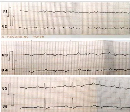
Se trata de un hombre de piel blanca y 54 años de edad, sin factores de riesgo car-diovascular, hábitos tóxicos, enfermedad valvular previa u otra cardiopatía, que fue remitido a consulta de arritmia por presen-tar episodios de pérdida de la conciencia, de breve duración, relacionadas con el es-fuerzo físico y situaciones de estrés, y final-mente taquicardia ventricular (TV), lo que degeneró en fibrilación ventricular recupe-rada tras cardioversión eléctrica. El ECG basal de doce derivaciones presentaba pa-trón de bloqueo de rama derecha incom-pleto con ondas T negativas y anchura del QRS mayor de 110 mseg; además, onda ép-silon y fragmentación del QRS en precor-diales derechas (Figura 1).
El ecocardiograma realizado demostró dilatación del ventrículo derecho (54 mm)
con pared libre engrosada y presencia de zonas de aspecto blanquecino, que pudieran estar en relación con infiltración fibroadiposa (Figura 2).
Se diagnosticó una DAVD, se indicó tratamiento con amiodarona y se le colocó un desfibrilador au-tomático implantable, con lo que evolucionó favora-blemente.
COMENTARIO
La DAVD es un trastorno heredable, que tiene clara incidencia familiar hasta en el 50% de los casos, con un patrón de transmisión autosómico dominante, diversos grados de penetración y una expresión fenotípica polimórfica. No obstante, también se ha descrito una forma autosómica recesiva1-5. Se puede asociar a una queratodermia palmo-plantar y a un pelo lanudo (enfermedad de Naxos)6. Este tipo de DAVD lo causa una mutación en el gen de la placo-globina, cuyo producto es un componente de los desmosomas y las uniones celulares adherentes (adherens), y el síndrome de Carvajal, con el mis-mo fenotipo cutáneo a predominio del ventrículo izquierdo en familias de la India y Ecuador6,7.
La primera mutación causante de una DAVD no sindrómica fue descrita por Rampazzo et al8 en 2002.
Dicha mutación se identificó en el gen de la plaquina, que codifica un componente del
soma. En 2004, Gerull et al9 describieron 25
muta-ciones del gen desmosómico cardíaco de la placofi-lina; posteriormente, se identificaron otras mutacio-nes presuntamente causantes de enfermedad en la placoglobina y la desmoplaquina, así como en otros genes desmosómicos (los de la desmocolina 2 y la desmogleína 2) en pacientes con DAVD no sindró-mica. Actualmente se considera que la disfunción desmosómica es la vía final común en la patogenia de la DAVD, ya que la integridad estructural y fun-cional del tejido cardíaco se basa en los desmoso-mas, las uniones adherentes (tipo adherens) y las uniones estrechas (tipo gap) situadas en los discos intercalares10.
En el mapa cromosómico se han localizado dife-rentes variantes genéticas de DAVD y se han descri-to más de 140 mutaciones causantes de enfermedad, la mayoría de ellas correspondientes a genes que codifican proteínas desmosómicas. Algunos genes no desmosómicos se han asociado también a una DAVD autosómica dominante, entre ellos el gen del factor de crecimiento transformador β-3 (TGFβ3)11, que modula la expresión de las proteínas de contac-to celular y el gen del recepcontac-tor de rianodina 2 (RyR2)11,12. El gen RyR2, que se describió primero
en ocho familias, codifica receptores que intervie-nen en la liberación del calcio del retículo sarco-plásmico; sin embargo, sigue habiendo opiniones divergentes en cuanto a si debe considerarse o no que los pacientes con mutaciones de este gen sufren una DAVD o una TV polimórfica catecolaminérgica. La mutación que se ha descrito de forma más re-ciente afecta al gen TMEM43 y causa una variante de DAVD de penetración completa, muy letal (DAVD tipo 5)13-15.
La degeneración y la muerte de los miocardioci-tos son la consecuencia anatomopatológica de estas mutaciones de proteínas de adhesión, con la consi-guiente sustitución progresiva por tejido adiposo y fibroadiposo. En la teoría inflamatoria, apoyada por la aparición de infiltrados inflamatorios en series necrópsicas, el daño miocárdico vendría explicado por un proceso continuado de daño y reparación que una miocarditis crónica13-15.
Las manifestaciones clínicas de la DAVD son va-riables y dependen de la inestabilidad cardíaca y la disfunción ventricular progresiva. Varían desde pa-cientes asintomáticos, MSC como primera manifes-tación, arritmias ventriculares y supraventriculares, hasta insuficiencia cardíaca derecha o biventricular. Se ha descrito la presencia de un desequilibrio en la inervación adrenérgica como posible coadyuvante en la génesis de las arritmias; de este modo, la pro-pensión a arritmias ventriculares aumenta en situa-ciones de exposición a las catecolaminas, especial-mente durante el ejercicio11.
Las alteraciones más frecuentes en el electrocar-diograma (ECG) son la inversión de la onda T (V1
-V3), presente hasta en el 50% de los sujetos. La
afec-ción más allá de V3 indica afección adicional del
ventrículo izquierdo12. Existen diferentes anomalías de la despolarización ventricular, el bloqueo de rama derecha incompleto es más frecuente (18%) que el completo (15%); la prolongación del QRS más de 110 milisegundos en V1 y V2 es un hallazgo más
específico, y pueden aparecer ondas épsilon que se observan al final del QRS y al inicio del ST, y co-rresponden a potenciales eléctricos retrasados de pequeña amplitud originados en las áreas de tejido sano rodeadas de infiltrado fibroadiposo13. La
chura del QRS y su fragmentación en las precordia-les derechas permiten predecir, de manera inde-pendiente, la presencia de dilatación y disfunción del ventrículo derecho e incluso la aparición de arritmias14.
Esta fragmentación del QRS se define como la presencia de muescas u ondas de bajo voltaje (R’) en la porción terminal del QRS o en el inicio del segmento ST, en al menos dos derivaciones conti-guas. Morita et al10 demostraron que constituyen un
marcador de la presencia de un sustrato propicio para el surgimiento de fibrilación ventricular espon-tánea, con una sensibilidad de 93% y especificidad de 90%.
El diagnóstico definitivo de la DAVD requiere la confirmación anatomopatológica de la sustitución fibroadiposa transmural, mediante muestras quirúr-gicas o necrópsicas. La naturaleza parcheada y pro-gresiva de la enfermedad hace que la biopsia endo-miocárdica tenga una utilidad limitada. No existe una única prueba para establecer el diagnóstico de DAVD3-7; éste se establece tras una evaluación
fun-cional, morfológica y electrocardiográfica, mediante la cual se determinan los criterios mayores y meno-res actualmente reconocidos15-17. En 2002 se propuso
una modificación de estos criterios para el diagnós-tico de la DAVD, en familiares de primer grado de un caso inicial16. En esta situación, la presencia de una inversión de la onda T en precordiales derechas (V2
-V3), potenciales tardíos en el ECG de promediación
de señal, TV con morfología de bloqueo de rama izquierda del haz de His (BRIHH), o la observación de cambios funcionales o morfológicos del ventrícu-lo derecho en las expventrícu-loraciones de imagen, deben considerarse criterios mayores con valor diagnósti-co para la DAVD familiar16. Posteriormente se publi-có una nueva modificación de estos criterios con la finalidad de aumentar la sensibilidad mediante el uso de las modalidades diagnósticas emergentes, los avances en la genética de la DAVD y la introducción de parámetros cuantitativos18.
La DAVD se manifiesta generalmente en forma de episodios de TV originada en el VD, por lo que tie-nen morfología de BRIHH, en adolescentes o adultos jóvenes aparentemente sanos. Las arritmias ventri-culares pueden ser asintomáticas y detectarse en un ECG sistemático o pueden causar palpitaciones, síncope o MSC. Se ha estimado que la DAVD explica hasta un 5-20% de los casos de MSC en individuos de menos de 35 años de edad18.
Aunque la información en relación con la historia natural es limitada, en general se admiten 4 esta
dios7,19,20:
I. La fase temprana o silente, generalmente asin-tomática, aunque el debut puede manifestarse con MSC.
II. Fase inestable con predominio de arritmias sin-tomáticas, generalmente con morfología de BRIHH, altamente sugestivas de origen ventricu-lar derecho.
III. Fase de fallo ventricular derecho con relativa conservación de la función del izquierdo.
IV. Fase final con progresiva dilatación biventricu-lar, a menudo indistinguible de la miocardiopatía dilatada. Las complicaciones más frecuentes en este estadio son las tromboembólicas y la fibrila-ción auricular.
En las últimas dos décadas, las arritmias origina-das en el VD, han atraído la atención del mundo científico por diversas razones, principalmente por-que suelen afectar a pacientes de menor edad y pueden conducir a la MSC. El mecanismo fisiopato-lógico de esas arritmias no se ha aclarado por com-pleto y a veces deja margen para diferentes interpre-taciones. Además, cada vez se involucra en mayor medida al intrigante mundo de la genética en los aspectos patogénicos, diagnósticos y pronósticos de algunas de estas arritmias. La infiltración por tejido fibroadiposo constituye un sustrato para la inestabi-lidad eléctrica y lleva a la arritmia ventricular que va desde las extrasístoles ventriculares aisladas hasta las TV sostenidas o la fibrilación ventricular1-4. Exis-ten múltiples predictores eléctricos para la aparición de las arritmias ventriculares y MCS10,21-23, el QRS de mayor duración, la fragmentación del QRS y la onda épsilon estuvieron presentes en nuestro paciente.
El objetivo principal de la estrategia terapéutica en este tipo de pacientes es la prevención de la MSC, para lo cual disponemos de 3 estrategias tera-péuticas principales: los fármacos antiarrítmicos, la ablación por catéter y el uso del desfibrilador auto-mático implantable24,25.
BIBLIOGRAFÍA
1. Dalla Volta S, Battaglia G, Zerbini E. “Auricularization” of right ventricular pressure curve. Am Heart J. 1961; 61:25-33.
2. Fontaine G, Guiraudon G, Frank R. Stimulation studies epicardial mapping in VT: Study of mechanisms and selection for surgery. En: Hulbertus HE, editor. Reen-trant arrhythmias. Lancaster: MTP Publishers; 1977. p. 334-50.
ner B, O'Connell J, et al. Report of the 1995 World Health Organization/International Society and Federa-tion of Cardiology Task Force on the DefiniFedera-tion and Classification of cardiomyopathies. Circulation. 1996; 93(1):841-2.
4. Brugada J, Brugada P, Brugada R. El síndrome de Bru-gada y las miocardiopatías derechas como causa de muerte súbita. Diferencias y similitudes. Rev Esp Car-diol. 2000;53(2):275-85.
5. Brugada J, Mont L, Brugada R. Displasia arritmogénica del ventrículo derecho. Rev Esp Cardiol. 1997;50(8): 541-7.
6. Burke AP, Farb A, Tashko G, Virmani R. Arrhythmo-genic right ventricular cardiomyopathy and fatty re-placement of the right ventricular myocardium: are they different diseases? Circulation. 1998;97(16):1571-80.
7. Corrado D, Thiene G, Nava A, Rossi L, Pennelli N. Sud-den death in young competitive athletes: clinicopatho-logic correlations in 22 cases. Am J Med. 1990;89(5): 588-96.
8. Rampazzo A, Nava A, Malacrida S, Beffagna G, Bauce B, Rossi V, et al. Mutation in human desmoplakin do-main binding to plakoglobin causes a dominant form of arrhythmogenic right ventricular cardiomyopathy. Am J Hum Genet. 2002;71(5): 1200-6.
9. Gerull B, Heuser A, Wichter T, Paul M, Basson CT, McDermott DA, et al. Mutations in the desmosomal protein plakophilin-2 are common in arrhythmogenic right ventricular cardiomyopathy. Nat Genet. 2004; 36(11):1162-4.
10.Morita H, Kusano KF, Miura D, Nagase S, Nakamura K, Morita ST, et al. Fragmented QRS as a marker of con-duction abnormality and a predictor of prognosis of Brugada syndrome. Circulation. 2008;118(17):1697-704. 11.Beffagna G, Occhi G, Nava A, Vitiello L, Ditadi A, Basso
C, et al. Regulatory mutations in transforming growth factor-beta3 gene cause arrhythmogenic right ventricu-lar cardiomyopathy type 1. Cardiovasc Res. 2005;65(2): 366-73.
12.Bassareo PP, Mercuro G. QRS Complex Enlargement as a Predictor of Ventricular Arrhythmias in Patients Affected by Surgically Treated Tetralogy of Fallot: A Comprehensive Literature Review and Historical Overview. ISRN Cardiol [Internet]. 2013 [citado 30 Jun 2018];2013:782508. Disponible en:
https://www.ncbi.nlm.nih.gov/pmc/articles/PMC35905 65/pdf/ISRN.CARDIOLOGY2013-782508.pdf
13.Marcus FI. Update of arrhythmogenic right ventricular dysplasia. Card Electrophysiol Rev. 2002; 6(1-2):54-6. 14.Shanmugam N, Yap J, Tan RS, Le TT, Gao F, Chan JX,
et al. Fragmented QRS complexes predict right ven-tricular dysfunction and outflow tract aneurysms in patients with repaired tetralogy of Fallot. Int J Cardiol. 2013;167(4):1366-72.
15.Merner ND, Hodgkinson KA, Haywood AF, Connors S, French VM, Drenckhahn JD, et al. Arrhythmogenic
right ventricular cardiomyopathy type 5 is a fully pen-etrant, lethal arrhythmic disorder caused by a mis-sense mutation in the TMEM43 gene. Am J Hum Genet. 2008;82(4):809-21.
16.Sen-Chowdhry S, Syrris P, McKenna WJ. Role of genet-ic analysis in the management of patients with arrhyth-mogenic right ventricular dysplasia/ cardiomyopathy. J Am Coll Cardiol. 2007;50(19): 1813-21.
17.Thiene G, Corrado D, Basso C. Arrhythmogenic right ventricular cardiomyopathy/dysplasia. Orphanet J Ra-re Dis [Internet]. 2007 [citado 5 Jul 2018];2:45. Disponi-ble en:
https://www.ncbi.nlm.nih.gov/pmc/articles/PMC22220 49/pdf/1750-1172-2-45.pdf
18.Marcus FI, McKenna WJ, Sherrill D, Basso C, Bauce B, Bluemke DA, et al. Diagnosis of arrhythmogenic right ventricular cardiomyopathy/dysplasia: proposed mod-ification of the task force criteria. Circulation. 2010; 121(13):1533-41.
19.Marcus FI, Zareba W, Calkins H, Towbin JA, Basso C, Bluemke DA, et al. Arrhythmogenic right ventricular cardiomyopathy/dysplasia clinical presentation and diagnostic evaluation: results from the North American Multidisciplinary Study. Heart Rhythm. 2009;6(7):984-92.
20.Prakasa KR, Dalal D, Wang J, Bomma C, Tandri H, Dong J, et al. Feasibility and variability of three dimen-sional echocardiography in arrhythmogenic right ven-tricular dysplasia/cardiomyopathy. Am J Cardiol. 2006; 97(5):703-9.
21.Dorantes Sánchez M, Ponce Paredes EF. Extrasístoles ventriculares con intervalo corto de acoplamiento: Su trascendencia. CorSalud [Internet]. 2015 [citado 20 Jun 2018];7(4):253-7. Disponible en:
http://www.revcorsalud.sld.cu/index.php/cors/article/ view/72/143
22.Dorantes Sánchez M, Ponce Paredes E, Falcón Rodrí-guez R. Extrasístoles ventriculares con intervalo corto de acoplamiento como detonantes de arritmias malig-nas. CorSalud [Internet]. 2016 [citado 28 Jun 2018]; 8(3):144-52. Disponible en:
http://www.revcorsalud.sld.cu/index.php/cors/article/ view/134/319
23.Dorantes Sánchez M. Despolarización y repolarización ventriculares para estratificar riesgo de arritmias ven-triculares malignas y muerte súbita. CorSalud [Inter-net].2018[citado10Jul2018];10(3):266-9.Disponibleen: http://www.revcorsalud.sld.cu/index.php/cors/article/ view/351/719
24.Buja G, Estes NA, Wichter T, Corrado D, Marcus F, Thiene G. Arrhythmogenic right ventricular cardiomy-opathy/dysplasia: risk stratification and therapy. Prog Cardiovasc Dis. 2008:50(4);282-93.
Cuban Society of Cardiology
_______________________Case Report
Fragmented QRS and sudden death in a patient with arrhythmogenic
right ventricular dysplasia/cardiomyopathy
Luis A. Rodríguez López, MD; Eliany Rodríguez Moreno, MD; Juan M. Cruz Elizundia, MD;
Yohan M. Díaz Sardiñas, MD; Reinaldo Gavilanes Hernández, MD; Dr. Ruben R. Quenta
Tarqui
, MD; Jhosely A. González Achacollo, MD; and Carlos Santana Santana, MD
Department of Cardiology, Cardiocentro Ernesto Che Guevara. Santa Clara, Villa Clara, Cuba.
Este artículo también está disponible en español
ARTICLE INFORMATION
Recibido: November 10, 2018 Aceptado: December 13, 2018
Competing interests
The authors declare no competing interests
Acronyms
ARVD: Arrhythmogenic right ventricular dysplasia
ECG: Electrocardiogram
SCD: Sudden cardiac death
VT: Ventricular tachycardia
RR Quenta Tarqui
Cardiocentro Ernesto Che Guevara Calle Cuba 610
e/ Barcelona y Capitán Velasco. Santa Clara 50200. Villa Clara, Cuba. E-mail address:
rubenqt@ucmex.vcl.sld.cu
ABSTRACT
The arrhythmogenic right ventricular dysplasia or cardiomyopathy is a genetic heart disease whose diagnosis is often a challenge for the clinician. It is one of the most common causes of sudden cardiac death in adolescence and in young adults. We present the case of a patient with a history of malignant ventricular arrhythmi-as and recovered sudden cardiac death due to arrhythmogenic right ventricular dysplasia, with QRS fragmentation in the right precordial leads, as a marker of the presence of a suitable substrate for the emergence of spontaneous ventricu- lar fibrillation. The pathogenesis, diagnosis and treatment of this disease are dis-cussed.
Keywords: QRS fragmentation, Sudden death, Arrhythmogenic right ventricular dysplasia
Fragmentación del QRS y muerte súbita en paciente con displasia
arritmogénica del ventrículo derecho
RESUMEN
La miocardiopatía o displasia arritmogénica del ventrículo derecho es una cardio-patía de origen genético cuyo diagnóstico supone, a menudo, un reto para el clíni-co. Es una de las causas más comunes de muerte súbita cardíaca en la adolescen-cia y en los adultos jóvenes. Se presenta el caso de un paciente con historia de arritmias ventriculares malignas y de muerte súbita cardíaca recuperada, por dis-plasia arritmogénica del ventrículo derecho, con fragmentación del QRS en las derivaciones precordiales derechas, como marcador de la presencia de un sustra-to propicio para el surgimiensustra-to de la fibrilación ventricular espontánea. Se comen-ta la patogenia, el diagnóstico y el tracomen-tamiento de escomen-ta enfermedad.
Palabras clave: Fragmentación del QRS, Muerte súbita, Displasia arritmogénica del ventrículo derecho
INTRODUCTION
Since the arrhythmogenic right ventricular dysplasia (ARVD) was first de-scribed by Dalla Volta et al1 in 1961, and subsequently characterized by
Fontaine et al2 in 1977, up to the present, that it has been included in the
contributions to the literature on this disease has been numerous4,5. Initially, the descriptions focused on the arrhythmic substrate of certain areas of the right ventricle, the so-called "triangle of dysplasia" ; but now the spectrum has expanded to give way to diffuse manifestations in the aforementioned ventri-cle, to the only left ventricular and biventricular disease in the advanced phase of the disease, often indistinguishable from dilated cardiomyopathy6. The prevalence varies widely according to the series described and there is controversy about the geo-graphical distribution of the disease. In the Veneto regions of Italy, a prevalence of 1 case per 1000 or 10000 people is estimated. Corrado et al7 point out
that it can cause up to 20% of sudden cardiac deaths (SCD) in young adults and Italian athletes, and it affects males most often. In USA it represents 5% of the SCD in under 65 years old and 3-4% in athletes8,9.
The ARVD is a disease of the cardiac muscle of genetic origin whose diagnosis is often a challenge for the clinician. The classic description usually re-fers to the final stage of the disease, in which the myocardium, fundamentally from the right ventricle, has been replaced by fibroadipose tissue; for this reason, the initial phases of the disease, which are not as vast in semiology, usually go unnoticed. Un-fortunately, the risk of fatal outcome is not low10.
CASE REPORT
This is the case of a 54-year-old man with white skin and without cardiovascular risk factors, toxic habits, previous valvu-lar disease or other heart disease, which was sent to the arrhythmia department presenting loss of consciousness, of short duration, related to physical exertion and stress, and eventually ventricular tachy-cardia (VT), which degenerated into ven-tricular fibrillation recovered after elec-trical cardioversion. The 12- lead basal electrocardiogram (ECG) showed an in-complete right bundle branch block pat-tern with negative T waves and QRS width bigger than 110 msec; also, epsilon wave and QRS fragmentation in right pre-cordial (Figure 1).
The echocardiogram showed right ventricular dilatation (54 mm) with thick-ened free wall and presence of whitish appearance in some areas, which could
be related to fibroadipose infiltration (Figure 2). An ARVD was diagnosed, a treatment with amiodarone was indicated and an implantable cardioverter de-fibrillator was placed; the evolution was favorable.
COMMENTS
The ARVD is a heritable disorder, having family in-cidence until 50% of cases, with an autosomal domi-nant pattern of transmission, varying degrees of pen-etration and polymorphic phenotypic expression. However, an autosomal recessive form has also been described1-5. It can be associated to a palmo-plantar keratoderma and to a wooly hair (Naxos dis-ease)6. This type of ARVD is caused by a mutation in the plakoglobin gene, whose product is a component of desmosomes and adherens junctions, and the Carvajal’s syndrome, with the same cutaneous phe-notype prevalence of left ventricular in families from India and Ecuador6,7.
The first mutation causing a non-syndromic ARVD was described by Rampazzo et al8 in 2002.
This mutation was identified in the desmoplakin gene, which codes a component of the desmosome. In 2004, Gerull et al9 described 25 mutations of the
chimeric desmosomal or plakophilin gene; later, there were identified other mutations that
bly caused disease in the plakoglobin and desmo-plakin, as well as other desmosomal genes (the ones of desmocollin 2 and desmoglein 2) in patients with non-syndromic ARVD. Nowadays, the desmosomal dysfunction is considered to be the final common pathway in the pathogenesis of the ARVD, since the structural and functional integrity of the brain tissue is based on desmosomes, adherens junctions and gap junctions located on the intercalary disks10.
In the chromosomal map there have been locat-ed different genetic variants of the ARVD and over 140 disease-causing mutations have been described, most of them corresponding to encoding desmoso-mal proteins. Some non-desmosodesmoso-mal genes have also been associated with an autosomal dominant ARVD, including the gene of the transforming growth factor β-3 (TGFβ3)11, which modulates the expres-sion of cellular contact proteins and the ryanodine 2 receptor gene. (RyR2) 11,12. The RyR2 gene, which was first described in eight families, encodes recep-tors that mediate the release of calcium from the sarcoplasmic reticulum; however, there are still di-vergent views as to whether or not it should be con-sidered in patients with mutations of this gene that they suffer ARVD or catecholaminergic polymorphic VT. The mutation that has been described more recently is the TMEM43 gene, which causes an ARVD variant of complete penetration, very lethal (ARVD type 5) 13-15.
The degeneration and death of the miocardio-cytes are the anatomopathological consequence of these adhesion protein mutations, with the conse-quent progressive substitution by adipose and fi-broadipose tissue. In the inflammatory theory, sup-ported by the appearance of inflammatory infiltrates
in necropsy series, the myocardial damage would be explained by a continuous process of damage and repair as a chronic myocarditis13-15.
The clinical manifestations of the ARVD are vari-able and depend on cardiac instability and progres-sive ventricular dysfunction. They will range from asymptomatic patients, SCD as the first manifesta-tion, ventricular and supraventricular arrhythmias, to right or biventricular heart failure. The presence of an imbalance in adrenergic innervation has been described as a possible coadjuvant in the genesis of arrhythmias; in this way, the propensity for ventricu-lar arrhythmias increases in situations of exposure to catecholamines, especially during exercise11.
The most frequent alterations in the ECG are the inversion of the T wave (V1-V3), present in up to 50%
of the individuals. The condition beyond V3
indi-cates additional involvement of the left ventricle12. There are different anomalies of ventricular depolar-ization, incomplete right branch block is more fre-quent (18%) than complete (15%); the prolongation of the QRS more than 110 msec in V1 and V2 is a
more specific finding, and epsilon waves are ob-served at the end of the QRS and at the beginning of ST, which correspond to delayed electric potentials and small amplitude originated in areas of healthy tissue surrounded by infiltrated fibroadipose13. The QRS width and fragmentation in the right precordials allow to predict, independently, the presence of dilation and right ventricular dysfunction and even dysrhythmias14.
This QRS fragmentation is defined as the pres-ence of notches or low voltage waves (R') at the terminal portion of the QRS or ST segment start, at least two contiguous leads. Morita et al10 showed
that they represent a marker of the presence of a substrate conducive to the emergence of spontane-ous ventricular fibrillation, with a sensitivity of 93% and specificity of 90%.
The definitive diagnosis of the ARVD requires the pathological confirmation of the transmural fibroadi-pose substitution, by means of surgical or necropsy samples. The patchy and progressive nature of the disease means that the endomyodocardial biopsy has limited utility. There is no single test to establish the diagnosis of the ARVD3-7; this is established after a functional, morphological and electrocardiograph-ic evaluation, whereby currently recognized major and minor criteria are determined15-1. In 2002, a mod-ification to these criteria for the diagnosis of the ARVD was proposed, in first-degree relatives of an index case16. In this situation, the presence of a re-versal of the T wave in right precordials (V2-V3), late
potentials in the ECG of signal averaging, VT with morphology of the left bundle branch block (LBBB), or the observation of functional or morphological changes of the right ventricular in imaging scans must be all considered major criteria with higher diagnostic value for family ARVD16. Subsequently, a new modification of these criteria was published, in order to increase the sensitivity by using emerging diagnostic modalities, advances in genetics of the ARVD and introducing quantitative parameters18.
The ARVD is usually manifested as episodes of VT originated in the RV, i.e., they have LBBB mor-phology in adolescents or young adults apparently healthy. Ventricular arrhythmias may be asympto-matic and detected by routine ECG or may cause palpitations, syncope or SCD. It has been estimated that the ARVD accounts for up to 5-20% of cases of SCD in individuals under 35 years of age18.
Although the information regarding natural histo-ry is limited, in general four stages are registered7,19,
20:
I. The early or silent phase, usually asymptomatic, although the debut may present with SCD. II. Unstable phase with predominance of
sympto-matic arrhythmias, usually with LBBB morphol-ogy, highly suggestive of right ventricular origin. III. Phase of the right ventricular failure with relative
conservation of the left’s function.
IV. Final phase with progressive biventricular dila-tion, often indistinguishable from dilated cardio-myopathy. The most frequent complications at this stage are the thromboembolic and atrial fi-brillation.
In the last two decades, the arrhythmias originat-ed in the RV have attractoriginat-ed the attention of the sci-entific world for several reasons, mainly because they tend to affect younger patients and can lead to SCD. The pathophysiological mechanism of these arrhythmias has not been fully clarified and some-times allows different interpretations. In addition, the intriguing world of genetics is increasingly in-volved in the pathogenic, diagnostic and prognostic aspects of some of these arrhythmias. The fibroadi-pose tissue infiltration is a substrate for electrical instability leading to ventricular arrhythmia ranging from ventricular extrasystoles isolated to sustained VT or ventricular fibrillation1-4. There are multiple electrical predictors for the occurrence of ventricu-lar arrhythmias and SCD10,21-23, the QRS of longer du-ration, QRS fragmentation and epsilon wave were present in our patient.
The main objective of the therapeutic strategy in these patients is prevention of the SCD, to which we have three main therapeutic strategies: antiarrhyth-mic drugs, catheter ablation and using implantable cardioverter defibrillator24,25.
REFERENCES
1. Dalla Volta S, Battaglia G, Zerbini E. “Auriculariza-tion” of right ventricular pressure curve. Am Heart J. 1961;61:25-33.
2. Fontaine G, Guiraudon G, Frank R. Stimulation studies epicardial mapping in VT: Study of mech-anisms and selection for surgery. En: Hulbertus HE, editor. Reentrant arrhythmias. Lancaster: MTP Publishers; 1977. p. 334-50.
3. Richardson P, McKenna W, Bristow M, Maisch B, Mautner B, O'Connell J, et al. Report of the 1995 World Health Organization/International Society and Federation of Cardiology Task Force on the Definition and Classification of cardiomyopathies. Circulation. 1996;93(1):841-2.
4. Brugada J, Brugada P, Brugada R. El síndrome de Brugada y las miocardiopatías derechas como causa de muerte súbita. Diferencias y similitudes. Rev Esp Cardiol. 2000;53(2):275-85.
5. Brugada J, Mont L, Brugada R. Displasia arritmo-génica del ventrículo derecho. Rev Esp Cardiol. 1997;50(8):541-7.
7. Corrado D, Thiene G, Nava A, Rossi L, Pennelli N. Sudden death in young competitive athletes: clinicopathologic correlations in 22 cases. Am J Med. 1990;89(5):588-96.
8. Rampazzo A, Nava A, Malacrida S, Beffagna G, Bauce B, Rossi V, et al. Mutation in human des-moplakin domain binding to plakoglobin causes a dominant form of arrhythmogenic right ventricu-lar cardiomyopathy. Am J Hum Genet. 2002;71(5): 1200-6.
9. Gerull B, Heuser A, Wichter T, Paul M, Basson CT, McDermott DA, et al. Mutations in the desmo-somal protein plakophilin-2 are common in ar-rhythmogenic right ventricular cardiomyopathy. Nat Genet. 2004;36(11):1162-4.
10.Morita H, Kusano KF, Miura D, Nagase S, Naka-mura K, Morita ST, et al. Fragmented QRS as a marker of conduction abnormality and a predic-tor of prognosis of Brugada syndrome. Circula-tion. 2008;118(17):1697-704.
11.Beffagna G, Occhi G, Nava A, Vitiello L, Ditadi A, Basso C, et al. Regulatory mutations in transform-ing growth factor-beta3 gene cause arrhythmo-genic right ventricular cardiomyopathy type 1. Cardiovasc Res. 2005;65(2):366-73.
12. Bassareo PP, Mercuro G. QRS Complex Enlarge-ment as a Predictor of Ventricular Arrhythmias in Patients Affected by Surgically Treated Tetralogy of Fallot: A Comprehensive Literature Review and Historical Overview. ISRN Cardiol [Internet]. 2013 [citado 30 Jun 2018];2013:782508. Disponible en:
https://www.ncbi.nlm.nih.gov/pmc/articles/PMC 3590565/pdf/ISRN.CARDIOLOGY2013-782508.pdf 13.Marcus FI. Update of arrhythmogenic right
ven-tricular dysplasia. Card Electrophysiol Rev. 2002; 6(1-2):54-6.
14.Shanmugam N, Yap J, Tan RS, Le TT, Gao F, Chan JX, et al. Fragmented QRS complexes predict right ventricular dysfunction and outflow tract aneurysms in patients with repaired tetralogy of Fallot. Int J Cardiol. 2013;167(4):1366-72.
15.Merner ND, Hodgkinson KA, Haywood AF, Con-nors S, French VM, Drenckhahn JD, et al. Ar-rhythmogenic right ventricular cardiomyopathy type 5 is a fully penetrant, lethal arrhythmic disor- der caused by a missense mutation in the TMEM 43 gene. Am J Hum Genet. 2008;82(4):809-21. 16.Sen-Chowdhry S, Syrris P, McKenna WJ. Role of
genetic analysis in the management of patients with arrhythmogenic right ventricular dysplasia/ cardiomyopathy. J Am Coll Cardiol. 2007;50(19): 1813-21.
17.Thiene G, Corrado D, Basso C. Arrhythmogenic right ventricular cardiomyopathy/dysplasia. Or-phanet J Rare Dis [Internet]. 2007 [citado 5 Jul 2018];2:45. Disponible en:
https://www.ncbi.nlm.nih.gov/pmc/articles/PMC 2222049/pdf/1750-1172-2-45.pdf
18.Marcus FI, McKenna WJ, Sherrill D, Basso C, Bauce B, Bluemke DA, et al. Diagnosis of arrhyth-mogenic right ventricular cardiomyopathy/dys-plasia: proposed modification of the task force criteria. Circulation. 2010;121(13):1533-41.
19.Marcus FI, Zareba W, Calkins H, Towbin JA, Bas-so C, Bluemke DA, et al. Arrhythmogenic right ventricular cardiomyopathy/dysplasia clinical presentation and diagnostic evaluation: results from the North American Multidisciplinary Study. Heart Rhythm. 2009;6(7):984-92.
20.Prakasa KR, Dalal D, Wang J, Bomma C, Tandri H, Dong J, et al. Feasibility and variability of three dimensional echocardiography in arrhythmogen-ic right ventrarrhythmogen-icular dysplasia/cardiomyopathy. Am J Cardiol. 2006;97(5):703-9.
21.Dorantes Sánchez M, Ponce Paredes EF. Extrasís-toles ventriculares con intervalo corto de aco-plamiento: Su trascendencia. CorSalud [Internet]. 2015 [citado 20 Jun 2018];7(4):253-7. Disponible en:
http://www.revcorsalud.sld.cu/index.php/cors/ar ticle/view/72/143
22.Dorantes Sánchez M, Ponce Paredes E, Falcón Rodríguez R. Extrasístoles ventriculares con in-tervalo corto de acoplamiento como detonantes de arritmias malignas. CorSalud [Internet]. 2016 [citado 28 Jun 2018];8(3):144-52. Disponible en:
http://www.revcorsalud.sld.cu/index.php/cors/ar ticle/view/134/319
23.Dorantes Sánchez M. Despolarización y repolari-zación ventriculares para estratificar riesgo de arritmias ventriculares malignas y muerte súbita. CorSalud [Internet]. 2018 [citado 10 Jul 2018]; 10(3):266-9. Disponible en:
http://www.revcorsalud.sld.cu/index.php/cors/ar ticle/view/351/719
24.Buja G, Estes NA, Wichter T, Corrado D, Marcus F, Thiene G. Arrhythmogenic right ventricular cardiomyopathy/dysplasia: risk stratification and therapy. Prog Cardiovasc Dis. 2008:50(4);282-93. 25.Arruda M, Armaganijan L, Fahmy T, Di Biase L,