Electrochemical and optical nanoparticlebased biosensors for point-of-care applications
217
0
0
Texto completo
(2) 2.
(3) The present thesis titled “Electrochemical and optical nanoparticle-based biosensors for point-of-care applications” presented by Lourdes Rivas Torcates to obtain the degree of Doctor, has been performed at the laboratories of the Nanobioelectronics and Biosensors Group at the Institut Català de Nanociència i Nanotecnologia (ICN2), under the supervision of Prof. Arben Merkoçi, Dr.Alfredo de la Escosura Muñiz and Prof. Josefina Pons Picart. Bellaterra, 25th September 2014 Directors. __________________________. ____________________________. Prof. Arben Merkoçi. Prof. Josefina Pons Picart. ICREA Professor. Department of Chemistry. Nanobioelectronics & Biosensors Group Institut Català de Nanociència i Nanotecnologia. Faculty of Science Universitat Autònoma de Barcelona. _________________________ Dr. Alfredo de la Escosura Muñiz Senior Researcher Nanobioelectronics & Biosensors Group Institut Català de Nanociència i Nanotecnologia. ___________________________ Lourdes Rivas Torcates Nanobioelectronics & Biosensors Group Department of Chemistry (UAB) Institut Català de Nanociència i Nanotecnologia (ICN2). 3.
(4) 4.
(5) Acknowledgment for the financial and logistical support Universitat Autònoma de Barcelona for the given grant in the framework of the “Programa de beques per a personal Investigador en Formació (PIF) per a departaments de la Universitat Autònoma de Barcelona” and Nanobioelectronics and Biosens ors Group of the Institut Català de Nanociència i Nanotecnologia (ICN2) for the financial support to carry out all the research work here presented. Acknowledgments are also given for the financial supports from sever al institutions / programs: MEC (Ma drid) for the projects MAT2011-25870 and IT2009-0092; NATO Science for Peace and Security Prog ramme for the project S fP98380; EU for the FP7 contracts number 246513 ‘‘NADINE” and 315653 “POC4PETS”.. i.
(6) ii.
(7) Acknowledgments / Agradecimientos Finalmente, llegó la hora de agradecer a quienes han hecho posible la realización de esta tesis y hacer de mi estancia en Barcelona, una de las etapas más provechosas a nivel académico y sobre todo, a nivel personal. A la Prof. Fina Pons, quien me recibió con entusiasmo en su grupo en la Unidad de Química Inorgánica para continuar con mi aprendizaje en el mundo de la síntesis y química de coordinación. Un día, la crisis tocó a nuestra puerta y a pesar de esta indeseable visitante, con el mismo entusiasmo y apoyo moral me condujo hasta el grupo de Nanobioelectronics and Biosensors, en aquel entonces ubicado en la ETSE. El cambio a una nueva línea de investigación supuso para mí muchas interrogantes, pero que fueron disueltas rápidamente por el apoyo y confianza ofrecidos por el Prof. Arben Merkoçi, quien siempre ha estado dispuesto a tender una mano amiga (y académica) cuando se necesita. Sin duda alguna, el ritmo frenético de información que recibía a diario y en cada seminario, debía ser dosificada y detallada por el Dr. Alfredo de la Escosura, que con su paciencia infinita, conocimiento y experiencia, ha aclarado muchas de mis inquietudes (y que aún continúa en ello). A mis tres Directores, gracias su apoyo, motivación y paciencia durante todo este tiempo, dentro y fuera del laboratorio. A mis compañeros de camino del Nanobioelectronics and Biosensors Group, por compartir los buenos momentos y los no tan buenos, por las discusiones científicas, por las tardes (casi noches) de café en la que podíamos liberar las presiones académicas y reírnos de nuestros asuntos más “mundanos”. A Anna, el pulpo de nuestro laboratorio, que con mil cosas en la cabeza y tres mil por hacer, siempre atiende nuestras peticiones, definitivamente sin ella, el grupo no sería lo que es. A Gemma, quien fue el puente de conexión entre el grupo de Inorgánica y el NBS & B Group, gracias por ser mi mentora en el tema de síntesis y por introducirme en el nuevo grupo. A mis tres postdocs favoritas: Carmen, Sandrine y Briza, por su ayuda no sólo en la parte académica, sino también por los buenos momentos compartidos, las tardes artesanales de “chic@s”, por las comidas deliciosas del país que cada una representaba y por muchas cosas más. Ahora mis compañeros de despacho. A las tres Mar…María, Marisol y Mariana, las otras participantes de las tardes de “chic@s”, a las dos primeras por su energía contagiosa y sus escandalosas risas en el laboratorio que me hicieron desconcentrar unas iii.
(8) cuantas veces, pero a las que sucumbí segundos más tarde. Y a la última, a quien admiro profundamente por su metódica forma de trabajar y concentración máxima en el despacho del “Cotillas group”, por sus valiosísimos aportes científicos y por su incondicional apoyo en todo momento. A Miquel Cadevaaaaaal, por nuestras discusiones científicas, por siempre comentarme curiosidades y también un poco de la historia de Catalunya. A Flavio, porque aunque siempre peleábamos por (literalmente) cualquier cosa, creo que por problemas de comunicación entre flaviano y español, aprendí a ser más tolerante y ya después me reía de tus cosas. Ahora, en el despacho más silencioso, agradezco a Claudio, mi gurú del Lateral Flow, por iniciarme a distancia en este mundillo que sin duda, ha sido la conexión perfecta entre el papel y la Ciencia. También por su disposición a responder cuanta cosa se me ocurriera, por confiar en mi gusto artesanal para llevar consigo su diario de viajes y por traerme de sus viajes a Italia, lo que alegraba mis tardes, mi debilidad achocolatada: Pan di Stella. A Helena, siempre discreta y enérgica, por nuestras conversaciones sobre el futuro de nuestros trabajos y por no abandonar nunca las ganas de ordenar el laboratorio de tal manera, que ni daban ganas de tocarlo. A Alex Chamorro el próximo Alcalde de Cerdanyola, por su empatía, por su manera naif de ver el acontecer político de mi país y por su pesimismo ocasional, que me permitió explotar en algunos momentos, mis dotes de coach motivacional. A Luisinho, el portugués más internacional que conozco, por su energía explosiva, por su manera relajada de ver la vida, por hacerme reír con sus ocurrencias y por permitirme enseñarle en sus inicios qué no debía hacer para explotar el laboratorio…ahh…y por dejar un agujereado recuerdo en mi bata de laboratorio. A Adaris, la reina del Caribe, y a Edén, por sus terapias alternativas, a ambos, gracias por los momentos compartidos en ambos grupos. Ahora, un merecido agradecimiento a “mis niños”, los “lourdinhos” y calificativos varios: Alex Zamora y Daniel Quesada. Recuerdo la época en la que Dani, muy temeroso, hacía sus primeros experimentos y no tenía ni idea de todo lo que le explicaba. Claro!... él estaba exactamente como yo cuando llegué al grupo unos años atrás. Pero pronto aprendió y altamente motivado, pudo “captar” a su amigo de carrera, Alex, para iniciarlo en el mundo de la nanotecnología. Mis alumnos con el pasar del tiempo, se convirtieron en mi mano derecha, en quien confiaba mis experimentos (las primeras veces con recelo maniático) y con quienes pasé meses, cortando y ensamblando tiras de lateral flow….ya hemos perdido la cuenta de cuantas hemos hecho hasta la fecha, pero estábamos a punto de convertirnos en una ensambladora. De mi experiencia docente tanto en mi país como en iv.
(9) la UAB, esta es la más enriquecedora que he tenido hasta la fecha, porque la enseñanza fue directa y exclusiva y el hecho de verlos ahora discutiendo con bases los problemas científicos de su propia investigación y enseñando lo que aprendieron conmigo a otros nuevos investigadores, me hace entender que enseñando también se aprende y que he hecho un buen trabajo. Estoy infinitamente orgullosa de “mis niños” y estoy segura que llegarán lejos por su determinación y ganas de seguir aprendiendo. A Lorena Serrano, de Vetgenomics por preparar las infinitas muestras de ADN y por “rezar” conmigo a través de Whatsapp para que todo saliese bien en los análisis. Después de muchos intentos, valió la pena! A las personas que hicieron posible mi breve estancia en el Laboratori de Sensori e Biosensori en la Università degli Studi di Firenze, en especial a la Prof. Giovanna Marraza, por permitirme aprender nuevas técnicas en su laboratorio. A mis compañeros Andrea, Diego, Anca y Vasi, quienes hicieron de los calurosos días de laboratorio, una amena experiencia. A todos aquellos que han pasado por el grupo y que tuve el placer de haber aprendido de sus trabajos y de su cultura: Marisa, Sergi, mi querida Tiziana, André, Irene, Lenka 1, Lenka 2, Serdar, Erika, Laura, Deniz, Hassan, Abdelmoneim, Amal, la dulce Jihane, Yuki, Sevinç, Thiago, Anna F., Natalia, Andrezj y en especial, Gisele, mi compañera del Lateral Flow, por sus nuestras charlas vespertinas y por su apoyo. A los técnicos del ICN2 responsables de la caracterización de materiales: Belén y Marcos, por su experticia en el manejo de muestras y por responder cada pregunta que hacía en las sesiones de TEM/SEM. A Guillaume y Pablo por atender diligentemente mis inquietudes en el XPS y rayos X. A mi familia, por su apoyo durante todo este tiempo, por escuchar mis lamentos científicos, por respetar mi tiempo de escritura y animarme en esos días grises. Por último, pero no menos importante, a la persona quien me ha acompañado enteramente en esta aventura: Ian. Por hacerme reaccionar y hacerme ver lo maravillosa que es la Ciencia, por creer en mí y por motivarme para que siga haciendo lo que me gusta: aprender. Gracias por vivir esta experiencia conmigo. TQF!. v.
(10) vi.
(11) Thesis Overview. This Thesis describes the study and development of different strategies based on novel properties of nanoparticles and other micromaterials for the improvement of the performance of two biosensing platforms with interest for analytical applications. The first one consists in screen printed carbon electrodes (SPCEs) used in combination with iridium oxide nanoparticles (IrO2 NPs). These nanoparticles are employed both as novel electrocatalytic labels for biomarkers detection, and also as electrode surface modifiers for impedimetric detection of small molecules such as toxins. The second platform consists in lateral flow assay (LFA) strips used in combination with gold nanoparticle (AuNP) labels, where different amplification strategies are developed for the improvement of the assay sensitivity, applied for protein and DNA detection. In Chapter 1, a general overview about the use of nanoparticles in electrochemical and paper-based platforms is presented. The first part gives a general overview on electrochemical biosensors and the different related applications of nanoparticles either as labels or modifiers of the surface of the working electrode of screen-printed carbon electrodes (SPCE) used as electrotransducer. The second part is focused on the use of nanoparticles in paper-based sensors, including basic concepts and the state-of-the-art of their application in this type of sensors. In Chapter 2 the objectives of this Thesis are presented. The third chapter (Chapter 3) presents the evaluation of the electrocatalytic activity of citrate-capped IrO2 NPs toward the water oxidation reaction and their use as novel electrochemical labels in protein diagnostics. Magnetic beads modified with antibodies are used as platform for the immunoassay which is applied for the detection of Apolipoprotein E (ApoE), a biomarker of Alzheimer disease. The main feature of the developed device is the signal generation in the same medium where the immunoassay takes place avoiding the use of additional reagents, which is expected to allow a simpler and user-friendlier methodology for protein detection. vii.
(12) In Chapter 4, a novel aptasensor based on SPCEs modified with conductive films of thionine and adsorption of citrate-capped IrO2 NPs is presented. This system is explored for label-free impedimetric detection of ochratoxin A (OTA), a highly toxic compound which represents a public health problem due to its presence in foods above the limit established by worldwide organizations. Each fabrication step has been characterized by electrochemical impedance spectroscopy (EIS) in the presence of a redox probe. The developed device is able to detect low concentrations of OTA showing also good specificity and reproducibility. A new strategy for improving the sensitivity of AuNPs-based LFAs by using printed barriers deposited onto the nitrocellulose membrane by wax printing technique is presented in Chapter 5. Pillars of different designs and distributions were printed, in order to create hydrophobic barriers that can cause flow delay. The controlled delays in microfluidics increase the binding time between the immunocomplex and the detection antibody, in addition to the generation of pseudo turbulences in the pillars zone that improves mixing between the analyte and the labeled antibody. This microfluidics delay in certain zones (incubation areas) combined with the generation of the pseudo turbulences directly affects the analytical performance of the LFA being transduced to a better sensitivity and detection limit. In Chapter 6, a novel LFA design with enhanced sensitivity able to detect very low quantities of isothermal amplified Leishmania DNA sequences with interest for animals diagnostics is presented. The enhanced methodology takes advantage of the use of AuNPs tags connected with polyclonal secondary antibodies which recognize primary ones. The polyclonal nature of the secondary antibodies allowed their multiple connections with primary ones, giving rise to the enhancement of the AuNP signal. Furthermore, an endogenous control was introduced so as to avoid false negatives and the analysis accordingly performed. Finally, general conclusions and the future perspectives are discussed in Chapter 7.. viii.
(13) Additionally, Annex A describes preliminary studies related to the size- and shapeeffect on the electrochemical response of AuNPs (including nanospheres and concave nanocubes) using differential pulse voltammetry (DPV) on SPCEs. The Annex B lists the publications, book chapter and manuscript submitted that resulted from this thesis.. ix.
(14) x.
(15) Resumen Esta Tesis describe el uso y desarrollo de diferentes estrategias basadas en propiedades novedosas de nanopartículas y otros micromateriales para la mejora en la sensibilidad de dos plataformas biosensoras para aplicaciones analíticas. La primera consiste en electrodos serigrafiados de carbón en combinación con nanopartículas de óxido de iridio (IrO2 NPS). Estas nanopartículas son empleadas tanto como novedosas etiquetas electrocatalíticas en la detección de biomarcadores, así como también en la modificación de la superficie de los mencionados electrodos para la detección impedimétrica de pequeñas moléculas tales como las toxinas. La segunda plataforma consiste en tiras de flujo lateral en combinación con nanopartículas de oro (AuNP) como etiquetas ópticas, en la cual diferentes estrategias de amplificación son desarrolladas para mejorar la sensibilidad de estos ensayos y su aplicación para la detección de proteínas y ADN. En el Capítulo 1, se presenta una introducción general acerca del uso de nanopartículas en plataformas electroquímicas y las basadas en papel. La primera parte contempla los conceptos generales de biosensores electroquímicos y las diferentes aplicaciones de nanopartículas ya sea como etiquetas o como modificadores de superficies electrotransductoras. La segunda parte está enfocada en el uso de nanopartículas en sensores basados en papel, incluyendo conceptos básicos y el estado del arte de sus aplicaciones en este tipo de sensores. En el Capítulo 2 se presentan los objetivos de esta Tesis. El tercer capítulo (Capítulo 3) presenta la evaluación de la actividad electrocatalítica de IrO2 NPs estabilizadas por iones citrato, hacia la reacción de oxidación de agua y su uso como nueva etiqueta electroquímica en el diagnóstico de proteínas. Esferas magnéticas modificadas con anticuerpos son usadas como plataformas para immunoensayos la cuales son aplicadas en la detección de Apolipoproteina E (ApoE), un biomarcador asociado a la enfermedad de Alzheimer. La principal característica del dispositivo desarrollado es la generación de señal en el mismo medio donde toma lugar. xi.
(16) el immunoensayo evitando así el uso de reactivos adicionales,. ofreciendo una. metodología más simple y amigable para la detección de proteínas. En el Capítulo 4, se describe un nuevo aptasensor basado en la modificación de electrodos serigrafiados de carbón modificados con películas conductoras de tionina y la adsorción de IrO2 NPs estabilizadas en citrato. Este sistema libre de etiquetas (labelfree) es empleado en la detección impedimétrica de ocratoxina A (OTA), un compuesto altamente tóxico que representa un problema de salud pública debido a su presencia en alimentos por encima de los límites establecidos por organizaciones alrededor del mundo. Cada paso de fabricación es caracterizado por espectroscopía de impedancia electroquímica en la presencia de un mediador redox. El dispositivo desarrollado es capaz de detectar bajas concentraciones de OTA mostrando también buena reproducibilidad y especificidad. Una nueva estrategia para mejorar la sensibilidad de ensayos de flujo lateral basados en AuNPs mediante barreras depositadas sobre la superficie de nitrocelulosa empleando la técnica de impresión de cera, es reportada en el Capítulo 5. Diferentes diseños de pilares fueron impresos para crear barreras hidrofóbicas que puedan causar retrasos en el flujo de la muestra. Los retrasos controlados en la microfluídica, incrementan el tiempo de reconocimiento entre el inmunocomplejo y el anticuerpo de detección, además de la generación de pseudo-turbulencias en la zona de los pilares mejoran el reconocimiento entre el analito y el anticuerpo etiquetado. Estos retrasos microfluídicos en las áreas de incubación combinados con la generación de pseudo-turbulencias afectan el rendimiento analítico de los ensayos de flujo lateral siendo traducidos en mejor sensibilidad y límite de detección. En el Capítulo 6, se describe un nuevo diseño de ensayo de flujo lateral. con. sensibilidad mejorada capaz de detectar muy bajas cantidades de secuencias de ADN de Leishmania isotérmicamente amplificadas. Esta metodología toma ventaja del uso de AuNPs como etiquetas ópticas conectadas con anticuerpos secundarios (de tipo policlonal) los cuales reconocen anticuerpos primarios. La naturaleza policlonal de los anticuerpos secundarios permite múltiples conexiones con los primarios, dando lugar a una mejora en la señal producidas por las AuNPs. Además de la introducción de control endógeno para evitar falsos negativos. xii.
(17) Finalmente, las conclusiones generales y las perspectivas futuras son discutidas en el Capítulo 7. Adicionalmente, en el Anexo A, se describen estudios preliminares relacionados con el efecto producido en la señal electroquímica por el cambio del tamaño y forma de AuNPs (incluyendo nanoesferas y nanocubos cóncavos) analizadas usando voltametría de pulso diferencial y electrodos serigrafiados de carbón. En el Anexo B se incluyen las publicaciones, un capítulo de libro y manuscritos enviados que resultaron de esta tesis.. xiii.
(18) xiv.
(19) List of acronyms. Acronyms Ab AChE AD AFB1 AMI ApoE ASSURED. AuNPs BSA CanL CCD CEA CFU CL CRP CV DNA DON DPV e.g. EA EDC EDTA EIA EIS ELISA et al. FAM FITC f-PSA hCG HER HIgG HIgM HIV HPLC. Definition Antibody Acetylcholinesterase Alzheimer Disease Aflatoxin B1 Acute Myocardial Infarction Apolipoprotein E Affordable, Sensitive, Specific, User-friendly, Rapid and robust, Equipment-free and Deliverable to endusers Gold Nanoparticles Bovine Serum Albumin Canine Leishmaniasis Charged Couple Devices Carcinoembryonic Antigen Colony-Forming Units Control Line C-reactive protein Cyclic Voltammetry Deoxyribonucleic acid deoxynivalenol Differential Pulse Voltammetry Exempli gratia (for example) Ethanolamine 1-Ethyl-3-(3-dimethylaminopropyl)carbodiimide Ethylenediaminetetraacetic Acid Enzymatic Immunoassay Electrochemical Impedance Spectroscopy Enzyme-Linked ImmunoSorbent Assay Et alii (and collaborators) Fluorescein Amidite Fluorescein Isothiocyanate Free Prostate Specific Antigen Human Chorionic Gonadotrophin Hydrogen Evolution Reaction Human Immunoglobulin G Human Immunoglobulin M Human Immunodeficiency Virus High Performance Liquid Chromatography xv.
(20) Acronyms HRP ICAT ICP-OES IgG IgG IGSS IrO2 NPs ISDPR ITO IUPAC LED LF LFA LFIA LoD MAR™ MB miRNA NADH NALF NALFIA NASBA NC NPs OC OTA OVA PAP PBS PCR pKi POC POCT PSA QDs Rct RIA RIgG RNA xvi. Definition Horseradish Peroxidase Immunochromatographic Assay on Thread Inductively Coupled Plasma-Optical Emission Spectrometry Immunoglobulin G Immunoglobulin G Immunogold Silver-Staining Iridium Oxide Nanoparticles Isothermal Strand-Displacement Polymerase Reaction Indium Tin Oxide International Union of Pure and Applied Chemistry Ligh-Emitting Diode Lateral Flow Lateral Flow Assay Lateral Flow Immunoassay Limit of Detection Magnetic Assay Reader Magnetic Beads Micro-ribonucleic acid Dihydronicotinamide Adenine Dinucleotide Nucleic Acid Lateral Flow Nucleic Acid Lateral Flow Immunoassay Nucleic Acid Sequence Based Amplification Nitrocellulose Nanoparticles Oligochromatography Ochratoxin A Ovalbumin Prostatic Acid Phosphatase Phosphate Buffer Saline Polymerase Chain Reaction Isoelectric Point Point-of-care Point-of-care testing Prostate Specific Antigen Quantum Dots Charge-transfer Resistance Radioimmunossay Rabbit Immunoglobulin G Ribonucleic acid.
(21) Acronyms RPA rRNA RSD SEM SPCE SPIA SPMNP ssDNA Sulpho-NHS TCP TEM TiO2 TL TL1 TL2 TMB t-PSA UV-Vis WHO WOR XPS. Definition Recombinase Polymerase Amplification Ribosomal Ribonucleic Acid Relative Standard Deviation Scanning Electron Image Screen-Printed Carbon Electrodes Sol Particle Immunoassay Superparamagnetic Nanoparticles Single-stranded Deoxyribonucleic acid N-hydroxysulfosuccinimide Trichloropyridinol Transmission Electronic Microscopy Titanium oxide Test line Test line 1 Test line 2 Tetramethylbenzidine Total Prostate Specific Antigen Ultraviolet-Visible World Health Organization Water Oxidation Reaction X-ray Photoelectron Spectrometry. xvii.
(22) xviii.
(23) Index. Acknowledgment for the financial and logistical support .............................................. i Acknowledgments / Agradecimientos ............................................................................ iii Thesis Overview ............................................................................................................. vii List of acronyms.............................................................................................................. xv Chapter 1 ..................................................................................................................... 1 Introduction ................................................................................................................. 1 1.1. Point-of care biosensors ................................................................................. 3 1.2. Nanomaterials-based electrochemical biosensors .......................................... 4 1.2.1. Nanomaterials as labels........................................................................... 5 1.2.1.1. Direct electrochemical detection ......................................................... 6 1.2.1.2. Indirect electrochemical detection: electrocatalytic determination ..... 7 1.2.2. Nanomaterials as modifiers of electrotransducing surfaces.................. 10 1.3. Lateral flow technology ............................................................................... 13 1.3.1. General introduction: Historical perspective ........................................ 13 1.3.2. Lateral Flow technology and its format assays ................................. 14 1.3.2.1. Operation Principles .......................................................................... 14 1.3.2.2. Components....................................................................................... 16 1.3.2.2.1. Assay membranes ........................................................................ 16 a) Sample pad ........................................................................................ 16 b) Conjugate pad.................................................................................... 17 c) Detection pad .................................................................................... 17 d) Absorbent pad ................................................................................... 19 1.3.2.2.2. Labels ........................................................................................... 20 a) Latex beads........................................................................................ 21 b) Liposomes ......................................................................................... 21 c) Carbon nanoparticles ......................................................................... 21 d) Fluorescent labels .............................................................................. 21 1.3.3. Lateral Flow Assays advantages and limitations .................................. 22 1.3.4. Gold nanoparticles based LF biosensors .............................................. 23 1.3.4.1. Protein detection................................................................................ 25 1.3.4.2. Nucleic acids ..................................................................................... 26 1.3.4.2.1. Infectious agents detection ........................................................... 29 1.3.4.3. Mycotoxins ........................................................................................ 31 1.3.4.2. Heavy metals ..................................................................................... 36 1.3.5. New trends on lateral flow technology ................................................. 39 1.3.5.1. Amplification strategies .................................................................... 39 1.3.5.2. Multianalyte detection ..................................................................... 43 1.3.5.3. Integration of novel materials ......................................................... 44 1.3.5.4. Integration with electrochemistry ................................................... 47 xix.
(24) 1.4. 1.5.. Conclusions and future perspectives ............................................................ 48 References .................................................................................................... 51. Chapter 2 ................................................................................................................... 61 Objectives .................................................................................................................. 61 Chapter 3 ................................................................................................................... 65 Alzheimer Disease biomarker detection through electrocatalytic water oxidation induced by iridium oxide nanoparticles.................................................................. 65 3.1. Introduction .................................................................................................. 69 3.2. Experimental section .................................................................................... 71 3.2.1. Reagents and apparatus ......................................................................... 71 3.2.2. Methods ................................................................................................ 72 3.2.2.2. Synthesis and characterization of iridium oxide nanoparticles (IrO2 NPs) 73 3.2.2.3. Calculation of IrO2 NPs concentration ............................................. 74 3.2.2.4. Conjugation of IrO2 NPs to ApoE antibodies ................................. 74 3.2.2.5. Magnetosandwich immunoassay for ApoE capturing and labelling with IrO2 NPs .................................................................................................. 75 3.2.2.6. Electrochemical measurements ......................................................... 76 3.3.2. Electrocatalytic activity of IrO2 NPs towards WOR............................ 78 3.3.3. Electrocatalytic detection of ApoE Alzheimer disease biomarker in human plasma ...................................................................................................... 80 3.4. Conclusions .................................................................................................. 83 3.5. References .................................................................................................... 84 Chapter 4 ................................................................................................................... 87 Label-free impedimetric aptasensor for ochratoxin-A detection using iridium oxide nanoparticles ................................................................................................... 87 4.1. Introduction .................................................................................................. 91 4.2. Experimental section .................................................................................... 93 4.2.2. Morphological Characterization ........................................................... 93 4.2.3. Methods ................................................................................................ 94 4.2.3.1. Screen-printed carbon electrodes (SPCEs) fabrication .................... 94 4.2.3.2. Synthesis and morphological characterization of iridium oxide nanoparticles (IrO2 NPs) .................................................................................. 94 4.2.3.3. Aptasensor development ................................................................... 94 4.2.3.4. Impedimetric measurements ............................................................. 95 4.3. Results and discussion.................................................................................. 95 4.3.2. Impedimetric studies of the modified SPCE surface ............................ 96 4.3.3. Impedimetric detection of OTA .......................................................... 101 4.3.4. Specificity of OTA aptasensor ............................................................ 102 4.4. Conclusions ................................................................................................ 104 4.5. References .................................................................................................. 104 xx.
(25) Chapter 5 ................................................................................................................. 107 Improving sensitivity of gold nanoparticles-based lateral flow assays by using wax-printed pillars as delay barriers of microfluidics ........................................ 107 5.1. Introduction ................................................................................................ 111 5.2. Experimental section .................................................................................. 113 5.2.2. Methods .............................................................................................. 114 5.2.2.2. AuNPs modification with antibodies .............................................. 114 5.2.2.3. Preparation of the strips .................................................................. 114 5.2.2.4. Lateral-flow assay procedure .......................................................... 115 5.2.2.5. Mathematical simulations ............................................................... 115 5.3. Results and discussion................................................................................ 116 5.4. Conclusions ................................................................................................ 127 5.5. References .................................................................................................. 128 Chapter 6 ................................................................................................................. 131 Enhanced lateral-flow assay using secondary antibodies: sensitive detection of isothermal amplified Leishmania DNA ................................................................ 131 6.1. Introduction ................................................................................................ 135 6.2. Experimental section .................................................................................. 137 6.2.2. Methods .............................................................................................. 138 6.2.2.2. Preparation of gold nanoparticles.................................................... 138 6.2.2.3. AuNPs modification with antibodies: preparation of the double antibody solution............................................................................................ 138 6.2.2.4. Preparation of the strips .................................................................. 139 6.2.2.5. Lateral-flow assay procedure .......................................................... 139 6.3. Results and discussion................................................................................ 140 6.3.2. Optimization of the enhanced lateral flow assay ................................ 142 6.3.2.2. Running buffer ................................................................................ 142 6.3.2.3. Concentration of primary antibody ................................................. 143 6.3.3. Semiquantitative assays: evaluation of the enhancement ................... 144 6.3.4. Second test line for endogenous control ............................................. 147 6.4. Conclusions ................................................................................................ 151 6.5. References .................................................................................................. 152 Chapter 7 ................................................................................................................. 155 Conclusions and future perspectives ..................................................................... 155 7.1. Conclusions ................................................................................................ 157 7.2. Future perspectives..................................................................................... 159 7.3. References .................................................................................................. 160 Annex A. Electrochemical studies of spherical gold nanoparticles and gold concave nanocubes Annex B. Publications and manuscript. xxi.
(26)
(27) Chapter 1. Chapter 1. Introduction Related publication Chapter 14 in press at “Gold Nanoparticles in Analytical Science and Technology” (Comprehensive Analytical Chemistry Series – Elsevier), Eds M. Valcárcel, A.I. LópezLorente) Lateral flow biosensors based on gold nanoparticles Lourdes Rivas1,2, Alfredo de la Escosura-Muñiz1, Josefina Pons2, and Arben Merkoçi1,3 1. ICN2-Nanobioelectronics. &. Biosensors. Group,Institut. Català. de. Nanociència. i. Nanotecnologia, Campus UAB, 08193 Bellaterra (Barcelona), Spain 2. Department of Chemistry, Universitat Autónoma de Barcelona, 08193, Bellaterra, Barcelona,. Spain 3. ICREA - Institució Catalana de Recerca i Estudis Avançats, 08010 Barcelona, Spain. 1.
(28) Chapter 1. 2.
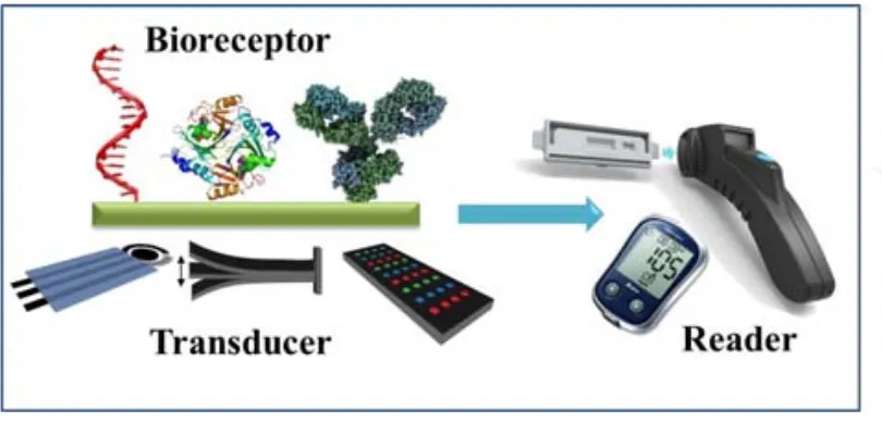
(29) Chapter 1 1.1. Point-of care biosensors During the last years, the development of analytical devices for detecting and quantifying a wide range of analytes with interest in food quality control, bio-security and especially, clinical diagnostics is continuously increasing. For example, monitoring of food and environment contaminants can lead to proper solutions that would avoid the effects produced in human health.1,2 In particular, biomarkers detection at low concentrations is extremely important in clinical diagnostics, even before any symptom of disease appears, since an early diagnosis is crucial for increasing medical treatment success and patient survival rates.3,4 In this regard, the development of the point-of-care (POC) testing devices, which can be used near to the patient or in resource-limited settings, has been growing in last years.5 The World Health Organization (WHO) has established the ASSURED criteria which have been widely applied in POC devices for their application in disease controls. The conditions POCs must fulfill are Affordable, Sensitive, Specific, User-friendly, Rapid and robust, Equipment-free and Deliverable to end-users.6 Biosensors represent interesting POC devices which fulfill the mentioned ASSURED criteria. According to the International Union of Pure and Applied Chemistry (IUPAC), a biosensor is an analytical device in which the recognition system is based on biochemical or biological mechanisms.7 A biosensor consists in mainly two integrated components: i) a biological element (such as enzymes, antibodies, ssDNA, tissues, microorganisms, etc.) or a synthetic receptor that recognizes and selectively binds to the target of interest and ii) the transducer or detector element that converts the binding reaction in a measurable quantity (see Fig. 1) which is displayed by a signal processor or reader device in a user-friendly way. Usually, biosensors can be divided according to the biological signaling mechanism or the signal transduction mode. Additionally, these transduction methods can be classified in two main groups: chemical or physical, in relation to the changes measured from the recognition event. In general, this classification leads to optical (surface plasmon resonance, chemiluminescence, fluorescence), electromechanical/mass (quartz crystal microbalance, surface acoustic wave), electrochemical (amperometry, potentiometry, conductimetry and voltammetry) beside other (ex. thermal change-based) biosensors.. 3.
(30) Chapter 1. Figure 1 .1. Schematic representation of m ain components of a biosensor ( different possible bioreceptors and transducers) that result in an integrated biosensing system.. Commonly bio sensors can be ref erred as label-based and label-free according to the transduction m ethod. A label-based biosensor needs a signaling label (e.g. enzymes, nanoparticles, quantum dots) for monitoring the event s produ ced on the transducer surface, since the recognition event doesn’t give rise to changes in any (physic)chemical property that can be d etected by th e transducer. On the other hand, in a label-free biosensor th e interaction between the tar get and the recognition element on the transducer surface can be detected directly without the use of a tag/label. An important part of the work presented in this thesis was devoted to the development of nano materials-based ele ctrochemical biosensors. Due to. the fact that several. comprehensive reviews have been addressing nanomaterials-based electr ochemical biosensors,8–12 the follo wing section 1.2 is bri efly describing this to pic followed by section 1.3. related to the sta te-of-the-art of the use of nanopar ticles in paper-based. sensors, a growing research a rea which represe nts the se cond main topic of th is PhD thesis.. 1.2. Nanomaterials-based electrochemical biosensors Electrochemical biosensors are very interesting for POC devices, since they allow the analysis of diff erent s pecies with high specificity and sensitivity, besides of their quickness, ease of use and the possibility of conducting the analysis “on-site”. A typical example of these electrochemical biosensors is the glucos e sensor,13 which has be en 4.
(31) Chapter 1 widely studied and improved through three generation of biosensors and also been commercialized since decades.In recent decades, nanomaterials have gained much interest due to their novel and unique properties which makes them useful for different fields. The term nanomaterial is referred to any material with size dimensions in nanoscale (1-100nm). Their physical and chemical properties depend on the size, as well as the percentage of atoms at the surface of the material that becomes significant as the size become smaller, making them more chemically reactive.14 In particular, nanomaterials such as nanoparticles, carbon-based nanomaterials and nanocomposites, have been used for the development of new and highly sensitive biosensors or for improving the performance of the existing ones, offering new methodologies for electrochemical signal transduction.15–18 Application of nanomaterials in electrochemical biosensing can be classified according to their two main functions either as labels or as modifiers of electrotransducing surfaces. Both strategies take advantage of the outstanding properties of nanomaterials (especially, nanoparticles) in order to enhance the electrochemical signal when used as label, or to improve the electron transfer rate when used as modifiers of electrochemical transducers. 1.2.1. Nanomaterials as labels The analytical performance of electrochemical assays has been improved thanks to the excellent catalytic activity, conductivity and biocompatibility of nanomaterials used as labels, that allows to obtain lower limits of detection (LoD) to in electrochemical bioassays.19 Different strategies for labelling biomolecules with nanomaterials have been reported. Briefly, these approaches include: i) covalent interactions, performed by direct chemical reaction, linker chemical reaction and click chemistry, forming stables bioconjugates; ii) non-covalent strategy make use of the electrostatic interactions, - stacking or Wan der Waals forces between the biomolecule and nanoparticle; and iii) affinity interactions such avidin/biotin which enable the effective conjugation of biomolecules to nanomaterials. A comprehensive review was assembled by Lei, where all these methods and their application for signal enhancement are explained.20. 5.
(32) Chapter 1 Detection of nanoparticles (NPs) as labels can be conducted directly by generation of electrochemical signals due to their own redox properties, or indirectly because of their electrocatalytic properties towards to other species. 1.2.1.1.. Direct electrochemical detection. In despite of the high sensitivity that offers the well-established stripping voltammetry, in which dissolution of metal NPs and subsequent voltammetric detection of metal ions in solution is achieved, this technique requires an extra analytical step and the use of toxic reagents. On the other hand, it is possible the metal detection without the dissolution of NPs, but it is necessary a direct contact between the electrode surface and superficial atoms of NPs. Thus, this strategy could result in a loss of sensitivity compared with stripping voltammetry. However, this type of detection offers advantages for sensing applications due to their fast response, procedure simplification and the reduced cost of analysis. The use of gold nanoparticles (AuNPs) as label on the direct electrochemical detection of anti-Human IgG was described for first time by Costa García et al. obtaining a LoD of 1.38 x 10⁻⁸ M.21 After this pioneer work, other important contributions emerged to the direct electrochemical detection. Pumera and co-workers described a new method for direct detection of AuNPs absorbed on the surface of graphite-epoxy composite electrode followed by their electrochemical oxidation in acidic media at fixed potential. The resulting tetrachloroaurate ions generated near the electrode surface were detected by differential pulse voltammetry (DPV), obtaining a LoD of 1.8 x 10⁸ AuNPs/cm3.22 Analysis of cadmium-sulfide quantum dots (CdS QDs) has been also reported, but in this case the Cd (II) ions present on the CdS QDs crystalline structure are electrochemical reduced to Cd (0) and immediately reoxidized to Cd (II), registering the oxidation peak.23 A double-codified nanolabel based on AuNPs modified with antihuman IgG horseradish peroxidase (HRP), has been reported for detection of human IgG.24 Thanks to the properties of this nanolabel, it was possible to measure the analyte concentration which was directly related to the AuNPs labels by using stripping voltammetry (Fig. 1.2A). The LoD for this electrochemical assay was significantly lower (260 pg/mL HIgG) than typical ELISA tests, showing to be a sensitive and rapid method. CdS QDs have been used as labels for the detection of carcinoembryonic antigen (CEA) in a disposable screen-printed graphite electrode by using square wave 6.
(33) Chapter 1 anodic stripping voltammetry to amplify the signal current response obtained from the dissolved a nti-CEA-CdS QD s (Fig. 1.2B). 25 The rep orted sens itive device showed a LoD of 32 pg/m L of CEA, co mparable to other elect rochemical assays and showed potential uses for POC diagnostics.. Figure 1.2. Direct electrochemical detection of nan oparticles. (A) Dire ct detection of HIgG based on a pre-oxidation of AuNPs as labels and later reduction of generated Au(III) ions. (B) Detection of QDs as labels in acidic solu tion by usin g square wave anodic strip ping voltammetry. Adapted with permission from references [24] and [25].. 1.2.1.2.. Indirect electrochemical detection: electrocatalytic determination. Alternatively to the direct electrochemical detection, electrocatalytic properties of NPs have been exploited to achieve high sensitive detection. By definition an electrocatalyst is a catalyst that participates in an ele ctrochemical reaction and contributes to th e electron transfer betwe en the electrode and th e rea ctants facilitating an int ermediate reaction. AuNPs are known also for their electrocatalytic properties , being their effect 7.
(34) Chapter 1 towards the silver deposition and the hydrogen ions reduction the most exploited in biosensing applications. In this regard, silver deposition on AuNPs has been reported for detection of DNA hybridization on magnetic beads used as immobilization platforms.26 This assay involves the hybridization of a target oligonucleotide to probecoated magnetic beads, followed by binding of the streptavidin-coated AuNPs to the captured target. Then, the DNA-linked aggregate is covered with silver, following the catalytic precipitation of silver on gold; these assemblies are magnetically collected on the electrode, leading a direct contact of the silver tag allowing the solid-state stripping detection. More recently, a novel magnetoimmunoassay with enhanced sensitivity due to the catalytic effect of AuNPs on the electroreduction of silver ions has been reported by our group.27 By choosing a suitable deposition potential, the direct electrocatalytic reduction of silver ions occurs onto the AuNPs surface and once a silver layer is already formed more silver ions are going to be reduced due to a self-enhancement deposition (Fig. 1.3A). Using this strategy, it was possible to obtain a low protein detection limit (23 fg/mL) which resulted to be 1000 times lower compared with direct electrochemical detection of AuNPs. On the other hand, electrochemical reduction of hydrogen ions by AuNPs was described for the first time by our group.28 Hydrogen ions present on the acidic medium are catalytically reduced to molecular hydrogen by the AuNPs on the surface electrode, generating a catalytic current which is related to the concentration of AuNP and enables their highly sensitive quantification (Fig. 1.3B). This method has been reported for the anti-hepatitis B virus antibodies detection29 and tumor cells detection.30. 8.
(35) Chapter 1. Figure 1.3. Indirect elec trochemical detectio n based on el ectrocatalytic pr operties of nanoparticles. (A) Left. Detection base d on th e catalytic pr operties of AuNPs on the silv er electroreduction in a magnetosandwich immunoassay and the further silver reoxidation. Right. Peak curren t obtained by th e re-oxidation of silv er ions that can be related with th e protein concentration. (B) Left. Hydrogen Evolution Rea ction in duced by AuNPs : In acid media, AuNPs are superficially oxid ized at +1.35 V gen erating Au (III) io ns, fo llowed by cata lytic reduction of hy drogen ions at -1.0 V p romoted by th e released gold io ns and AuNPs. Right: Detection of HIgG by i ndirect electrochemical method. Adapted with permission fro m references [27] and [28].. 9.
(36) Chapter 1 Catalytic activity of platinum nanoparticles toward oxygen reduction reaction has been reported by Polsky et al.31 In this work, the antibodies were previously immobilized onto AuNPs and then Pd was deposited onto the AuNPs surface, forming core-shell Au@Pd NPs. Detection of antibodies is achieved indirectly by measuring the catalytic current generated from this electrocatalytic reduction in glassy carbon electrodes.. 1.2.2. Nanomaterials as modifiers of electrotransducing surfaces Electrochemical performance of an electrode is strongly influenced by its size and composition that can be tailored in order to enhance the analytical output. Modification of electrotransducer surfaces can be achieved with NPs, promoting the electron transfer and improving the electrochemical response. AuNPs were the first example of nanoparticle-modified electrodes because of their electrocatalytic and conductive properties, and also their ease of synthesis. In addition, AuNPs offer stable immobilization platforms without degrading the activity of the biomolecules which is a major advantage in the design of biosensors.10,32 Different methods for incorporating NPs to conventional surface electrodes are described in literature such as: i) adsorption of nanoparticles to the bulk electrode surface; ii) addition of NPs in a composite; and iii) layer-by-layer.32 Ding et al. reported the electrodeposition of AuNPs to an ionic liquid carbon paste electrode for determination of -fetoprotein at clinical relevant levels.33 Determination of human chorionic gonadotrophin (hCG) with high sensitivity was reported by Chen and coworkers by using a highly hydrophilic, non-toxic and conductive colloidal AuNPs/titania sol–gel composite membrane prepared on a glassy carbon electrode.34 An ultrasensitive immunosensor for detecting prostate specific antigen was conducted in a pyrolytic graphite electrode modified with glutathione-decorated AuNPs assembled layer-by-layer, showing a LoD 8-fold better than other reported methods (Fig. 1.4A).35. 10.
(37) Chapter 1. Figure 1.4. Nanomaterials as modifiers of electrotransducing surfaces. (A) Left: Sche me of detection of pr ostate specific antigen using a m odified pyrolitic graphite ele ctrode with glutathione-decorated AuNPs as sembled layer by layer. Left: catalytic elec trochemical reduction of hydrogen peroxide on HRP/AuNP electrodes from rotating disk amperometry and atomic fo rce microscope i mages of AuNP platform aft er covalent linkage of anti -PSA antibodies onto the glutathion e carbo xylate gr oups of AuNPs. (B) Left: h ierarchically dr iven single crystalline IrO 2 on a platinum microwire, which is directly used as an elec tric signal transducer fo r hi ghly effec tive sensing of bi ologically relevant m olecules su ch as H 2O2 and NADH. Right: amperometric responses of the bare Pt microwire and IrO 2-platinum microwire electrodes to successive additions of H 2O2 aliquots in to a stirred PBS solution at neu tral pH. Adapted with permission from references [35] and [36].. Metal oxid es often exhibit semiconductor prope rties that may b e modified by decreasing the siz e up to the nanoparticulate state. Nanostructurated metal oxi des constitute exceptional immobilization plat forms and/or m ediators thanks to inherent properties o f nanoparticles, bes ides to chemical stability, nontoxi city also exhibit enhanced ele ctron-transfer kineti cs. and. biocompatibility, provi ding. suitable. 11.
(38) Chapter 1 microenvironments for the immobilization of biomolecules.37 In this regard, nanostructurated metal oxides are used in a wide range of analytical applications. Wen et al. reported a sensitive sensor for hydrogen peroxide (H2O2) detection by using Au/Pt hybrid nanoparticles self-assembled onto amino-functionalized titanium oxide (TiO2) colloid spheres obtaining a LoD at nanomolar levels.38. Moreover, the TiO2-. Au/Pt was employed on the photoelectrochemical catalysis of glucose, demonstrating the excellent performance-enhancing of this hybrid material. Recently, Shim and coworkers, reported the analytical application of hierarchically driven IrO2 nanowires on the surface of platinum microwires for the electrochemical detection of H2O2 and dihydronicotinamide adenine dinucleotide (NADH) without the aid of enzymes, achieving a LoD of 5 µM and a very short response time of 10 s (Fig. 1.4B).36 Detection of Mycobacterium tuberculosis has been reported by using a gold electrode modified with nanostructurated zirconium oxide film in which a ssDNA specific to the bacteria is immobilized, achieving a LoD of 0.065 ng/µL in less than 60 s.39 Electrochemical biosensors are significant tools for a wide range of analytical applications, especially in diagnostics, because of their good performance and sensitivity. New trends on POCT are leading to simple, portable and easy-tomeasurements biosensing devices, taking advantages of the nanotechnology and material science in order to develop a new generation of biosensors that satisfies the requirements of an ASSURED device.. 12.
(39) Chapter 1 1.3. Lateral flow technology 1.3.1. General introduction: Historical perspective The concept of rapid body fluid tests is documented in the history since thousand years ago. In ancient China, a saliva-based diagnostic called “the rice test” determined the guilt of an accused person.40 The earliest urine-based tests for determining the pregnancy was documented on Egyptian papyrus of 1350-1200 BC. 41,42. : if the urine of. the woman was able to provoke (or not) the growth of wheat, then it was possible to give a pregnancy diagnosis. In the Middle Age and Renaissance, pregnancy and some diseases could be diagnosed by the color of the urine. In nineteen century, physicians still practiced the examination of urine in a more rational way including scientific approaches but it was not until early 1900 that the term “hormones” was introduced by Ernest Starling.43 After this study, scientists began to explore in detail a particular hormone that can be found only in pregnant woman called human chorionic gonadotropin (hCG).. 44,45. In this context, many advances have been achieved until the. development of devices for rapid diagnostic methods being lateral flow (LF) devices one of the most important. The basic principles of the LF dated back middle 1950 starting with the work of Plotz and Singer using the latex agglutination test.46 At the same time, other applications in immunoassays with special importance for LF were developed. The radioimmunossay (RIA) by Berson and Yalow47 based on the detection of radioactive compounds, although being a very sensitive and specific technique due to the special equipment and precautions required, was replaced by a safer and nonhazardous-based technique such as the enzymatic immunoassay (EIA).48,49 In 1980 Leuvering et al.50 reported the sol particle immunoassay (SPIA) in which silver and gold colloids was used as labels for sandwich immunoassays obtaining limits of detection comparable to RIA and superior in comparison to EIA. This work became pioneer in the use of nanoparticles as labels in immunoassays. The main application for LF technology is the human pregnancy test developed in mid1970’s. The “early pregnancy test” (e.p.t.TM) developed by Warner-Chilcott was a complex system that involved the use of different tubes and reagents and the result was obtained about two hours making difficult and ambiguous the determination of the end point. Since the launching of this home-diagnostic method, lateral flow assay (LFA) gained more importance in the biosensors field and continued progressing becoming 13.
(40) Chapter 1 completely established in the late ’80s by the introduction of several patents related to LF devices for pregnancy test.51–53 In 1988, U nipath L td, launched the first one-step rapid LF assay for home pregnancy test known as Clea rblue brand. This test was specifically qualitative: if two blue lines are developed means that woman is pregnant being the result visualized in few m inutes. In a pre gnant woman, leve ls of rapidly rise and can be dete cted in urine f rom 9 -10 days. after conception. hCG 54. and. Clearblue™ is able to detect hCG protein in urine. Although pregnancy tests have been conceived for providing qualitative visua l results (yes/no response), it is possible to obtain semi-quantitative results in order to estimate the pregnancy stage. Pregnancy test market has also evolved for generating new and more accurate devices and nowadays it is possible to find digital pregnancy test which give semi-quantitative results. This is a new generation of devices where LF technology is integrated with electr onics in the same platform. 1.3.2. Lateral Flow technology and its format assays 1.3.2.1.. Operation Principles. The LFAs are based on chromatographic principle in which test sample flows through a solid sub strate v ia capillary action. Each strip is composed by dry pre-stored rea gents and different porous membranes that have specific functions.. Figure 1.5. (A) Components o f a l ateral f low strip. ( B) Scheme o f a t ypical l ateral f low assay (LFA).. 14.
(41) Chapter 1. Figure 1.5 shows a typical scheme of LF strip. As pre-requisite for the proper functioning of LFA, all the components of the strips must be previously treated for making them hydrophilic and overlapped between them over an adhesive backing card to guarantee the continuous flow of the sample. The fluid sample applied at the end of the strip (at the sample pad) migrates through the next region, the conjugate pad, where nano/microparticles conjugates are immobilized. This conjugate is composed by the biological components of the assay, e.g. antibodies, which are attached to the particles and previously have been deposited in the conjugated pad. In this step, the analyte interacts with the biological component of the conjugated particle forming a complex. There is a wide variety of particles used as labels (see Section 1.3.2.2.2), but the most commonly used are latex microparticles and AuNPs. The sample continues migrating by capillary forces through the detection pad, composed by a porous membrane in which the biological complementary part of the assay has been previously deposited. The capture lines, test and control line, are constituted by antibodies, antigens or proteins and their function is to capture the analyte and the excess of conjugated particle, respectively. The excess of reagents is absorbed by the wicking or absorbent pad located at the end of the LF strip. Visual results can be interpreted as the presence or absence of analyte in sample and can be detected by naked-eye or using a strip reader. According to the type of format used, the results can be interpreted in opposite way. In figure 1.6 details of two main LFA formats such as the sandwich (direct) and competitive assay are shown. For the detection of molecules with a high molecular weight, with two or more antigenic sites e.g. big proteins, the sandwich assay is used (figure 1.6A). In this case, the presence of analyte shows two colored zones: test and control lines. On the other hand, if the analyte is a low molecular weight molecule with one antigenic site (hapten) for binding with antibody, e.g. pesticides or toxins, the competitive assay is employed. In this type of assay, the analyte and the analyte immobilized at the membrane compete for binding with the detector antibody. The presence of analyte is indicated by the absence of test line, therefore, only one line at the control zone is observed (see figure 1.6B). In both cases, the control line is always visible and independent of the format used allowing to control the adequate migration of the sample through the strip. 15.
(42) Chapter 1. Figure 1.6. Typical formats of LFAs: (A) direct and (B) competitive assays.. 1.3.2.2.. Components. 1.3.2.2.1.. Assay membranes. The function as well the materials that constitute the different components of LFA are summarized in Table 1.1 and discussed in the following part. a) Sample pad Its purpose is to receive and then distribute the sample in a controlled and uniform way, releasing the analyte with high efficiency.55 Additionally, it controls the rate flow that enters to the conjugation pad preventing flooding of device. The most commonly used sam ple pads are porous materials such as cellulose . Cellulose has lar ge bed volumes (> 25 µL/cm 2) and are extensively used for testing various kinds of liquid samples (ex . uri ne). Du e to adsorption capability loadi ng of blocking a gents, pH and ionic strength modifiers and viscosity enhancers is easy to be performed. When pre-treated by impregnation with proteins, buffers and detergents, the sample pad can modulate the flow properties increasing the sample viscosity, preventing nonspecific adsorptions of both the co njugate and analyte, increasing the reaction time in the conjugate pad modulating in this way the interaction with the sample.. 16.
(43) Chapter 1 b) Conjugate pad The function of this pad is to keep the dried conjugated particles and hold them stable in the pad until the test is run and to uniformly transfer the signaling reagent (ex. gold nanoparticles conjugates) and sample onto the membrane.55 When the sample flows through the conjugate pad, the signaling reagent solubilizes, recognizes the analyte and the formed conjugate moves through the membrane. Assay sensitivity can be severely affected by the conjugate deposition, drying, inadequate conjugate mixing and releasing from the pad. Ideally, the conjugate pad must have low non-specific binding of signaling reagent or analyte to the pad, allow a suitable formation of the immunocomplex at the test line. In addition must ensure a constant flow and bed volume so as to avoid problems of uneven signals that can alter the sensitivity of assay. Glass fibers, cellulose filters, surface-treated (hydrophilic) polyester or polypropylene filters and rayon are the most used materials for conjugate pad. To ensure an optimal release of the sample, the pad must be pre-treated with a pH adjusted buffer containing proteins and surfactants, followed by a suitable drying. The deposition of conjugated particles can be achieved manually (batch mode) or by using a dispensing machine that delivers in a continuous mode solutions of reagents including suspension of nanoparticles modified with antibodies. c) Detection pad Its main purpose is to bind irreversibly the capture reagents at the test and control lines and facilitate the visualization of colored bands indicating the presence or absence of analyte in the sample.55 Nitrocellulose (NC) is the most widely used material for detection membranes in LF technology and other point-of-care (POC) devices. Depending on the application, other materials, e.g. nylon and polyethersulfone can be used as detection membrane. NC is a versatile polymer that has been used for years due to its unique as biomolecules adsorbing properties. For this reason, the use of NC has been extended to powerful blotting techniques for identifying. the presence of biomolecules such as proteins. (Western blot), DNA (Southern blot) and RNA (Northern blot).56–58 Based on these applications, over the past 25 years, NC has gained importance as a suitable porous support on LF technology.. 17.
(44) Chapter 1 Detection membrane is the most important part of a lateral flow assay because the binding and detection occurs within it. Therefore, the adsorption of biomolecules is an essential aspect for a good performance of LFAs. Even though the binding protein to NC has been reported and used for powerful and existing techniques, the binding mechanism is still not clear enough.57 Different mechanisms have been proposed for explaining the binding mechanism. According to one of the theories proteins are thermodynamically driven to adsorb to nitrocellulose by non-covalent interactions.56 Others claim that an initial attraction of protein to NC involves the interaction between the dipoles of the nitro groups on the NC and the carbonyl groups in peptide bonds of protein.59 Either way, different forces (hydrogen bonding, hydrophobic and electrostatic interactions) are involved in the adsorption process of protein to NC; as recommended LF manufacturers must consider all these interactions, materials and assay reagents for optimizing the binding of the proteins to NC.60 Membranes made of nitrocellulose are hydrophobic by nature. However, membranes can easily be wet due to the presence of surfactants added during membrane production55. Selection of type and amount of surfactant use to be recommended by the manufacturer but also adapted according to the application. Once the membrane is processed and ready to be used, depositing of the capture reagents at test and control lines, can be carried out using either contact or non-contact dispensing system. The best option is non-contact dispenser, typically inkjet system, because the membrane does not suffer damages that can impede a normal flow of the sample. Nevertheless, low-contact dispenser systems can be found in the market.61 In this case, the nozzles can dispense the sample with a minimal contact pressure avoiding any mechanical stress on the membrane. Flow rate is difficult to be measured since decays exponentially while the fluid front moves through the membrane.55 A common parameter to evaluate the NC membranes is the capillary flow time which is the time required for a liquid to move along and fill completely 4-cm long of membrane. The capillary flow time and the sensitivity of the assay increase when the pore size decreases, since the reagents spend more time on the membrane and the formation of immunocomplex between the analyte and the label is more effective at the test line.. 18.
(45) Chapter 1 Another parameter is the thickness of the membrane since it has a direct influence on bed volume and signal visibility. Bed volume is the volume of liquid required to saturate a given area of membrane and is related to the porosity and the thickness. As the thickness increases, the bed volume also increases. However, this consideration may be not important when an absorbent pad is used because the total volume of the sample is governed mainly by the bed volume of the absorbent pad.55 When capture reagents are dispensed onto the membrane the liquid penetrates downward into it and moves laterally. In a thin membrane, spreading of the reagents will be occurring since its insufficient depth provokes the diffusion of the liquid (including nanoparticles used as labels) through the whole membrane causing difficult to be visualized signals (diffused / undefined detection and control lines). d) Absorbent pad Located at the end of the strip, its main function is to drain off the fluid from the membrane. Using an absorbent pad, the volume of the sample can be increased, being the background signal reduced and the sensitivity of the assay improved. Most of absorbent pad are made of high-density cellulose and no treatments are required. However, once the experiment is achieved and a visual record is needed, it is recommendable to remove the absorbent pad because it can act as reservoir once the liquid has evaporated and it might release the excess of label reagent causing false positives.62. 19.
(46) Chapter 1. Table 1.1. Components of LFA and their functions. Components. Material. Function Distributes the sample in a controlled way. Sample pad. Cellulose. for releasing the analyte with high efficiency Retains the conjugate between its fibers. Conjugation pad. Glass fiber. until the test is running. Coupling of analyte and conjugate takes place in this pad Binds irreversibly the capture reagents and. Detection pad. Nitrocellulose. facilitates the visualization of test and control lines. Absorbent pad. 1.3.2.2.2.. Cellulose. Drains off the excess of fluid of the detection pad. Labels. Immunoassay conjugate consists of an antibody with an appropriate label which function is to generate a signal that correlates with the immunocomplex formed and its antigen, allowing the detection by using techniques such as colorimetry, fluorescence and luminescence.63 Most of the commercial LFAs use colored latex beads, carbon nanoparticles and gold nanoparticles which allow visual detection without the use of readers if the test provides a qualitative response. On the other hand, if LFA is intended to be quantitative, the use of a reading system is required depending on the type of label used, for example: charged couple devices (CCD) cameras for AuNPs or colored beads; readers based on ligh-emitting diode (LED) technology for fluorescent labels, and magnetic readers such as Magnetic Assay Reader (MAR™) for superparamagnetic labels. Applications of traditional labels will be briefly summarized in this section, while gold nanoparticles, will be discussed in detail in section 1.3.4. 20.
(47) Chapter 1 a) Latex beads Latex beads are spherical microparticles composed of an amorphous polymer (usually polystyrene). These beads have been used as the first labels in LF technology due to their high binding protein capacity.63 Latex beads with various sizes and with controlled surface chemistries and able to incorporate colored dyes, fluorescent dyes and also magnetic or paramagnetic components have been already developed and are available in the market. Sensitivity of LFA that employs colored latex beads is in principle lower compared with the achieved with gold nanoparticle labels since their smaller sizes (typically 20-40nm) and consequently their higher surface area to volume ratio allow them to bind more antibodies onto their surface. b) Liposomes Liposomes are colloidal vesicles formed by a lipid bilayer membrane capable to encapsulate part of the aqueous media in which they are dispersed.64 They can encapsulate i.e. visual and fluorescent dyes or enzymes inside their cores, improving the sensitivity of LFA. It is possible to add different functional groups to the lipid surface for coupling with biomolecules or chemicals.. Different studies with interest for. example for DNA65, allergenic agents66 and other analytes have been reported. However, liposomes have some disadvantages related to their high production cost, short shelf-life and propensity to leakage and fusion of encapsulated molecules. c) Carbon nanoparticles Carbon nanoparticles are the most inexpensive label for LFA and provide an excellent color contrast to the white NC membrane. These amorphous labels are suitable for physical adsorption because of their hydrophobic features and have been used for DNA67,68 and small molecules e.g. chemical contaminants69,70 detection. The main limitations of carbon nanoparticles are related to their low density, which difficult their visualization by electron microscopy (TEM or SEM), their irregular sizes and shapes and the requirement of surfactants addition to stabilize the carbon dispersions.71. d) Fluorescent labels Organic and inorganic fluorophores can also be used as labels. A typical organic fluorophore for labeling antibodies is fluorescein isothiocyanate (FITC) which is 21.
(48) Chapter 1 reactive to the primary amines of antibodies producing a fluorescent conjugate.72 LFA using fluorescent labels to detect for example influenza virus has been shown to be more sensitive that colorimetric assays.73 Organic fluorescent dyes (e.g. Ru(bipy)32+) have been used for coating of silica nanoparticles and their subsequent use in LFA demonstrating to be remarkably more sensitive than gold nanoparticles for nucleic acid detection.74 Inorganic fluorophores such as QDs and lanthanides also have been reported for LF applications. QDs are semiconductor nanoparticles in size range of 1-10 nm composed of GaN, GaP, ZnO, ZnS, CdS or CdSe75. As a consequence of their small sizes, they have unique properties such as the high quantum yield and molar extinction coefficients, broad absorption spectra and resistance to photobleaching and chemical degradation.76 Uses of QDs in biosensors have grown in last years because of their advantages over common fluorophores. QDs can be conjugated to different biomolecules, especially antibodies. However, the main limitation of QDs is related to their low colloidal stability in biological media.75 QDs have been used as labels in LFA in competitive assays for detection of a primary metabolite biomarker for pesticides77 toxins detection78 and also, in sandwich formats for cancer biomarkers.79 Lanthanides (e.g. Eu(III)) also have unique fluorescent properties that can be applied for their use as sensitive labels for prostate-specific antigen (PSA) detection in LFA, increasing 300-fold the sensitivity of assay.80. 1.3.3. Lateral Flow Assays advantages and limitations. LF is a well-established technology that has many advantages such as a) rapid qualitative or semi-quantitative tests; b) easy to use (results can be interpreted by untrained personnel); c) utility for in-field applications in remote areas where high-tech equipment is not available; d) prolonged shelf-life (about a year) and often without refrigeration; e) small volumes required; f) relatively low-cost production; g) good sensitivity and specificity and h) possibility of integration with electronics and electrochemical systems. 22.
Figure


+7


Documento similar
For this, the IAD algorithm provides an accurate estimation of the optical properties for biological tissues, such as artificial human oral mucosa and native rabbit oral
The purpose of the research project presented below is to analyze the financial management of a small municipality in the province of Teruel. The data under study has
Next, the electrochemical properties of the new μ‐azo derivatives were evaluated by cyclic voltammetry in THF solutions (Figure 3c,d). We
The redemption of the non-Ottoman peoples and provinces of the Ottoman Empire can be settled, by Allied democracy appointing given nations as trustees for given areas under
In order to study the closed surfaces with constant mean curvature in Euclidean space, Alexandrov proved in 1956 that any embedded closed CMC surface in R 3 must be a round sphere..
“ CLIL describes a pedagogic approach in which language and subject area content are learnt in combination, where the language is used as a tool to develop new learning from a
In the preparation of this report, the Venice Commission has relied on the comments of its rapporteurs; its recently adopted Report on Respect for Democracy, Human Rights and the Rule
H I is the incident wave height, T z is the mean wave period, Ir is the Iribarren number or surf similarity parameter, h is the water depth at the toe of the structure, Ru is the
