Animal test alternatives in metal toxicology research. A study by "in vitro" cellular systems
136
0
0
Texto completo
(2) This work has been carried out at the Joint Research Centre- Ispra (JRC) of the European Commission, Institute for Health and Consumer Protection (IHCP), ECVAM Unit (Professor Michael BALLS, Head of Unit)..
(3) Index Page Introduction. 1. 1.1. Alternative Testing Methods. 1. 1.2. The present research in the context of ECVAM activities: the IMETOX project. 2. 1.3. Metal toxicity and human health. 5. 1.4. Objectives of the present research. 9. Literature survey. 11. 2.1. “In vitro” tests and their growing importance in metal toxicology. 11. 2.1.1. Screening studies. 13. 2.1.2. Mechanistic studies. 14. 2.1.3. Contribution to risk assessment. 16. 2.2.“In vitro” models for metals in different toxicological areas. 17. 2.3. Metal dermatotoxicity. 17. 2.3.1.HaCaT cell line 2.4. Metal neurotoxicity 2.4.1. PC12 cell line 2.5. Metal embryotoxicity 2.5.1. Embryonic stem cells, D3 2.6. Metal carcinogenicity 2.6.1. Balb/3T3 cell line 2.7. Selected metals 2.7.1.Platinum as new potential environmental pollutant. 20 21 27 29 31 32 35 37 37. 2.7.2. Environmental levels and levels in diet, biological fluids and human tissues. 37. 2.7.3. Toxicological/carcinogenic effects and estimation of health risk. 39. 2.8. Methyl Mercury, a well known poisoning. 41. 2.8.1. Environmental exposure. 42. 2.9. Arsenic, as environmental pollutant 2.9.1. Toxicological/ carcinogenic effects. 43 45.
(4) Page 2.9.2. Biotransformations. 46. Materials and methods. 48. 3.1. Experiments on metal dermatotoxicity. 48. 3.1.1. Chemicals. 48. 3.1.2. HaCaT cell line. 49. 3.2. Experiments on metal neurotoxicity. 52. 3.2.1. Chemicals. 52. 3.2.2. PC12 cell line. 53. 3.3. Experiments on metal embryotoxicity. 56. 3.3.1. Chemicals. 56. 3.3.2. Embryonic Stem Cells D3 cell line. 56. 3.4. Experiments on metal carcinogenicity. 60. 3.4.1. Chemicals. 60. 3.4.2. BALB/3T3 cell line. 61. 3.4.3 Analytical determination of arsenic. 65. 3.5. Statistical treatment. 68. Results. 70. 4.1. HaCaT cell line. 70. 4.1.1. Study on different Platinum compounds. 70. 4.1.2. Systematic study of the cytotoxic effect of 51 metal compounds 4.1.3. Dose- effect study on selected metals 4.2. PC12 cell line. 73 77 79. 4.2.1. Systematic study of the cytotoxic effect of metal compounds. 79. 4.2.2. Dose- effect study on selected metals. 82. 4.3. Embryonic stem cells, D3. 88. 4.3.1. Study of the effect of methyl mercury by the ES Cell Differentiation Assay 4.4. BALB/3T3 cell line. 88 89. 4.4.1. Uptake and intracellular repartition of As. compounds in BALB/3T3 cells. 89.
(5) Page 4.4.2. Neoplastic morphological transformation of BALB/3T3 cells by As compounds. Discussion. 92 93. Conclusions and some priorities for future research. 102. Acknowledgements. 105. References. 106.
(6) INTRODUCTION. 1.1.. Alternative Testing Methods. Regulatory toxicity testing in animals raised a number of scientific, humanitarian, legislative, practical and economic questions and conflicts. For example, while the use of animal procedures in identifying the potential hazards represented by chemicals and products of many kinds is required by various laws and regulations, there are other laws, of no less importance, which are intended to protect laboratory animals from unnecessary pain, suffering, distress or lasting harm (Balls, 1995a). They stipulate that the use of laboratory animals should only be permitted when it can be justified as necessary on strong scientific grounds. However, the scientific basis of many animal tests themselves, and, in particular, of the ways whereby the data they provide are currently applied in human risk assessment, is weak. In addition, the present dependence of hazard prediction on animal tests requires considerable human and economic resources, as a result of which only a very small proportion of the chemicals which might threaten the well- being of humans, and of the environment in general, have been subjected to anything approaching a scientifically- satisfactory evaluation (Balls, 1995b). Non- animal tests and testing strategies can offer solutions to many of the problems caused by the current over- reliance on the routine, check- list application of animal tests. They can offer a more- mechanistic basis for understanding toxic effects at the systemic, organ, cellular and molecular levels (Sabbioni and Balls, 1995). When human cells and tissues are used, or modelling is based on human experience and responses, the problem of species differences, which severely limits the relevance of animal tests, can be avoided. The change of emphasis from “in vivo” to “in vitro”. 1.
(7) approaches must also embrace the increasing use of properly- obtained and safelymaintained “human” rather than “animal”, cells and tissues. Well- designed “in vitro” studies can also assist with the selection of the most- appropriate laboratory animal species for any subsequent, necessary use as a model for man (Fentem and Balls, 1994). Non- animal methods offer an escape from many of the ethical and legal conflicts inherent in current practices. Often, they also offer the possibility of moremanageable, less time- consuming and less expensive testing strategies, so that the practical and economic limitations which currently prevent the adequate testing of many chemicals and products can be overcome (Balls, 1995a).. 1.2 The present research in the context of ECVAM activities: the IMETOX project. Cell culture technology and accompanying “in vitro” cytotoxicology, is an important newly developing discipline of modern toxicology and is gaining increasing acceptance in the field. In this context, it is useful to separate cell culture methods in toxicology into two different areas of applications which complement each other: a) study of mechanisms of toxic effects, including effects on cell membranes, DNA, protein and lipid production as well as biotransformation of chemicals b) testing, where the qualitative and quantitative toxicity of previous unclassified chemicals is estimated (Sabbioni and Balls, 1995). These aspects are also key points of the research activities carried out in the context of IMETOX project (“In vitro” Metal TOXicology) (Sabbioni, 1998) at ECVAM (European Centre for the Validation of Alternative Methods) which foresees the use of “in vitro” toxicity testing of trace metals in different toxicological areas such as. 2.
(8) embryotoxicity, haematotoxicity, nephrotoxicity, neurotoxicity, metabolism-mediated toxicity (Balls and Fentem, 1997). Since this work is a part of the IMETOX herein few essentials on the aims and activities concerning ECVAM and IMETOX projects as reported below. ECVAM is a Unit of the JRC at Ispra created by the European Commission in October 1991 to implement the Directive 86/ 609/ EEC regarding the Protection of Animals Used for Experimental and Other Scientific Purposes (O. J. EEC, 1986) which is directed to reduce, refine and replace the use of laboratory animals in accordance with the Three Rs concept of Russel and Burch (Russel and Burch, 1959). The main task of ECVAM is the co-ordination of the validation of alternative testing methods at the EU level; and the promotion of dialogue among legislators, industries, biomedical scientists, consumer organisations and animal welfare groups with a view to the regulatory acceptance of non- animal, regulatory tests that is based on the formal validation which includes three phases (Figure 1.1):. The IMETOX project aims at integrating the aspect of toxicity of metal compounds with current ECVAM activities related to the in- house development of “in vitro” tests and prevalidation/ validation studies. Key aspect of the project is the study of the response to metal exposure on cellular level (uptake, intracellular distribution, biotransformation and cytotoxicity of metals) in relation to endpoints at molecular level. Exposure of cells to low doses of specific or combined mixtures of different metal compounds and long term cell culture testing are other main aspects considered (Sabbioni and Balls, 1995). Selection of metal compounds to be tested takes into account the existing “in vivo” knowledge on their metabolism, mechanisms of toxicity, induced toxicological effects. 3.
(9) which are relevant to environmental and occupational exposure, drug therapy, use of food and cosmetics, implantation of biomedical devices. The choice of cell lines for toxicity testing and the selection of metal compounds investigated in this work are placed in this context.. Figure 1.1 STAGES IN THE EVOLUTION OF REGULATORY TESTS. 1. TEST DEVELOPMENT (Laboratory of Origin) • Purpose and need for test • Derivation of method • Application to appropriate chemicals • Prediction model for application of results. 2. PREVALIDATION (Informal Interlaboratory Study) • Confirmation of interlaboratory transferability • Optimisation of prediction model. 3. VALIDATION (Formal, Interlaboratory Study, including a Blind Trial) • Study design • Selection of tests and laboratories • Selection and distribution of chemicals • Data collection and analysis • Assessment of outcome according to prediction model • Evaluation and proposal of next steps. 4. INDEPENDENT ASSESSMENT OF STUDY AND PROPOSALS. 5. PROGRESSION TOWARD REGULATORY ACCEPTANCE. a: from Balls and Fentem, 1997. 4.
(10) 1.3. Metal toxicity and human health There are several reasons for the great interest in the biological effects of trace metals from the aspect of their toxicity. The concept of the ”health impact” of trace metals can be discussed characterising the optimal doses of metals responsible for the essential actions in the organism, and on the other side, the doses able to induce toxic effects in the organism as a consequence of excessive expositions to trace metals. Bertrand (Bertrand, 1912) studied how high concentrations of trace metals present in the environment represent a potential health risk for the human body. In particular, the concentrations of certain trace metals or their compounds in the environment and their uptake by the organism may, under certain conditions, reach levels incompatible with human health. For some time, these adverse conditions were confined to some pre- determined areas of the geosphere, where they have been existing without any significant interference through human activity. It has been found important to consider the relation between the exposure to single chemical agents and the relative human risk. Some trace metals are essential elements for reproduction, development and growth of the organism (Mertz, 1981). Intake of low doses of these metals can alter the optimal biologic function of the body. The biological function can be restored when normal doses of these elements in the body occur (Sabbioni, 1984). On the other hand high exposure to these essential elements can produce harmful effects to human health. This concept has been expressed by the dose- relationship curve (Bertrand, 1912) and represents still the basis of the modern trace metal toxicology. For some time the hazards connected with man- made changes in the environment were limited to selected groups of individuals. Toxic effects were confined to relatively narrow groups of individuals near the source of the toxic metals, particularly at the workplace (Browning, 1969). During the past decades the problem of trace metal toxicity has exceed. 5.
(11) the limits of geochemically or professionally exposed subjects. The broad scope of environmental changes, mineral pollution of air, water and soil, connected with the growth of industry, urbanisation, development of transport and the use of agrochemicals and other changes affecting human and animal nutrition, have caused far- reaching changes in the exposure of large population groups to certain trace metals (Sabbioni et al., 1983; Sabbioni et al, 1985 and Sabbioni, 1985a). The situation can be serious not only because toxic metals can remotely act from their original source and affect large population groups, but also because the changed uptake is not necessarily confined to one metal only but could involve a simultaneous uptake of other trace metals (interrelation among elements). Change of the exposure of living organisms to mercury and selenium at the same time by human activities can lead to unexpected toxic results (Moffitt and Clary 1974). Taking into account the above mentioned considerations the setting of standard health criteria to regulate the emission of trace metals in the environment and to establish maximum levels of exposure compatible with human health is necessary. Although this action involves political decisions, the determination of the criteria on which these decisions are based is a scientific problem that requires a multidisciplinary approach (Sabbioni et al., 1990). The risk that large population groups could be exposed to trace metals in a form and dose incompatible with good health and that this could arise from human activity seems to reveal completely new problems connected with trace metal toxicology. First of all, in addition to clinical manifestations of acute illness, much more should be known about subclinical adverse effects of the inadequate exposure of trace metals. For example, it is growing evident that in highly developed countries occupational exposure to toxic metals is decreasing, in some cases approaching the levels of environmental exposure. Thus, certain trace metals and their compounds can be involved in some chronic human diseases, affecting systems which are sensitive to low dose exposure over long periods such as. 6.
(12) nervous-, reproductive-, immune- and cardiovascular systems (Alessio, 1992). In this contest The European Commission promoted an action programme concerning the environmental protection (O. J. EEC ,1977; O. J. EEC, 1980; Vagn H. 1993). The importance of the exposure of trace metals is demonstrated by cases of intoxication of general population by cadmium and mercury (Kitamura, 1978; Kiragishi, 1981). These events were caused by scientific ignorance related to toxicological effects induced by longterm low doses exposure of species of both metals. The health impact of trace metals is complicated by the fact that people can be exposed to different chemical forms of the same metal like simple inorganic forms, complex inorganic forms and metallorganic forms (Sabbioni et al., 1987). The different forms of the same compound interact differently with cell components inducing different toxicological effects (speciation) (Sabbioni et al. 1990). Metabolic- speciation studies on inorganic arsenic have proved clear evidence that no animal species, among those so far investigated, is an appropriate model for arsenic metabolism in humans (Sabbioni et al., 1985b). The different metabolic patterns and toxicological effects of arsenic “in vivo” and “in vitro” as inorganic forms (trivalent and pentavalent ions) compared to organo- arsenic species (e. g. arsenobetaine in seafood) are examples of how different metal species of a same metal affect its toxicity (Bertolero, 1987 and Sabbioni, 1991). Inorganic arsenic (As(III) and As(V)) were found to be more toxic than organo arsenic species such as (monomethylarsonic acid (MMA), dimethylarsinic acid (DMA) and trimethylarsenoxide (TMAO), arsenobetaine and arsenocholine) (Sabbioni and Balls, 1995; Cocco, 1999). Chromium is another example is essential when its oxidation state is (III) but is toxic when is present as chromium (VI) (Mertz, 1972). Moreover, it has become clear that several metals undergo biotransformations in mammalian tissues and that the metabolism has important implications in trace metal human toxicology, pharmacology and occupational. 7.
(13) health (Manzo et al., 1992). The knowledge of the metabolism of the different chemical species of the trace metals is essential to describe the detoxification process of trace metals. In this process the initial toxicity of a metallic compound is reduced before its eventual elimination. Two examples are methyl mercury and arsenic. Mammals are able to de- methylate Hg and methylate As generating low toxic metabolites (Sabbioni et al., 1990; Marafante et al., 1982). These results are important for the researches aimed at developing metabolic models that describes quantitatively the toxicokinetics of absorption, of distribution, the transformation and excretion of the different chemical forms of trace metals in the organism. These models (Lutz et al., 1987) are an important aspect to understand the link between exposition models and models for risk analysis in order to predict the accumulation of metals in target organs. Therefore, it can be concluded that some aspects of trace metal toxicology should have priority and that certain research needs can be formulated concerning the present status of the problem: a) there is a need to provide more systematic information on the toxicological effects of trace metals following chronic exposure to levels which are normally present in polluted environments (long term- low level exposure)(Sabbioni, 1981). b) there is a need for metabolic studies as a basis for interpretation of metal toxicity. The potential of metals to cause toxicity has traditionally be regarded as a function of dose and potency of the metal itself. Since the effects of metals are related to their concentration at specific target sites, metabolic data are also important in interpreting toxicological findings. This includes also metabolic- speciation studiesat intracellular and molecular level. c) the assessment of risk to human health associated with exposure to trace metals involves an integrated approach which is based on data from “in vivo” studies on animals, clinical. 8.
(14) studies on humans and “in vitro” toxicity testing methods. These latter can contribute to give fundamental information on the ranking of metals for their potency, the identification of both mechanisms of toxic action of metals and of potential target organ toxicity (Frazier, 1992).. 1.4. Objectives of the present research The purposes of this work are connected with the aim of the IMETOX project (“In vitro” Metal TOXicology). In particular, the research of this thesis has been carried out in the context of four different toxicological areas (Table 1.1) such as: a) Dermatotoxicity. Investigations carried out concern a screening of the cytotoxic effect of 51 metal compounds in human keratinocytes (HaCaT immortalised cell line) exposed to a fixed dose of 100µM of individual metal compound and the setting of dose- effect relationships for selected metal compounds including “newer” potential environmental pollutants such as different chemical forms of Pt. b) Neurotoxicity. A systematic study on the cytotoxic response of 14 metal compounds was performed on engineered modified rat pheochromocytoma PC12 cell line by exposure to a fixed dose (100µM) of cells to individual metal compounds. c) Embryotoxicity (cardiotoxicity). A preliminary study was focused on the effect of methyl mercury at different stage of differentiation of embryonic stem cells D3 into contracting myocardiac cells. d) Carcinogenic potential. Uptake, intracellular repartition, cytotoxic effects and morphological transformation of inorganic species (As (III) and As (V)) as well as organo- arsenic compounds (arsenobetaine (Asβ), arsenocholine (AsCh)) in BALB/3T3 cell lines.. 9.
(15) Table 1.1- Objectives and type of studies of the present research. Cell line. Type of end- point. Expected contribution as. Type of study carried out. “in vitro” model Development HaCaT. CFE Test. of. a. Screening. toxicity. cellular model for metal. testing at a fixed dose.. dermatotoxicity. Determination. of. the. IC50 for selected metals Development PC12. MTT Test. of. innovative and sensitive. Screening. toxicity. testing at fixed doses. cellular model for metal neurotoxicity Development ES- D3. Beating measurements. of. Effect. of. MeHg. innovative and sensitive. differentiation. cellular. contractile cells. model. for. on in. embryonic cardiotoxicity Development BALB/3T3. CFE- Morphological Transformation (Foci formation). of. “in. Effect of different metal. vitro” model for the. compounds. potential carcinogenicity. formation of type III foci. of metals. 10. on. the.
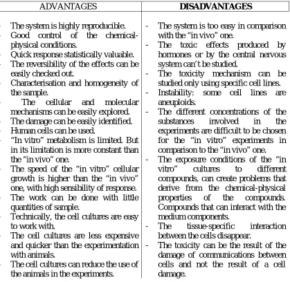
(16) LITERATURE SURVEY. 2.1. “In vitro” tests and their growing importance in metal toxicology There is growing and increasing interest in the use of cell cultures in metal toxicology research as simple and rapid “in vitro” assays that can act as preliminary screens, possibly reduce and replace the need for animal tests, and as a tool in understanding the mechanisms of metal- induced cytotoxicity at subcellular and molecular levels. Figure 2.1 summarises advantages and disadvantages of the use of cell cultures in toxicological studies connected with the exposure to xenobiotics, including trace metals. Figure 2.1- Advantages and disadvantages in the use of “in vitro” toxicity testing. ADVANTAGES -. -. -. -. The system is highly reproducible. Good control of the chemicalphysical conditions. Quick response statistically valuable. The reversibility of the effects can be easily checked out. Characterisation and homogeneity of the sample. The cellular and molecular mechanisms can be easily explored. The damage can be easily identified. Human cells can be used. “In vitro” metabolism is limited. But in its limitation is more constant than the “in vivo” one. The speed of the “in vitro” cellular growth is higher than the “in vivo” one, with high sensibility of response. The work can be done with little quantities of sample. Technically, the cell cultures are easy to work with. The cell cultures are less expensive and quicker than the experimentation with animals. The cell cultures can reduce the use of the animals in the experiments.. DISADVANTAGES -. -. -. -. 11. The system is too easy in comparison with the “in vivo” one. The toxic effects produced by hormones or by the central nervous system can’t be studied. The toxicity mechanism can be studied only using specific cell lines. Instability: some cell lines are aneuploids. The different concentrations of the substances involved in the experiments are difficult to be chosen for the “in vitro” experiments in comparison to the “in vivo” one. The exposure conditions of the “in vitro” cultures to different compounds, can create problems that derive from the chemical-physical properties of the compounds. Compounds that can interact with the medium components. The tissue-specific interaction between the cells disappear. The toxicity can be the result of the damage of communications between cells and not the result of a cell damage..
(17) The knowledge of the trace metal metabolism is not sufficient per se to establish doseeffect relationships as basis to estimate the toxicological risk connected to trace metal exposure. There is also the need of correlating pathological processes, deriving from metal exposures, to specific characteristics of tissues and cells from where these processes take place. In particular, the use of cellular systems of mammalian origin in combination with nuclear and radioanalytical techniques such as neutron activation analysis and the use of radiotracers of high specific radioactivity can be considered of particularly utility for investigating the correlations between the metabolic pathways of the metals (uptake, intracellular distribution, binding with molecular components) and the toxicological effects induced by trace metals “in vitro” (Sabbioni and Balls, 1995). The use of radiotracers with high specific radioactivity is a potent tool in analytical toxicology related to research on trace metals carried out by cell cultures. Main advantages are: a) possibility to label “low doses” of trace metals under conditions adapted for biochemical purposes, e. g. to follow the metabolic patterns of low levels of metals in different cell fractions and molecular components. b) their addition to cell culture medium that does not induce significant alteration of the concentration of the stable metal already present in the system, and permits to delineate the “normal physiological metabolism” in the cell, e. g. to establish the distribution among cellular components and the binding with biomolecules carried out at concentrations far from saturation phenomena at binding sites. c) the simultaneous administration, detection and measurement in cellular systems allow the study of combined mixtures of trace metals, gaining information on synergistic and antagonistic effects between essential and toxic elements.. 12.
(18) 2.1.1. Screening studies “In vitro” screening investigations on the cytotoxicity of metals are important not only to establish the ranking of toxicity of metal species present in the environment in wide sense (general environment, workplace, industrial processes and use of metals in biomedical field as drugs). Such kind of studies are of great scientific value because of the possibility of performing experiments exposing cells to a mixture of metal compounds (aspect of exposure to combined exposure). For example, biomaterials implanted in the organism (e.g. dental implants) can release metals when such devices come into contact with biological fluids, generating mixtures of metals. A second example of combined exposure concerns hard metal exposure. Its cytotoxicity mechanisms can be studied with alternative methods (cell cultures). Hard metal is a metal alloy made of tungsten carbide (W, 70%95%) with different percentages of other metals like titanium, tantalum and vanadium as carbides with cobalt as binder (5%- 25%). The composition of these alloys can change according to their programmed use, in particular in the metal mechanic industry, in automobile and in military field. Professionally exposed workers to those powders react with asthma and pulmonary fibrosis (or “hard metal disease”) (Sabbioni et al., 1994). This latter disease was found not to be correlated to the anagraphic age but correlated to the time spent working and the exposure dose. The origin of this disease is immunotoxic: the immune sequence is primed by cobalt that acts as hapten, although synergistic/ antagonistic effects are also possible due to the simultaneous exposure to other metals (Sabbioni et al., 1994). Thus, since the extrapolation of animal data to humans is problematic (e.g. due to qualitative and quantitative interspecies differences in the metabolism and toxicity of a metal compound (Blaauboer, 1995). Screening studies by cellular models seem suitable as complements to risk assessment.. 13.
(19) 2.1.2. Mechanistic studies The mechanisms of action of metals are related to their biokinetics inside the cells. Many of the characteristics of metals that determine their kinetics and dispositions are quite different form those that control the kinetics and disposition of volatile organic chemicals. Key aspects include binding to macromolecules with respect to distribution, sequestration and mechanisms of action of a metal; mechanisms of metal uptake by cells; the possibility of metal- metal interactions; and, for some metals, the long biological half life in the body. Since this matter is too wide and a deep review of the aspects related to the mechanisms of action of metals is not the scope of this work we limit our considerations to some bioinorganic aspects and to basic processes involved in the detoxication mechanisms involving some metals. Bioinorganic aspects. Some metals form oxyanions in biological solution. Those that resemble phosphate structurally (for example vanadate and arsenate), use the phosphate transport system to penetrate cells (Huang and Lee, 1996), while those that resemble sulfate (for example chromate, molybdate and selenate), use the sulfate transport system (Cardin and Mason, 1975; Wetterhahn- Jennette, 1981). Certain cations can penetrate into cells through voltage- sensitive calcium channels. Also neutral, lipid- soluble complexes diffuse into cells. Diffusion does not preclude the parallel operation of a carrier uptake mechanism. Certain other metals may enter cells by facilitated transport in the form of organic complexes (Clarkson, 1993). In this context, toxic and essential metals may compete with each other for binding sites on proteins, including membrane transport proteins and enzymes. Interactions of metals at enzyme active and structural sites, and at metabolic control points, are varied, complex,. 14.
(20) and often difficult to predict “a priori”. Nevertheless, such interactions may be integral to the mechanism(s) of action of a metal (Fridovich, 1975). Since the binding of a metal to a protein involves specific binding sites, it is always potentially capacity- limited (O’ Flaherty et al., 1996). The simplest way to characterise capacity- limited binding is by means of a Michaelis- Menten type of expression, with binding determined by the maximum binding capacity and the half- saturation constant. Inducibility of binding proteins may be age dependent. A study on high- affinity metalbinding proteins has shown that they are important not only in control of tissue distribution but also of target organ toxicity (Squibb, 1996). Detoxication mechanisms involving trace metals. The cell protects itself against metal toxicity by different mechanisms. Binding of metals to proteins may represent a detoxication mechanism. The protein metallothionein is one of the most intensively studied metal- binding protein, particularly because it is able to bind essential elements such as zinc and toxic elements such as cadmium and mercury, in liver and kidney (Palmiter, 1995). Binding to metallothionein is at least partly responsible for the extended residence time and accumulation of cadmium in the kidney (Squibb, 1996). The binding to proteins includes also low- molecular- weight ligands including carboxyl-, amino-, and sulphydryl- containing organic compounds and chloride, water, hydroxyl, and bicarbonate groups. Many of these complexes are quite labile. The pH, ionic strength, and composition of body fluids determine the range of metal species present. The structure of and net charge on the metal complex will, in turn, determine the ease of its transfer into cells as well as the route(s) and mechanisms of its excretion (Palmiter, 1995). In addition to the introduction of specific sequestring proteins (e.g. metallothionein) main processes involved in detoxication processes include oxidation/ reduction or alkylation/. 15.
(21) dealkylation of toxic metal compounds. For example, chromium (Cr6+) is reduced to Cr3+ intracellularly (De Flora and Wetterhahn, 1989), while inorganic arsenic, As5+, is reduced to As3+, which in turn is methylated to monomethyl, dimethyl, and trimethyl derivatives. Not all species are able to methylate arsenc at the same extent (Vahter, 1994). In addition, most experimental animal species excrete arsenic as dimethylarsinic acid (DMA), while humans excrete also monomehtylarsonic acid (MMA).. 2.1.3. Contribution to risk assessment One major goal of “in vitro” metal toxicological research addressed to regulatory work is to provide scientific information on exposure and health risk assessment (Salama et al., 1999). Risk assessment can be defined as a process used to determine the probability that adverse or abnormal effects are associated with exposure to a chemical (e.g. heavy metals), physical or biological agent under particular conditions of exposure. The risk assessment process usually involves hazard identification, dose- response assessment, exposure assessment, and risk characterisation. Risk management, on the other hand, is the process that applies information obtained through the risk assessment process to determine whether the assessed risk could be reduced and, if so, to what extent (Stingele, 2000). In this context, the “in vitro” metal toxicology by cell cultures represents an important contribution to the risk assessment process. For example, many approaches and techniques have been developed for monitoring human populations that have been exposed to environmental mutagens (Hulka et al., 1990). The traditional approach has been to use readily available cells to document biomarker of effects (Salama et al., 1999). However, biomarkers studies are still not generating the type of reliable information needed for precise risk assessment. Some of the problems are due to inconsistent observation of biological effects from similarly exposed populations, lack of predictable dose- response. 16.
(22) relationship and existence of interindividual variations in response to exposure (Au et al., 1998). “In vitro” studies by c<ell cultures can contribute to elucidate whether the doseresponse to specific exposure conditions are reproducible, to identify the more sensitive and specific tissue target, and to gain more precise prediction of health effects (Salama et al., 1999). The tests done in our study were not only developed as “toxicological tests” for specific toxicological area (dermatotoxicity, neurotoxicity, embryotoxicity and carcinogenicity) but as a first “crude contribution” to risk assessment.. 2.2.“In vitro” models for metals in different toxicological areas. 2.3. Metal dermatotoxicity The skin is a target organ susceptible to the action of metal compounds absorbed locally and/ or systemically. Adverse reactions may be caused by both direct toxic effects and by the perturbation of natural levels required for homeostasis (e. g., cell- mediated immunity can be depressed by an excess or a deficiency of key metal species). Although skin is a generally efficient barrier to the penetration of metals and metal compounds, it can function as a reservoir or sink for some dermally contacting materials, and it also plays a significant role in the excretion of others (Hostynek et al., 1993). For example, in the case of iron, skin is the only significant elimination pathway once the metal is absorbed; indeed, conditions causing severe chronic epidermal desquamation can result in iron deficiency. The skin is clearly an important organ in the clinical manifestation of allergy (Epstein et al., 1963). Although the skin is an excellent barrier to the passive penetration of allergens, it also actively processes invading haptens through the Langerhans cells. Thus, the skin is a main organ expressing the state of hypersensitivity. Although immunogenic metal. 17.
(23) compounds may cause contact dermatitis as the main reaction, they also have the potential to induce type I, immediate sensitisation (Turk et al., 1987). The percutaneous absorption of metal compounds appears to be influenced by multiple factors including their molecular size, charge, electropositivity, chemical reactivity, and oxidation state. The nature of the metal compound (salt vs, organometallic) also affects profoundly the rate of skin penetration. Thus, a simple algorithm for predictive purposes is not yet on hand, and only limited, quantitative penetration data are available for interpolation and extrapolation. In this context, metal compounds are able to penetrate the stratum corneum with a different penetration rates (Hostynek et al., 1993). Transepidermal (primarily intercellular) and transappendageal permeation pathways have been also identified for metal compounds. Penetration via hair follicles, sebaceous glands, and sweat glands has been established. However, “a priori” prediction of the major route of transport for any particular compound is not presently possible. Concerning lipophilic organometallic compounds, it has been shown that they are more easily absorbed across the skin, in relation to metal coordination complexes and electrolytes (Hostynek et al., 1993). Metal- ion size and charge also influence penetration. Transition metals in groups Vb, Vib, and VIII (e. g., Cr, Cd, Hg) have a particular capacity to coordinate with nucleophilic S-, N-, and O- containing functional groups, which are abundantly found in epidermal proteins. Certain metals, on contacting the skin in their elemental state (e. g., Pb, Ni, Cr, Cu), are oxidised by fatty acids present on the skin surface and then penetrate the stratum corneum as the derivative salts (Schwarz et al., 1959). Several physicochemical factors, affect penetration route and pathway of metal compounds in skin: molecular size, electropositivity, oxidation state, chemical reactivity, chemical nature (including lipophilicity), concentration, pH, and water and oil solubilities. In terms of biological functions, metal compound penetration is influenced by: (a) the role of the metal in homeostasis, (b) the formation and/ or existence. 18.
(24) of a reservoir, (c) the presence of specific sequestrants (e. g. metallothioneins), and (d) metabolism (oxidation/ reduction).. Keratinocyte cell cultures seem a suitable “in vitro” model to study metal dermatotoxicity. For many years toxicological studies in animals have been carried out in order to assess the risk of dermal exposure to metals. Direct effects on the skin include inflammation, oedema, epidermal hyperplasia, and induction of skin tumors. In this context, while toxicological research involving living animals is continuing, the importance of complementary studies by cell cultures is becoming increasing. Epidermal keratinocytes (Holtbrook and Hennings, 1983) or “in vitro” systems which include dermal components (Bell et al., 1983) are caluable “in vitro” models in understanding the response of skin cells to external stimuli. Many reports of the successful organ, explant, and cell cultures of epidermal keratinocytes from various species have appeared in the past 25 years (Holtbrook and Hennings, 1983). The extent of maintenance of the differentiated phenotype varies with culture techniques, but final verification by grafting to the backs of athymic mice (Strickland et al., 1988) has been reported with each type of culture. Recently, the development and validation of “in vitro” toxicity testing methods for regulatory purposes have found an important place in the European legislation. These methods, validated under the umbrella of ECVAM (3T3 Neutral Red Uptake photoxicity test, Epi Skin skin corrosivity test, rat TER skin corrosivity test and EpiDerm corrosivity test), represent alternatives non- animal test methods of relevant interest to replace animal testing in cosmetic industry, a field that involves also the inorganic aspect which has been , however, neglected until now (Table 2.1). Table 2.1 summarises some chemicals used in cosmetology to which man is currently exposed.. 19.
(25) Table 2.1- Metals used in cosmetology. Metal. Metal- based additives (FDA, Federal Drug Administration). Ag. Powder. Al. Powder. Bi. Citrate/ oxychloride. Co. Cr- Co- Al hydroxide. Cr. Cr- Co- Alh, Cr oxide/ hydroxide (green). Cu. Cu- Na2EDTA/ Chlorophillin complex. Fe. Oxide/ ferrocyanide. Mn. Violet. Pb. Acetate. Sn. Powder. Sr. Chloride/ acetate/ lactate. Ti. Dioxide. Zn. Oxide. 2.3.1. HaCaT cell line HaCaT (Human adult, low Ca2+, high Temperature) is the abbreviation that defines the origin of these keratinocytes. They originate from the skin of a human adult isolated with low concentration of Ca2+, at high temperature (Boukamp et al., 1988). Although this established cell line maintains the characteristic of epidermal differentiation, is immortal (> 140 passages) and has a transformed “in vitro” phenotype, it remains nontumorigenic. In addition, HaCaT cells from passage 80 or higher show a comparable induction of suprabasal keratins in response to vitamin A depletion in the culture medium. In this way, the HaCaT cell line is closely approximated to normal keratinocytes, and thus offers a. 20.
(26) suitable model to study regulatory mechanisms in the process of differentiation of human epidermal cells. Despite the altered and unlimited growth potential, HaCaT cells, similar to normal keratinocytes, reform and orderly structured and differentiated epidermal tissue when transplanted onto nude mice. The specific keratins and other markers are regularly expressed. The karyotype is aneuploid with chromosomal stabilised markers that indicate the monoclonal origin of this cell line (Jefrey et al., 1985).. 2.4. Metal neurotoxicity The nervous system is one of the most complex organs in terms of structure and function and it is particularly sensitive to toxic insult as nerve cells are often not capable of regenerating after exposure to neurotoxic insult (Costa, 1998). In particular, interest in metal neurotoxicology has increased in response to the growing recognition that: -. Some environmentally and occupationally metals are known to be neurotoxic and may have a role in neuro degeneration and ageing, while even more are of suspected or unknown neurotoxicity.. -. Little is known about the mechanisms involved in metal neurotoxicity.. -. Many different chemical forms of metals have never been adequately tested for neurotoxic potential.. -. There is a need to test metal compounds for neurotoxic potential (Atterwill et al., 1991; Walum et al., 1992; Binding et al., 1996; MacPhail et al., 1997).. The complexicity, and diversity of the nervous system has precluded the rapid development of “in vitro” alternatives for metal neurotoxicity testing (Williams et al., 1994). In fact, the use of “in vitro” models has been mostly used for mechanistic studies in neurobiology and neurotoxicology, (Atterwill et al., 1994; Costa, 1998).. 21.
(27) Systems involving primary cultures offer the possibility of studying the neurotoxic effects trace metals on different cell types, like neurons, astrocytes and oligodendrocytes, but cannot provide the cytoarchitecture of the nervous system and neuronal circuitry of the specific brain area (Costa, 1998). In general, primary cultures express the more- normal properties of neurons or glial cells relative to their respective, transformed counterparts. However, they contain multiple cell types and they may require longer differentiation times. Moreover, primary cultures require more- complex nutrient media for survival and they need to grow on a substratum to support attachment, and this can lead to experimental variability (Veronesi et al., 1997). It has been suggested, for example, that primary astrocyte- enriched cultures may have a potentially valuable role in “in vitro” neurotoxicology (Cookson and Pentreath, 1994). Useful hybrid cell lines can result from a fusion of primary cells with, for example, neuroblastoma cell lines (Veronesi et al., 1997). From the point of view of reducing the numbers of animals, cell lines represent the best alternative, as this model is the only one not using cells or tissues directly derived from animals (Costa, 1998). Representatives of neurons (neuroblastomas), and glia (oligodendrocytes, schwannoma, astrocytomas) from various species are commercially available (Veronesi, 1992). In addition to cell lines derived from tumors, more recently, the techniques of genetic engineering have resulted in the production of cell lines with specific desired characteristics. This has been accomplished by the insertion of desired genes into preexisting immortal cell lines or, alternatively, by immortalising primary cells. In the nervous system, these techniques have produced immortal cell lines with neuronal and glial properties (Geller et al., 1991). The advantages of using cell lines are: -. commercial availability, self- propagation;. -. growth in chemically- defined media;. 22.
(28) -. the ability to maintain frozen cell stocks that can be used for the inter- laboratory validation of methods;. -. the large amount of information on their physiology and biochemical composition; and. -. the reproducibility of results is often better in cell lines than in primary cell cultures (Veronesi et al., 1997; Costa, 1998).. For example the human neuroblastoma derived cell line, SH- SY5Y, has been suggested for use as an “in vitro” model in the pre- clinical screening of potentially neurotoxic agents and in studying the pathophysiology of drug- induced neurotoxicity . The disadvantage of cell lines, besides those cited also for primary cells, is that they are transformed and lack many of the specific characteristics of their cells of origin. Chemicals which disrupt the integrity of the blood- brain barrier (BBB) and gain access to the neuropile can be especially devastating (Veronesi, 1996). “In vitro” systems that could mimic the BBB have been characterised, for example consisting of co- cultures of glial cells and endothelial cells (Balls and Walum, 1999; Stannes et al., 1996; Veronesi, 1996). Their potential value for mechanistic neurotoxicological studies is great (Pentreath, 1999), and they are being tested for their suitability for detecting neurotoxins. Table 2.2 shows endpoints used in “in vitro” neurotoxicity testing and the corresponding measurements of the endpoints. If nervous system- specific endpoints are affected at concentrations lower than those producing cytotoxic effects, a chemical can be considered to be a potential neurotoxin (Atterwill et al., 1994).. 23.
(29) Table 2.2- Some endpoints used in “in vitro” neurotoxicology. Modified from Atterwill et al. (1994). Endpoints. Measurements of endpoints. General endpoints Necrosis. Neutral red uptake, MTT reduction ,fluorescein hydrolysis,lactate dehydrogenase leakage, ethidium bromide. diacetate. Apoptosis. ELISAa for determination of nucleosomes DNA laddering, TUNELd , flow cytometrical/ morphological evaluation after dying with fluorescent dyes. Proliferation. Cell counting, [H3]- thymidine incorporation, flow cytometry. Differentiation Glia. Glial fibrillary acidic protein (GFP), monoamine oxidase B, myelin basic protein. Neurons. Neurite- like outgrowth, neurofilament protein (NFP), transmitter metabolism, uptake and content, microtubule- associated proteins. Cell homeostasis. Voltage sensitive and ion- sensitive fluorescent dyes. Specific endpoints Receptors. Radioligand binding. Ionotropic. Electrphysiology, dye measurements (Fluo- 3, Fura- 2). Metabotropic. Cyclin nucleotides (RIAb), Inositol phosphates (radio- labelled or mass measurement), Intracellular pH (cytosensor). Ion channels. Electrophysiology, ion fluxes 86Rb, 22Na, 45Ca, 36Cl), dye measurements. Signal transduction. Protein phosphorilation, [32P]ATP incorporation, blotting. Enzymes. Acetylcholinesterase, choline acetyltransferase, monoamine oxidase, neurotoxic esterase (NTE). Uptake systems. Radio- labelled ligand uptake. Release. Radio- labelled tracers, electrocapacitance, endogenous release (HPLCECDc,RIAb, bioassays for cytokines). Energy metabolism. ATP levels (luciferin/ luciferase assay, HPLC). a= enzyme- linked immunoabsorbent assay b= radioimmunoassay c= high perfprmance liquid chromatography- alectrochemical detection d= terminal deoxyuridine nucleotide and labelling. 24.
(30) Before “in vitro” can begin to gain industrial and regulatory acceptance, they have to validated in order to correlate “in vitro” results with neurotoxicological responses in whole humans or animals (Sobotka et al., 1996). At present, no validation has been done in the field of neurotoxicity testing. Only studies have been performed in which metals were tested and different testing bacteries were used (Costa, 1998). At present, “in vitro” studies on neurotoxic effects of metals included different “in vitro” systems and are mainly limited to Pb, Cd and Mn. It is known that some trace metals induce neurotoxic effects due to disruption of ionic mechanisms involved in neurotransmission, including calcium translocation across the neuronal membrane (Bressler, 1991). Studies on the interaction of lead with voltage- gated calcium channels have uniformly reported a reduction in current amplitudes following acute lead exposure in invertebrate neurons (Audesirk, 1993; Buesselberg, 1993), mouse and human neuroblastoma cells (Audesirk, 1991; Oortgiesen, 1993; Reuveny, 1991; Vijverberg, 1994 and Evans, 1991)., rat hippocampal neurons (Audesirk, 1993), and rat dorsal root ganglion neurons (Buesselberg, 1993, 1994). The effects of lead in these widely different experimental models were qualitatively similar, although cell types were unequally sensitive to trace metals action (Hegg et al., 1996). The mechanisms of metal neurotoxicity have poorly been investigated and limited to fewmetals. Cadmium and methyl mercury disrupt neuronal function by altering the function of multiple cellular proteins and calcium homeostatic mechanisms, including voltage- gated calcium channel function (Shafer, 1998). Voltage- gated calcium channels have been shown to be sensitive targets for trace metals. Cadmium (Chow, 1991; Thevenod and Jones, 1992) and methyl mercury (Shafer and Atchison, 1991a; Leonhardt et al., 1984; Sirois and Atchison, 1997) block N- and L- type calcium channel currents with IC50 values < 10 µM (total metal). Cadmium permeability through calcium channels is. 25.
(31) very poor relative to that of calcium, and cadmium is an effective blocker of calcium uptake (Lansman et al., 1986; Tsien et al., 1987). Despite this, the amount of cadmium entering through voltage- sensitive calcium channels can be significant in cells with high calcium channel activity, and organic calcium channel blockers protect such cells from the toxic effects of cadmium (Hinkle et al., 1987; Flanagan and Friedman, 1991; Blazka and Shaikh, 1991; Borowitz and McMaughlin, 1992). Manganese is another neurotoxic element able to promote neurite outgrowth in rat pheochromocytoma (PC12) cells (Walowitz and Roth, 1999). Classic neurotoxic symptoms from overexposure to manganese (Mn) consist of psychomotor excitement, irritability, and compulsive behaviour in the early stages of the disorder to more pronounced and severe extra pyramidal symptoms upon prolonged exposure (Huang et al., 1993; Shukla and Singhal, 1984; Rodier, 1955). Chronic exposure manifests itself in neuronal degeneration characterised by dyskinesias resembling Parkinson’s disease including tremors and difficulty in walking. In contrast (Shukla , 1984; Chandra and Shukla, 1981; Chandra et al., 1979), acute exposure to manganese paradoxically causes hyperactivity accompanied by elevated brain levels of catecholamines and their metabolites. Manganese intoxication in humans is usually identified with prolonged occupational exposure to dangerously high levels of this metal. However, several recent articles (Pomier- Layragues et al., 1995; Krieger et al., 1995) have indicate that patients with chronic liver failure may also be at particular risk to manganese poisoning. These latter studies suggest that some of the behavioural deficits and neurodegenerative feature observed in patients with liver failure are typical of patients with manganese neurotoxicity (Walowitz and Roth, 1999). Table 2.3 summarises the neurotoxicity induced in humans by trace metals to which man is currently exposed.. 26.
(32) Table 2.3- Neurotoxicity of metals observed in humans Type of exposure. Metal Environmental. Occupational. Iatrogenic. Au. (+). Al. (+). As. (+). (+). B. (+). Bi Hg. (+) (+). (+). Mn Pb. (+) (+). (+). Pt. (+). Sn. (+). Te. (+). Tl. (+). (+). (+). (+). (+). 2.4.1. PC12 cell line The rat pheochromocytoma cell line, PC12, was originally cloned from a transplantable rat pheochromocytoma (Green and Tischler, 1976). PC 12 cells exhibit the phenotypic properties associated with pheochromocytomas and their non- neoplastic counterparts, adrenal chromaffin cells. The cells synthetise, store and can release catecholamines (principally dopamine and norepinephrine; these cells do not express detectable amounts of epinephrine), and respond reversibly to nerve growth factor (NGF) by induction of the neuronal phenotype and the acquisition of a number of properties characteristic of sympathetic neurons. For instance, the NGF- treated cells cease proliferation, extended. 27.
(33) long, branching neuronal- like processes, become electrically excitable, express new receptor proteins, and show a number of changes in composition associated with enhanced neuronal differentiation. PC 12 cells display a polygonal morphology, are poorly adherent to plastic, grow in small clusters, and display a doubling time of 92h. The PC 12 cell line is frequently used in neurobiology as it is relatively stable, homogeneous, has a high degree of differentiation, shows a vigorous response to NGF, and has the potential for genetic manipulation (Greene et. Al., 1988). The disadvantage of this cell line is that it lacks functional N- methyl- D- aspartate (NMDA) channels (Rossi et al., 1997). PC12- p53 engineer modified cell line contain the gene that codes for protein p53. p53 is a protein with a wide array of physiological and biological function, including safeguarding the integrity of the genome, and in cell cycle regulation, apoptosis, differentiation and angiogenesis (Stingele, 2000). p53 is expressed at very low levels in normal cells. It has a half- life of only about 20- 30 minutes, but accumulates (Huges et al., 1997) in response to DNA damaging agents such as UV- irradiation or γ- irradiation and genotoxic compounds (Kastan et al., 1991; Maltzman and Czyzyk, 1984 and Johnson et al., 1998), or physiological stress conditions, such as hypoxia and heat (Graeber et al., 1994; Ohnishi et al., 1996). Interestingly, the modification of the growth medium influences the expression of p53 gene. Metal solutions in a medium tetracycline free (Tet- on) induce p53 expression, while a medium with addition of tetracycline (Tet- off) leads to a lack of p53 expression.. 28.
(34) 2.5. Metal embryotoxicity Very little is known about the role of metals in embryonic development and on their teratogenic effects. Methyl mercury (MeHg) is certainly one of the more investigated by “in vivo” and “in vitro” methods metal ocmpound. It crosses the mammalian placenta and has been shown to reach a 30% higher concentration in foetal erythrocytes than in maternal red blood cells (Suzuki et al., 1967). In this study on mice, the reported average foetal erythrocyte levels were 0.115 µg/ gram red blood cell (RBC), while comparable average maternal RBC levels were 0.0086 µg. Newborn plasma levels were lower than red cell levels by a factor of 100, suggesting that methyl mercury may selectively bind to foetal RBC’s. but in this study only 14 mother and newborn pairs were examined, limiting the interpretation of this animal data. For mice, placental transfer and increased foetal erythrocyte binding of mercury suggest that the greater risk of mercury to the foetal nervous system may be due, in part, to foetal mercury “trapping”. There are no comparable human data available which simultaneously measure maternal and foetal mercury levels. Methyl mercury has been found giving reproductive effects (Burbacher et al., 1984) when animals were exposed at levels that do not produce overt toxicity. It has been shown that reproductive dysfunction is one of the earliest effects of MeHg administration in adult females. Increased blood Hg concentrations were associated with decreased fertility and increased early spontaneous abortion (Burbacher et al., 1984). The embryopathic effects of congenital MeHg administration include alterations in behaviour (Spyker et al., 1972) and morphology (Fuyuta et al., 1978) as well as a reduction in the size of the offspring (Mottet, 1974; Fuyuta et al., 1978). Behavioural alterations which included motor in coordination and increased activity and decreased offspring size were demonstrated in prenatally exposed mice at doses of MeHg which did not affect maternal morphology or behaviour (Burbacher et al., 1984).. 29.
(35) Different experimental studies have been reported on the embryotoxic and teratogenic effects of MeHg in rats (Ramel, 1967; Matsumoto et al., 1967; Moriyama, 1967; Nonaka, 1969; Inoue et al., 1972; Casterline and Williams, 1972; Nolen et al., 1972a, b; Khera and Tabacova, 1973; Scharpf et al., 1973; Ware et al., 1974; Mottet, 1974; Chang and Sprecher, 1976), mice (Spyker and Smithberg, 1972; Khera and Tabacova, 1973; Su and Okita, 1976a, b) cats (Moriyama, 1967; Khera, 1973) and hamsters (Harris et al., 1972). Interestingly, in general selenium compounds seem to have a protective effect on the toxicity of MeHg in rats (Iwata et al., 1973; Johnson and Pond, 1974; Potter and Matrone, 1974; Stilling et al., 1974; Ueda et al., 1975; Ohi et al., 1975a, b, 1976) and in Japanese quail (Ganther et al., 1972; Stoewsand et al., 1974)., while an excess of selenium has an adverse effect on reproduction in some experimental animals (Rosenfeld and Beath, 1954; Schroeder and Mitchner, 1971a). However, the effects of selenium on the embryotoxicity and teratogenicity of MeHg have not been investigated. Table 2.4 summarises the reproductive toxicity of some metals and their site of damage.. Table 2.4- Reproductive toxicity of metals.. Metal. Site of damage. Cd. Non spermatogenic tissue. Cr. Interstitial tissue. Ni. Spermatozoa. Pb. Chromatin. of. spermatozoa;. seminal. vesicles; prostate; endothelial cells of testis. 30.
(36) 2.5.1. Embryonic stem cells, D3 The ES cell line ES- D3 was derived from eight 129/ Sv+/ + 4- day blastocysts, day of plug detection being set at 1 day of embryonic development (Doetschman et al., 1985). Blastocyst- derived embryonic stem (ES) cells are established “in vitro” from substrateattached blastocysts without passage of the cells through tumors. They are maintained in an undifferentiated pluripotent state by culturing on an embryonic fibroblast feeder layer and spontaneously differentiate in the absence of feeder layer cells (Doetschman et al., 1985). All blastocyst- derived ES cell lines so far described spontaneously differentiate and form cystic embryoid bodies (Evans and Kaufman, 1981; Martin, 1981; Robertson et al., 1983). The degree to which organised development similar to that of the embryo occurs within them, however, has not been described. The investigation reported here has done this by analysing the most advanced embryonic- like structures developed by a blastocyst- derived cell line, ES- D3. It has compared the extent of this development, as well as that of several other ES cell lines from 129 and C57 mouse strains, to the postimplantation embryo. It is shown that the blastocyst- derived cells can differentiate at a remarkably high frequency to form heart and blood cell- containing cystic structures similar to the visceral yolk sac of the embryo (Doetschman et al., 1985). It has been shown that ES cells can be genetically manipulated to generate transgenic or “knockout” mice (Thomas and Capecchi, 1987), and “in vitro” cell culture models were established to study myogenesis, angiogenesis, hematopoiesis, neurogenesis and cardiogenesis in the mouse (Wiles and Keller, 1991; Wobus et al., 1991; Heuer et al., 1994; Maltsev et al., 1994; Rohwedel et al., 1994; Struebing et al., 1995; Kolossov et al., 1998). The differentiation of ES cells into cardiac cells (Doetschman et al., 1985) has been used in investigations on prenatal pharmacology, electrophysiology and molecular genetics. 31.
(37) (Wobus et al., 1991; Maltsev et al., 1993; Metzger et al., 1996; Kolossov et al., 1998, Scholz et al., 1999).. 2.6. Metal carcinogenicity 1. The carcinogenic potential of trace metals is a matter of European regulation. Considering European regulations there is a Directive 87/302/EEC that establish guide lines for “in vitro” morphological transformation in relation to the classification, preparation and marking of dangerous substances (Yamasaki, 1995; O. J. EEC, 1988). In this context, some metal compounds show a carcinogenic activity classified as “moderate vs strong” in laboratory animals (Gold, 1984- 1992; Sunderman et al., 1987; Sunderman, 1971; Sunderman, 1975; Sunderman, 1977; Sunderman, 1984). It is known that some of these compounds are linked to human carcinogenicity (Lee, 1965; Furst, 1977; IARC, 1980; Woo et al., 1981; Leonard et al., 1984; Rall, 1991; IARC, 1973; IARC, 1993; NTP, 1994; Landsdown, 1995). In fact IARC (International Agency for Research on Cancer, Lyon) established an evaluation grade of the carcinogenic potential of some metal compounds in human (Boffetta, 1992) (Tables 2.5, 2.6) Table 2.5- IARC (International Agency for Research on Cancer, Lyon) criteria to establish an evaluation grade of the carcinogenic potential of metal compounds. Group 1. Carcinogenic potential. N° of evaluated chemicals. Carcinogenic for humans. 57. 2A. Probable human carcinogen. 50. 2B. Possible human carcinogen. 191. Non human carcinogen. 443. 3. 32.
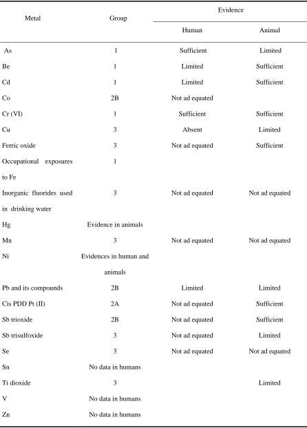
(38) Table 2.6- IARC classification of the carcinogenic potential of metals and their compounds (Boffetta, 1992). Evidence Metal. Group Human. Animal. As. 1. Sufficient. Limited. Be. 1. Limited. Sufficient. Cd. 1. Limited. Sufficient. Co. 2B. Not ad equated. Cr (VI). 1. Sufficient. Sufficient. Cu. 3. Absent. Limited. Ferric oxide. 3. Not ad equated. Sufficient. Occupational exposures. 1. Not ad equated. Not ad equated. Not ad equated. Not ad equated. to Fe Inorganic fluorides used. 3. in drinking water Hg. Evidence in animals. Mn. 3. Ni. Evidences in human and animals. Pb and its compounds. 2B. Limited. Limited. Cis PDD Pt (II). 2A. Not ad equated. Sufficient. Sb trioxide. 2B. Not ad equated. Sufficient. Sb trisulfoxide. 3. Not ad equated. Limited. Se. 3. Not ad equated. Not ad equated. Sn. No data in humans. Ti dioxide. 3. V. No data in humans. Zn. No data in humans. Limited. 33.
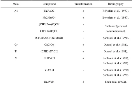
(39) Concerning the possible mechanisms relative to transforming action induced by metal compounds, it is known that, being cancer a multi- stadium process, the toxic action of metals can be the result of interference or damage at different step of the process. For example chromosomic damage, DNA repair, inhibition of DNA repair, DNA linkage, wrong synthesis of DNA, stress proteins activation, inhibition of cellular communication. All these processes can occur dependently from the oxidation state of the different metal compounds (Mazzotti, 1999). Table 2.7 shows studies relative to the carcinogenic potential of metal compounds in BALB/3T3 systems, the cellular model used in the present work.. Table 2.7- Metal compounds tested by morphological transformation assay using BALB/3T3 cell linea. Metal. Compound. Transformation. Bibliography. As. NaAsO2. +. Bertolero et al. (1987).. Na2HasO4. +. Bertolero et al. (1987).. (CH3)2As(O)OH. -. CH3Has(O)OH. -. communication).. (CH3)3AsCH2C(O)OH. -. Sabbioni et al. (1991).. Cr. CaCrO4. +. Dunkel et al. (1981).. Ti. (C5H5)2TiCl2. +. Dunkel et al. (1981).. V. NH4VO3. +. Sabbioni et al. (1991);. Sabbioni (personal. Sabbioni et al. (1993). VOSO4. -. Sabbioni et al. (1991); Sabbioni et al. (1993).. Na3VO4. +. Sheu et al. (1992).. a: += transformation activity; +/-= weak transformation activity; -= no observed transformation activity.. 34.
(40) 2.6.1. BALB/3T3 cell line BALB/3T3 cell line derives from subclones (Saaronson et al. (1968)) of the A31 clone, originally isolated from embryonic mouse cells. (Saffiotti et al., 1984). The BALB/3T3 Cl A31- 1- 1 cell line of mesenchimal origin was chosen since it is one of the recommended system to study basic aspects of the response of mammalian cells to carcinogens and since it is one of the most valuable short term tests for bioassays permitting both a qualitative as well as quantitative dose- dependent response to carcinogens including trace metals (Bertolero et al., 1987). This cell line is heterogeneous and the different subclones differ from each other by the different way of spontaneous transformation and different sensibility to induce transformation. The clone has been chosen because shows a spontaneous incidence of transformation included between those two values 10-5 and 10-6. The transformation frequency increases of some order of magnitude with known carcinogens (es. to >10-2 for benzo(α)pyrene) (Saffiotti et al., 1984). Is also known that the spontaneous transformation frequency is established for those cells with a low passage (subculture, cells are transferred from initial flask to another) and becomes higher with the age of the cell line. To interpret the results is important to remember that cells of this line are not comparable with normal cells because these cells have acquired the phenotypic characteristics of transformed cells like immortalisation, aneuploid number of chromosomes and high plating efficiency (5060%). In the meantime they maintain characteristics of normal cells like contact inhibition (if put in culture they are able to form a monolayer), like the dependence of the growth from the anchorage and the non tumorigenicity. They have also conserved the possibility to be furtherly transformed in “in vitro” cultures (Saffiotti et al. 1989). Clone A31- 1- 1 has a number of chromosomes subtetraploids. The karyotypic analysis revealed the 98 % of the. 35.
(41) metaphases contained between 70 to 80 chromosomes. This number is increasing (100140) when the cells are incubated with a carcinogenic substance (Saffiotti et al. 1984). A quantitative dose- response relationship between carcinogen concentration and morphological transformation rate is obtainable with these cell lines while untreated controls or cultures treated with noncarcinogens do not show transformation or produce tumors. Transformation is calculated on the basis of either transformed colonies or transformed foci on a background of normal cells. The transformation foci consist of cell colonies that are able to grow in non organised and invasive layers in comparison to the surrounding monolayer. The foci are of three types: Type I: focus of lightly group together cells. Type II: focus made of mass of cells grown in multilayer. Type III: focus made of cells strictly linked one over another. The peripheral part of the colony is usually characterised by criss- cross cells grown in a disorganised and invasive way. In the evaluation of the transforming potential effect, only types 3 foci are counted as malignant transformations. An elevated percentage (85 %) of these foci are able to origin neoplasia when injected in nude mice (Saffiotti et al. 1984). Although BALB/3T3 system was reported to be susceptible to transformation by a range of different organic chemical carcinogens, data on metal- induced transformation were lacking and only recently a systematic study was carried out on several metal compounds.. 36.
(42) 2.7. Selected Metals. 2.7.1. Platinum as new potential environmental pollutant Platinum, palladium and rhodium are required for “three- way catalyst” of cars to control the three noxious exhaust car emissions, namely carbon monoxide, hydrocarbons and nitrogen oxides (Damiano, 1990). Since automobile catalysts are mobile sources of platinum, palladium and rhodium, (some loss of the elements can be released into the environment due to mechanical and thermal impact) emissions from these car catalysts suggest concern the platinum could represent a “new” potential environmental pollutant.. 2.7.2. Environmental levels and levels in diet, biological fluids and human tissues Platinum is a metal present on earth crust at parts for billion level. The more common oxidation states are (+2) and (+4). The known deposits are localised principally in Siberia, in South Africa, (the major Pt producer), and in Canada (Loebenstein, 1988). Data on platinum emissions from automobile catalysts are very limited. Engine test stand experiments were carried out in Germany (Fraunhofer Institute of Toxicology and Aerosols Research, Hannover) in 1990 as part of a programme of the Ministry of Research and Technology for assessing the health risk of this man- made environmental source. The results showed platinum emissions from few to some tens ng m-3 (Hill et al. 1977 and H. P. Koenig et al., 1989) depending on the temperature. It has been suggested that platinum is released as metal or oxide (an important aspect for toxicological considerations). Air concentrations of platinum were calculated between 0.005 to 1.3 ng Pt m-3 near and on roads, and 0.9 ng m-3 in tunnels (Tab. 2.8).. 37.
(43) Table 2.8- Quantity of Pt present in air of different sites. Site of exposure. Pt concentration (ng/m3). Personal garage. 0.8-6.7. Silos. 0.4-5.6. Galleries. 0.4-0.9. On roads. 0.07-0.16. Near roads. 0.004-0.13. (Rosner et al., 1990).. Studies in Germany gave workplace concentrations of 0.08-0.01 µg Pt m-3 for soluble Pt compounds. There are little information concerning Pt level in water. Typical values are: 0.1-0.25 ng Pt L-1 in oceanic water and 110ngPt L-1 in mineral water. Few data on Pt levels in human tissues and biological fluids are reported in literature (Pietra et al., 1994). Vaughan and Florence (1992) studied Pt in Australian diet. The daily ingestion don’t exceed 1.5 µg Pt/person. The intestinal absorption of Pt, estimated time ago was near 42 %, a value which today is not realistic. The absorption of PtCl4 is much lower (less than 1%) (Filov, 1977). Some scientific reports show that Pt is increasing in blood/ urine of general population. Anyway this is not fully demonstrated due to technical difficulties in establishing the “normal” reference values of Pt in human fluids and tissues. In the context of the EURO TERVIHT project (Sabbioni, 1992) (Trace Element Reference Values In Human Tissues) 40- 50 ng L-1 of platinum were found in blood/ urine of people living in Northern Italy (Unpublished results). It has been suggested that Pt would be mobile in the different compartments of the ecosystem. This is may be due methylation by vitamin B12 (Wood, 1974).. 38.
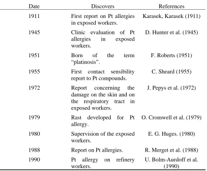
(44) 2.7.3. Toxicological/ carcinogenic effects and estimation of health risk The main health hazard of Pt compounds in asthma and allergy as observed in occupational workers. However, the data are extremely limited to believe that Pt emissions from car catalyst can induce hypersensitivity and so, at present, any attempt to link the allergenic effect of Pt salts with Pt mobilised from car catalyst is not based on a scientific evidence. Until recently, the exposure to Pt salts was confined to occupational settings (Pt refinery and catalyst production plants). In this case the main health hazard is asthma (Linnet, 1987). These allergic symptoms are characteristic of a pathology described since 1977 as “Platinosis” (Parrot et al., 1969) (Tab. 2.9). Table 2.9- Reports on Pt allergy.. Date. Discovers. References. 1911. First report on Pt allergies in exposed workers.. Karasek, Karasek (1911). 1945. Clinic evaluation of Pt allergies in exposed workers.. D. Hunter et al. (1945). 1951. Born of “platinosis”.. term. F. Roberts (1951). 1955. First contact sensibility report to Pt compounds.. C. Sheard (1955). 1972. Report concerning the damage on the skin and on the respiratory tract in exposed workers.. J. Pepys et al. (1972). 1979. Rast developed allergy.. 1980. Supervision of the exposed workers.. 1988. Report on Pt allergies.. R. Merget et al. (1988). 1990. Pt allergy workers.. U. Bolm-Aurdoff et al. (1990). the. on. 39. for. Pt. refinery. O. Cromwell et al. (1979) E. G. Huges. (1980).
(45) An incidence of asthmatic symptons of about 50 % at a workplace air concentration of 0.1 µg Pt m-3 has been estimated. This air concentration should be higher by a factor 1,000 compared to the concentration of platinum of 0.1 ng m-3 calculated at busy roadsides. In addition, the insoluble chemical form of Pt emitted from car catalysts may result in even much lower ambient air concentration of total Pt emitted. So, it is unlikely that platinum emitted from car catalysts causes asthma.. Platinum compounds are not allergenic “per se” because they have a low molecular weight. They would act as haptens able to bind carriers with a high molecular weight like blood proteins. Some studies demonstrate that Pt (II) is able to react with –SH groups of albumin (Trynda et al., 1994) which is the most abundant plasmatic protein and with human transfer proteins (Trynda et al., 1994). An important aspect which is the subject of newspaper articles is the potential carcinogenic of Pt compounds. There is a lack of information to assess the carcinogenicity/ mutagenicity of Pt compounds. The alarmism which appeared on this subject (Pt emission from car catalysts could cause cancer) is completely unjustified. When we speak on carcinogenicity of metals we must take into account their chemical form, because it is known that different chemical species of an element can lead to completely different toxicological/ carcinogenic effects. Most of the small amounts of Pt emitted from car catalyst is presumably in form of metal or oxide. These chemical species must not be confused with the cis- Platinum which is used as an antitumor drug at pharmacological doses (Pooly and Lohman, 1980) (cis- Pt has a proper neoplastic activity and it is classified as “ probably carcinogenic to human, (category 2A0 by the International Agency of Research of Cancer (IARC)) (Boffetta, 1993). Thus, the extrapolation of the moderate neoplastic action of the Pt and cis- Pt to the. 40.
(46) problem of Pt emitted in mostly insoluble metal (or oxide) chemical species from car catalyst has little sense and can be considered a speculation. At present, it is considered that the health risk arising from the emission of Pt from car catalysts should be “low”. This conclusion, however, is based more on lack of available data rather than on a scientific evidence. So, research in this field is necessary and should include: a) analytical work (geochemical maps showing concentration of Pt in top soil and sediments; extent and chemical form of Pt released to the environment; establishment of current levels of Pt in body fluids and tissues of different subjects, e. g motorway maintenance workers, population living near motorways and general unexposed population) b) toxicological studies to set dose- effect relationships of Pt in experimental and human models which should also include studies at workplace as well as in “in vitro” cell culture models.. 2.8. Methyl mercury, a well known poisoning Organic mercury compounds have been characterised as hazardous, especially the alkylated compounds. Because of the alkylated mercury compounds’ long retention by the human body, because of their toxic effect on developing tissue and furthermore, because of their known propensity for central nervous system damage, alkylated mercury compounds pose particular and significant hazards for the fetus. Toxicologically and environmentally the most dangerous alkyl compound is methyl mercury (Grass, 1969). Mercury and its congeners are extremely toxic substances which exist in several physical and chemical forms: inorganic mercury, organic mercury and mercury vapour. Sources of human exposure to mercury include seafood, seeds, foodstuffs, water and dental amalgam (Shenker et al., 2000).. 41.
Figure


+7

Documento similar
(2007) Accumulation of arsenic in tissues of rice plant (Oryza sativa L.) and its 974. distribution in fractions of
Keywords: Metal mining conflicts, political ecology, politics of scale, environmental justice movement, social multi-criteria evaluation, consultations, Latin
For instance, (i) in finite unified theories the universality predicts that the lightest supersymmetric particle is a charged particle, namely the superpartner of the τ -lepton,
Given the much higher efficiencies for solar H 2 -generation from water achieved at tandem PEC/PV devices ( > 10% solar-to-H 2 energy efficiency under simulated sunlight) compared
Plotinus draws on Plato’s Symposium (206c4–5) – “procreate in what is beautiful” – 3 in order to affirm that mixed love (which is also a love of beauty) is fecund, although
Although some public journalism schools aim for greater social diversity in their student selection, as in the case of the Institute of Journalism of Bordeaux, it should be
In the “big picture” perspective of the recent years that we have described in Brazil, Spain, Portugal and Puerto Rico there are some similarities and important differences,
We consider different methods aim to evaluate cellular uptake and localization in cells and tissues, and in vitro methods for the study of the toxicity induced by MNPs
