Thyroid-like cholangiocarcinoma of the liver:
an unusual morphologic variant
with follicular, trabecular and insular patterns
Fredy Chablé-Montero,*,** Amy Shah BS,*** Daniel Montante-Montes de Oca,*Arturo Ángeles-Ángeles,* Donald E. Henson,*** Jorge Albores-Saavedra*,**
* Department of Pathology, Instituto Nacional de Ciencias Médicas y Nutrición Salvador Zubirán, Mexico City, Mexico. ** Department of Pathology of Medica Sur Clinic and Foundation, Mexico City, Mexico.
*** George Washington University Cancer Institute, Washington DC. USA.
ABSTRACT
We report the case of a 26-year-old woman with a 19 cm malignant hepatic neoplasm with morphological features that closely resembled a follicular thyroid carcinoma. Despite this, it was interpreted as a cholan-giocarcinoma due to the absence of a primary thyroid tumor and the lack of thyroglobulin and TTF-1 im-munoreactivity by the hepatic tumor. The left hepatic lobectomy specimen showed an encapsulated and multinodular gray-white mass with cystic and hemorrhagic areas. Microscopically, it displayed predominant macro and microfolicullar patterns with focal solid, trabecular and insular areas. The small and distended follicles contained a colloid-like secretion and were lined by low cuboidal cells with scant cytoplasm, round or oval hyperchromatic nuclei with fine chromatin. The solid areas, trabecular and insular structu-res were similar to those of follicular or papillary thyroid carcinomas. In addition, some of the neoplastic cells had clear nuclei with occasional grooves. The tumor was positive for cytokeratin (CK) 7, CK 19 and CD138, and negative for TTF-1, thyroglobulin, Hepar-1, Glypican-3, alpha-fetoprotein and neuroendocrine markers. A thyroid neoplasm was excluded clinically and by ultrasound and computed tomography. Al-though, the residual hepatic parenchyma was initially not cirrhotic, the patient eventually developed cryptogenic cirrhosis. The patient received adjuvant chemotherapy and died of metastatic disease 18 mon-ths after surgery. The thyroid-like pattern broadens the morphologic spectrum of cholangiocarcinoma.
Key words. Thyroid-like pattern. Hepatocellular carcinoma. Hepar-1. TTF-1. Thyroglobulin.
Correspondence and reprint request: Fredy Chablé Montero, MD.
Department of Pathology, Instituto Nacional de Ciencias Médicas y Nutrición Salvador Zubirán.
Vasco de Quiroga, Núm. 15, Col. Sección XVI, Tlalpan, México D.F. C.P. 14000
E-mail: [email protected]
Manuscript received: February 03, 2012. Manuscript accepted: April 30, 2012.
INTRODUCTION
Cholangiocarcinoma (CHC) is a malignant epithe-lial tumor arising in the intrahepatic bile ducts ac-counts for 8 to 10% of all malignant hepatic tumors. Cholangiocarcinomas are more frequent in men than in women (60-70%) and their mean age at pre-sentation is in the seventh decade.1 A variety of mi-croscopic patterns have been described in CHC.2
The purpose of this report is to describe the se-cond case of a new morphological variant of CHC
that resembles a thyroid carcinoma. In addition, we compare the incidence of hepatocellular carcinoma and cholangiocarcinoma recorded in the Surveillance, Epidemiology, and End Results (SEER) Program of the National Cancer Institute from 1992 through 2008.
CLINICAL HISTORY
bilirrubines, viral hepatitis antibodies and auto-anti-bodies were normal or negative. The alpha-fetopro-tein (AFP) serum levels were normal. Because of the age of the patient the neoplasm was clinically diag-nosed as a probable hepatoblastoma. A left hepatic lobectomy was performed.
GROSS AND
MICROSCOPIC FEATURES
The hepatic lobectomy specimen showed an en-capsulated and multinodular gray-white mass with
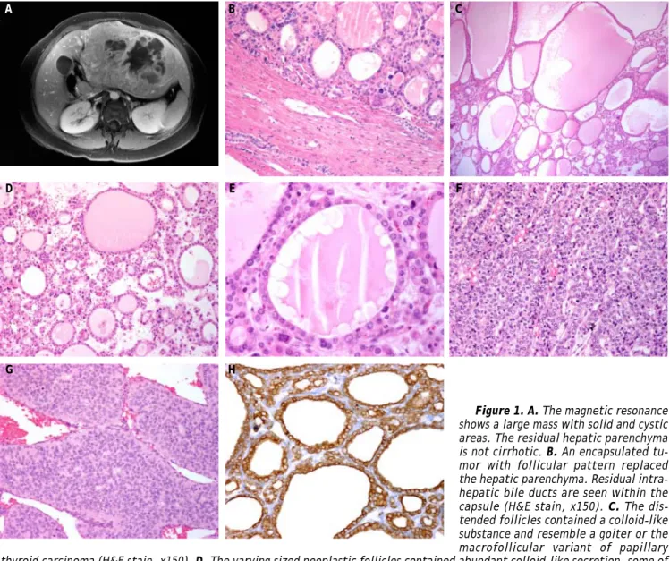
cystic and hemorrhagic areas measuring 19 cm in greatest dimension. Microscopically, it had a thicke-ned capsule and was composed of a predominant ma-cro and mima-crofolicullar pattern (70%) with solid, trabecular and insular areas (Figures 1B-1C). The follicles were lined by low cuboidal cells with scant cytoplasm, round or oval hyperchromatic nuclei with fine chromatin, resembling a thyroid neoplasm with follicular phenotype (Figure 1D). The vast majority of neoplastic follicles contained an abundant colloid-like secretion, some of which showed peripheral vacuolation and foamy macrophages that mimicked
Figure 1. A. The magnetic resonance shows a large mass with solid and cystic areas. The residual hepatic parenchyma is not cirrhotic. B. An encapsulated tu-mor with follicular pattern replaced the hepatic parenchyma. Residual intra-hepatic bile ducts are seen within the capsule (H&E stain, x150). C. The dis-tended follicles contained a colloid-like substance and resemble a goiter or the macrofollicular variant of papillary thyroid carcinoma (H&E stain, x150). D. The varying sized neoplastic follicles contained abundant colloid-like secretion, some of which showed a few foamy macrophages (H&E stain, x150). E. The follicles are lined by low cuboidal cells some with clear nuclei. The intrafollicular colloid-like secretion showed peripheral vacuolation mimicking a follicle of goiter (H&E stain, x250). F. The trabecular areas showed cells with clear nuclei, some with grooves similar to those of papillary thyroid carcinoma (H&E stain, x150). G. Insular areas with mitotic figures similar to those of papillary and follicular thyroid carcinomas (H&E stain, x250). H. The neoplastic cells are diffusely and strongly positive for cytokeratin 7 (Immunostain, x150).
A B C
D E F
a hyperplastic goiter (Figure 1E). The interfollicular stroma had numerous microcalcifications but no psammoma bodies. The solid areas were composed of cords, trabecular and insular structures similar to those that occur in follicular or papillary thyroid carcinomas (Figures 1F-1G). Moreover, some of the cells had clear nuclei with occasional grooves simi-lar to those of papilsimi-lary carcinoma. However, no nuclear pseudoinclusions were present. Mitotic figu-res were seen only in the insular areas. Lympho-vascular invasion was also seen. The neoplasm extended to the surgical margin and the portal vein. The residual hepatic parenchyma was not cirrhotic. However, portal spaces showed moderate fibrosis and a chronic inflammatory infiltrate composed pre-dominantly of lymphocytes. Moreover, non-alcoholic steatohepatitis was not seen.
MATERIAL AND METHODS
Multiple sections of the hepatic tumor were avai-lable for review. From representative paraffin blocks additional sections were obtained for immunohisto-chemical analysis. The following antibodies were used:
• Hepar-1 (Biocare, Concord CA, 1:200).
• Glypican-3 (BioSB, Santa Barbara CA, 1:500). • AFP (Dako, Carpinteria CA, 1:500).
• CK 7 (Biocare, Concord CA, 1:100). • CK 19 (Biocare, Concord CA, 1:200).
• CD138 (Syndecan-1) (Biocare, Concord CA, 1:100).
• Chromogranin (Biocare, Concord CA, 1:300).
• Synaptophysin (Biocare, Concord CA, 1:100). • CD56 (Biocare, Concord CA, 1:50).
• Thyroid Transcription Factor 1 (TTF-1). (Cell marque, Rocklin CA, 1:200), thyroglobulin (Bio-genex, San Ramon CA, 1:500).
• HBME-1 (Dako, Carpinteria CA, 1:100).
Data were obtained from the SEER Program, which is a population-based registry that collects demogra-phic, anatomic, morphologic, extent of disease, and survival information on patients with cancer. For this study data from de SEER program on cho-langiocarcinoma and hepatocellular carcinoma from 1992 through 2008 were analyzed.
Immunohistochemical profile
The neoplastic cells expressed CK 7 (Figure 1H), CK 19 and CD138, and were negative for TTF-1 and thyroglobulin. The tumor also lacked reactivity for Hepar-1, Glypican-3, AFP, chromogranin, synapto-physin and CD56. This immunophenotype was con-sistent with cholangiocarcinoma and excluded a thyroid carcinoma.
Follow-up
A metastatic thyroid carcinoma was excluded by physical examination of the neck, ultrasound and CT of the thyroid gland. The patient developed CHILD C stage cirrhosis and additional serological tests for viral hepatitis and autoimmune hepatic di-sease were performed, but were all negative. The diagnosis of cryptogenic cirrhosis was established.
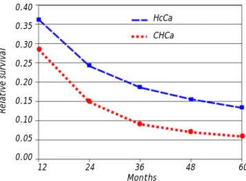
Figure 3. Five year relative survival rate for hepatocellular carcinoma and cholangiocarcinoma (SEER, 1992-2008).
Relative survival
0.40
0.35
0.30
0.25
0.20
0.15
0.10
0.05
0.00
12 24 36 48 60
Months Figure 2. Age frequency distribution for hepatocellular
carcinoma and cholangiocarcinoma (SEER, 1992-2008). 0.040
0.035 0.030 0.025 0.020 0.015 0.010 0.005 0
20 25 30 35 40 45 50 55 60 65 70 75 80 Age
Frequency
HcCa
CHCa HcCa 42,808
The patient received adjuvant chemotherapy for 14 months, but she developed local recurrence. A re-peat determination of serum AFP was normal. She died with massive hepatic recurrence and metastasis in the right adrenal gland and regional lymph no-des, 18 months after surgery. The clinical stage was considered as IVB (pT2a, N1, M1). An autopsy was not performed.
Seer data
From 1992 through 2008, there were 47,289 cases of carcinoma of the liver of which 42,808 (90.5%) were hepatocellular carcinoma and only 4,481 were cholangiocarcinoma (9.5%). The ratio of hepato-cellular carcinoma to cholangiocarcinoma was 10 to 1. The age at presentation was 54 years-old for hepa-tocellular carcinoma and 74 years-old for cholangio-carcinoma. Figure 2 summarizes these findings. The 5-year relative survival rate of patients with hepato-cellular carcinoma was 13% and for patients with cholangiocarcinoma it was 6% (Figure 3).
DISCUSSION
Cholangiocarcinoma is the second most common malignant neoplasm of the liver. The SEER data shows that the hepatocellular carcinoma/CHC ratio is 10:1. Both tumors are associated with a poor prognosis. The 5-year survival rate of patients with hepatocellular carcinoma is 13% and 6% for patients with CHC (Figures 2 and 3). The data reveal that the incidence of hepatocellular carcinoma has been increasing since 2000. This increase in incidence of hepatocellular carcinoma is attributed to a rise in the incidence of viral hepatitis C and related cirrho-sis, whereas the incidence of CHC has remained re-latively stable.
In contrast to hepatocellular carcinoma, CHC is usually not associated with cirrhosis. Only 5% of CHC arise in a background of chronic hepatitis C.1,2 Moreover, carcinomas of the extrahepatic bile duct are not associated with chronic hepatitis.3 A number of histologic patterns have been described in CHC in-cluding tubular, mucinous,2 adenosquamous,4 with clear cells,5 spindle-sarcomatoid6 and lymphoepithe-lioma-like.7,8 However, only one case reported previo-usly had a thyroid-like growth pattern.9
The microscopic features of this hepatic tumor clo-sely resemble a thyroid carcinoma. It had a predomi-nant follicular pattern (70%) with trabecular and insular areas as described in papillary and follicular thyroid carcinomas.10,11 In some areas the distended
follicles resembled those of the macrofollicullar va-riant of papillary carcinoma.10,12,13 In other areas, the tumor had features of papillary thyroid carcinoma with small follicles lined by low columnar cells with clear nuclei and occasional nuclear grooves but without nuclear pseudoinclusions. Moreover, the in-terfollicular tissue showed numerous microcalcifica-tions but no psammoma bodies. Initially, the morphologic diagnosis of a metastatic well-differen-tiated thyroid carcinoma was strongly considered, but the absence of a primary tumor and the lack of thyroglobulin and TTF-1 immunoreactivity excluded such a diagnosis and favored the interpretation of cholangiocarcinoma. Thyroglobulin is a specific mar-ker for benign and malignant tumors with follicular cell phenotype while TTF-1 is a homeodomain trans-cription factor normally expressed by thyroid follicu-lar cells and pneumocytes and a sensitive but not specific marker for benign and malignant thyroid neoplasms with follicular phenotype. Moreover, the diagnosis of a metastatic thyroid carcinoma was ex-cluded by ultrasographic and tomographic studies. Few cases of hepatic metastasis from malignant thyroid neoplasms had been reported.14-16 The diffe-rential diagnosis includes intrahepatic thyroid ecto-pic tissue, but in these cases the ectoecto-pic thyroid tissue is normal.14,17,18 Both metastastic tumors and ectopic thyroid tissue expressed TTF-1 and thyroglo-bulin.14-18
Other tumors included in the differential diagno-sis were hepatocellular carcinoma with acinar or pseudoglandular pattern and neuroendocrine carci-noma.2 However, the lack of reactivity for Hepar-1, AFP and Glypican-3 ruled out a hepatocellular car-cinoma.2,19 The lack of neuroendocrine markers (chromogranin, synaptophysin and CD56) excluded the possibility of neuroendocrine carcinoma.20,21
Cholangiocarcinoma is not the only neoplasm that shows a like pattern. Tumors with thyroid-like morphology had been described in the breast and kidneys. In fact, some carcinomas of the breast su-perficially resemble a metastatic tall-cell variant of papillary thyroid carcinoma.22,23 Likewise, some re-nal cell carcinomas are similar to well-differentiated follicular thyroid carcinomas.24-26 In these organs, thyroid carcinoma metastases were excluded by the lack of reactivity for TTF-1 and thyroglobulin.22-27 Finally, a case of plasmacytoma with thyroid-like fea-tures has been reported, but morphologically this tu-mor was not similar to ours.28
reported a 52-year-old male with an 18 cm hepatic tu-mor with a pure follicular pattern that lacked thyroid follicular markers and expressed CK 7, CK 19, CAM 5.2 and CK AE1&AE. In addition, the thyroid gland did not harbor a malignant primary tumor.28
In conclusion, CHC represents the second most common malignant hepatic tumor, usually not asso-ciated with cirrhosis or hepatitis C, with variable morphological patterns and associated with poor prognosis. We report a CHC with microscopic featu-res closely featu-resembling a metastatic well-differentiated thyroid carcinoma with follicular, solid, trabecular and insular areas. The absence of a primary thyroid tumor, knowledge of the morphological features and the lack of thyroid markers in the liver tumor are useful in the diagnosis of this rare and new morpho-logical variant of cholangiocarcinoma.
ACKNOWLEDGMENTS
This paper was supported by La Fundación Clínica Médica Sur.
REFERENCES
1. Shaib Y, El-Serag HB. The epidemiology of cholangiocarci-noma. Sem Liver Dis 2004; 24: 115-24.
2. Goodman ZD. Neoplasms of the liver. Mod Pathol 2007; 20(Suppl. 1): S49-S60.
3. Albores-Saavedra J, Henson DE, Klimstra DS. Tumors of the gallbladder, extrahepatic bile ducts, and ampulla of Vater. Atlas of tumor pathology. 3rd. series. Fascicle27. Was-hington: Armed Forces Institute of Pathology; 2000. 4. Nakanuma Y, Sato Y, Harada K, Sasaki M, Xu J, Ikeda H.
Pathological classification of intrahepatic cholangiocarci-noma based on a new concept. World J Hepatol 2010; 2: 419-27.
5. Albores-Saavedra J, Hoang MP, Murakata LA, Sinkre P, Ya-ziji H. Atypical bile duct adenoma, clear cell type: a pre-viously undescribed tumor of the liver. Am J Surg Pathol 2001; 25: 956-60.
6. Sato K, Murai H, Ueda Y, Katsuda S. Intrahepatic sarco-matoid cholangiocarcinoma of round cell variant: a case report and immunohistochemical studies. Virchows Arch 2006; 449: 585-90.
7. Adachi S, Morimoto O, Kobayashi T. Lymphoepithelioma-like cholangiocarcinoma not associated with EBV. Pathol Int 2008; 58: 69-74.
8. Huang Y, Tsung JS, Lin CW, Cheng TY. Intrahepatic cho-langiocarcinoma with lymphoepithelioma-like carcinoma component. Ann Clin Lab Sci 2004; 34: 476.
9. Fornelli A, Bondi A, Jovine E, Eusebi V. Intrahepatic cho-langiocarcinoma resembling a thyroid follicular neoplasm. Virchows Arch 2010; 456: 339-42.
10. Albores-Saavedra J, Wu J. The many faces and mimics of papillary thyroid carcinoma. Endoc Pathol 2006; 17: 1-18.
11. Rosai J, Carcangiu ML, De Lellis R. Tumors of the thyroid gland, Atlas of Tumor Pathology. 3rd. series. Fascicle 5. Armed Forces Institute of Pathology; 1992.
12. Albores-Saavedra J, Gould E, Vardaman C, Vuitch F. The ma-crofollicular variant of variant of papillary thyroid carcino-ma. A study of 17 cases. Hum Pathol 1991; 22: 1195-205. 13. Albores-Saavedra J, Housini I, Vuitch F, et al.
Macrofo-llicular variant of papillary thyroid carcinoma with minor insular component. Cancer 1997; 80: 1110-16.
14. Kondo T, Katoh R, Omata K, et al. Incidentally detected li-ver metastasis of well-differentiated follicular carcinoma of the thyroid, mimicking ectopic thyroid. Pathol Int 2000; 50: 509-13.
15. Kouso H, Ikegami T, Ezaki T, et al. Liver metastasis from thyroid carcinoma 32 years after resection of the primary tumor: report of a case. Surg Today 2005; 35: 480-2. 16. Kraft O. Hepatic metastasis of differentiated thyroid
car-cinoma. Nucl Med Rev Cent East Eur 2005; 8: 44-6. 17. Ghanem N, Bley T, Altehoefer C, et al. Ectopic thyroid gland
in the porta hepatis and lingua. Thyroid 2003; 13: 503-7. 18. Strohschneider T, Timm D, Worbes C. Ectopic thyroid
gland tissue in the liver. Chirurg 1993; 64: 751-3.
19. Chan ES, Yeh MM. The use of immunohistochemistry in li-ver tumors. Clin Liver Dis 2010; 14: 687-703.
20. Frankel WL. Update on pancreatic endocrine tumors. Arch Pathol Lab Med 2006; 130: 963-6.
21. Capelli P, Martignoni G, Pedica F, et al. Endocrine neoplas-ms of the pancreas: pathologic and genetic features. Arch Pathol Lab Med 2009; 133: 350-64.
22. Eusebi V, Damiani S, Ellis IO, et al. Breast tumor resembling the tall cell variant of papillary thyroid carcinoma. Am J Surg Pathol 2003; 27: 1114-18.
23. Tosi AL, Ragazzi M, Asioli S, et al. Breast tumor resembling the tall cell variant of papillary thyroid carcinoma: report of 4 cases with evidence of malignant potential. Int J Surg Pathol 2007; 5: 14-19.
24. Amin MB, Gupta R, Ondrej H, et al. Primary thyroid-like fo-llicular carcinoma of the kidney: report of 6 cases of a his-tologically distinctive adult renal epithelial neoplasm. Am J Surg Pathol 2009; 33: 393-400.
25. Jung SJ, Chung JI, Park SH, Ayala AG, Ro JY. Thyroid folli-cular carcinoma-like tumor of the kidney: a case report with morphologic, immunohistochemical and genetic analy-sis. Am J Surg Pathol 2006; 30: 411-15.
26. Sterlacci W, Verdofer I, Gabriel M, Mikuz G. Thyroid folli-cular carcinoma-like renal tumor: a case report with mor-phologic, immunophenotypic, cytogenetic and scintigraphic studies. Virchows Arch 2008; 452: 91-5. 27. Angeles-Angeles A, Chablé-Montero F, Martínez-Benítez B,
Albores-Saavedra J. Unusual metastases of papillary thyroid carcinoma: report of 2 cases. Ann Diagn Pathol 2007; 13: 189-96.