Prognostic implication of serum vascular endothelial growth
factor in advanced hepatocellular carcinoma staging
Ender G. Yegin,*,§ Aydos Siykhymbayev,**,§ Fatih Eren,*** Nural Bekiroglu,**** Osman Cavit Ozdogan** Department of Gastroenterology, Marmara University, Faculty of Medicine, lstanbul, Turkey. ** Department of Internal Medicine, Almaty Sema Hospital, Almaty, Kazakhstan.
*** Department of Medical Biology and Genetics, Marmara University Faculty of Medicine, Istanbul, Turkey. **** Department of Biostatistics and Bioinformatics, Marmara University Faculty of Medicine, Istanbul, Turkey.
§ Both authors have equal contribution.
ABSTRACT
Background. Staging systems have considerable impact on hepatocellular carcinoma (HCC) treatment approaches and outcomes. There is an unmet need to improve their stratification ability. We have evalua-ted four commonly used staging systems and assessed whether angiogenic biomarker vascular endothelial growth factor (VEGF) could improve their prognostic stratification. Material and methods. Four staging sys-tems; Okuda, Cancer of the Liver Italian Program (CLIP), Barcelona Clinic Liver Cancer (BCLC), and Child-Pugh were evaluated in 78 HCC patients; their stratification abilities were detected by Kaplan-Meier curves and log-rank test; their accuracies of predicting survival were compared with the concordance in-dex. Serum VEGF levels were measured using ELISA method. Recursive partitioning was used to determine the optimal VEGF cutoff. The prognostic significance of VEGF cutoff and other parameters were analyzed using univariate and multivariate models. Results. None of the staging systems demonstrated better discri-minatory ability in predicting survival. The four staging systems did not reveal significant differences in pro-bability of survival across their intermediate-advanced stages. Optimal cutoff identified for VEGF was 445 pg/mL. In advanced HCC, VEGF level (p = 0.004) and in early HCC, bilirubin level (p = 0.009) were identified as the independent prognostic factors. Survival comparison with high and low VEGF levels was significant for advanced HCC, while insignificant for early disease. Conclusion. Staging systems with conventional pa-rameters did not provide good prognostic stratification for survival in advanced HCC population. Serum VEGF level was an independent predictor of survival in advanced HCC, and provided more survival homoge-neity within the advanced stages of conventional staging systems.
Key words. Biomarker. Stratification. Survival. Tumor angiogenic activity. Intrinsic tumor feature.
Correspondence and reprint request: Ender G. Yegin
Fevzi Çakmak mahallesi, Mimar Sinan caddesi, no: 41, Üst Kaynarca, Pendik, Istanbul.
Ph.: +90 5326718247
E-mail: [email protected]
Manuscript received: June 10, 2013. Manuscript accepted: July 02, 2013.
INTRODUCTION
Hepatocellular carcinoma (HCC) is the fifth most common type of cancer affecting approximate-ly one million people every year and the third most frequent cause of annual cancer-related deaths worldwide.1-3 In many parts of the world, the inci-dence and mortality from HCC continues to rise as
patients with cirrhosis are expected to have a lon-ger survival due to improved medical manage-ment.4-8 The similarity between mortality and incidence rates is indicative of poor survival of this disease.9 Because of these, in recent years, many prognostic factors of HCC have been evaluated, particularly, angiogenesis-related markers have been a subject of interest.
Angiogenesis plays a crucial role in growth and progression of HCC, which is one of the most vascularized solid tumors.10 Vascular endothelial growth factor (VEGF) is a critical mediator regula-ting angiogenesis in HCC.10-12 There have been many studies suggesting a relationship between VEGF and prognosis of HCC.13-20
In the management of HCC disease, the role of a staging system is to estimate prognosis, to define the suitable patient population to be recruited in
clinical trials and to provide a common terminology to compare outcomes of these trials. Importantly, by guiding the treatment decisions, they have conside-rable impact on the outcomes.21 HCC is a heteroge-neous disease with distinct risk factors, underlying liver function, clinical presentation, natural history, and response to therapeutic modalities, which leads to very different clinical outcomes. Widely accepted HCC staging systems are Barcelona Clinic Liver Cancer (BCLC), Cancer of the Liver Italian Pro-gram (CLIP) and Okuda systems.22-24 Tumor-node-metastasis (TNM) staging system which is commonly used in other malignant diseases, is based on the anatomic tumor extension and does not in-clude many important parameters affecting HCC prognosis such as underlying liver function status. Child-Pugh classification is a widely used clinical measure of hepatic functional reserve, but lacks pa-rameters representing the tumor itself. Ideal staging systems for HCC should necessarily involve many prognostic parameters that accurately stratify pa-tients with regard to survival outcome. Among the many staging systems introduced for HCC, there is no worldwide consensus on which one is the best in predicting prognosis yet, and it is unclear whether integration of additional prognostic variables can improve their stratification ability.21,25-27
The aim of this study is to compare the accura-cies of four commonly used staging systems at predicting survival and to investigate whether angiogenic factor VEGF can be a prognostic measure, and hence, improve prognostic stratification of these staging systems in a series of HCC patients treated with different therapeutic modalities.
MATERIAL AND METHODS
Patients and data collection
From January 2008 to January 2009, a total of 89 eligible HCC patients attending to our Hepato-logy Cirrhosis Clinics were identified at Marmara University Faculty of Medicine Hospital in Istanbul, Turkey. HCC was diagnosed by radiologic criteria or by histological confirmation as described by the American Association for the Study of Liver Diseases.28
Patients with incomplete information required for the analysis, who were lost during the follow up and patients with a history of other concur-rent malignancies were excluded from the study. We obtained approval of the Central Research Ethics Committee for the study protocol and
informed consent form of the patients prior to participation.
The primary outcome for the analysis was to evaluate the correlation of biomarker VEGF with overall survival and its role in the prognostic strati-fication of HCC disease. Required data were collected to stage patients according to Child-Pugh, Okuda, BCLC, CLIP systems and to perform statistical analysis, and the data included demographics; clinical, etiological, laboratory, and therapeutic variables; and tumor data determined by available imaging. Patients were also classified as having early or advanced disease; advanced HCC was defined as disease that surgical or locoregional therapies (radiofrequency ablation, alcohol ablation, chemo-embolization) were not suitable, or that have recur-red after therapy.29
Serum samples were obtained from 78 HCC pa-tients, 20 cirrhotic patients with no evidence of HCC, and 20 healthy adults as controls. Venous blood samples were drawn into a serum separator tube and centrifuged at 5,000 rpm for 10 min, then, samples were stored at -20 °C until analysis. VEGF serum level was quantitatively measured by Chemi-Kine sandwich ELISA kit (Chemicon International, Billerica, MA, USA) by following the company’s instructions.
Statistical analysis
Overall survival was calculated by using the Ka-plan-Meier method. Survival comparison among the stages of each prognostic system was performed by using the log-rank test. We also compared median survival of patients with high and low VEGF levels in each stage of four prognostic systems, and in ear-ly and advanced HCC patient groups using log-rank test.
Recursive partitioning method was used to search for the optimal VEGF cutoff value. Recursive parti-tioning is known as a tree analysis method, which creates a decision tree based on the likelihood ratio test to examine all possible binary splits of HCC pa-tients and selects the VEGF value that maximally discriminates between those who survive and those who do not survive.
To identify independent prognostic factors, we fit-ted variables that demonstrafit-ted a p value < 0.1 in the univariate analysis to the multivariable Cox re-gression model.
other. Concordance index (c-index) is measured by calculating the area under the receiver operating characteristic (ROC) curve, and varies between 0.5 and 1.0 with a higher value for a system indicating a better predictive ability for survival.
We used independent-samples t test, one-way Ano-va test, and Pearson correlation test to correlate VEGF levels with various parameters and prognos-tic scoring systems.
Continuous data were expressed as median and range. Comparisons between groups were performed by the χ2 test (or Fisher exact test where appropriate) for categorical variables, and the Mann-Whitney U-test for continuous variables.
Statistical analysis was performed using SPSS 15.0 for Windows statistical software. P values < 0.05 were considered statistically significant.
RESULTS
Patient characteristics
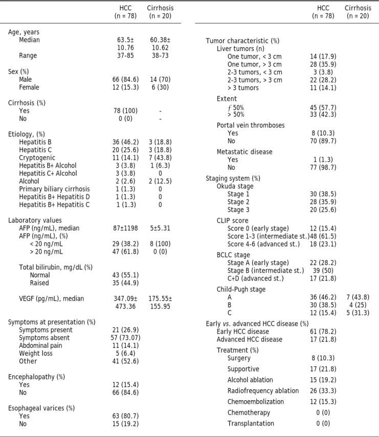
Among the 89 HCC patients identified as eligible, 78 patients were enrolled in the study; 11 patients were excluded due to incomplete data or no follow-up. The diagnosis of HCC required liver biopsy in 4/ 78 (5%) cases. The majority of the HCC patients were male (84.6%) and the median age was 63.5 (37-85) years. Median duration of follow-up was 4 ± 7 (1-60 mo) months. All HCC patients had underlying cirrhosis and 46.2% had preserved hepatic functio-nal reserve (Child-Pugh A). The study population included 17 (21.8%) patients with an advanced tu-mor. Of the 78 patients, 49 (62.8%) did not survive by the time of data analysis. Table 1 summarizes pa-tient characteristics.
Evaluation of staging systems
Four prognostic systems were analyzed separate-ly for their ability to stratify patients into stages with survival differences by Kaplan–Meier curves. Child-Pugh and Okuda systems stratified patients more effectively into different prognostic risk groups than the BCLC and CLIP systems by log-rank comparison (p = 0.006 for Child-Pugh, p = 0.04 for Okuda, p = 0.064 for BCLC, and p = 0.112 for CLIP).
Pairwise log-rank comparison between stages within each prognostic system revealed significant survival differences between Child-Pugh stages with the exception of stages B and C (Child-Pugh A vs. B
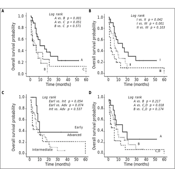
p = 0.001; A vs. C p = 0.051; B vs. C p = 0.571), and between Okuda stages with the exception of stages II and III (Okuda I vs. II p = 0.042; I vs. III p = 0.001; II vs. III p = 0.103). Within the stages of CLIP system, early CLIP stage tended to have a longer survival than the intermediate and the advanced stages, while no survival difference was demonstrated among intermediate-advanced stage comparison (CLIP early vs. intermediate p = 0.054; early vs. advanced p = 0.074; intermediate vs. advanced p = 0.537). When BCLC system stages were compared, stage A had a better prognosis than stage C-D, while no significant survival difference were found among A and B stages, among B and C-D stages (BCLC A vs. B p = 0.217; A vs. C-D p = 0.018; B vs. C-D p = 0.174) (Figure 1).
Comparison of staging systems
By c-statistic analysis, the discriminatory abilities at predicting survival were ranked as Okuda (0.694; 95% CI, 0.574 to 0.814), Child-Pugh (0.690, 95% CI, 0,568 to 0,812), BCLC (0.689; 95% CI, 0.569 to 0.808), and CLIP (0.614; 95% CI, 0.481 to 0.747) with no statistically significant difference among each other (p > 0.05 for paired comparisons) (Table 2).
Survival results and predictors of outcome
At the end of follow up, 49/78 (62.8%) patients had died. The estimated median overall survival du-ration of 78 HCC patients was 8.0 months (95% CI, 6.2-9.8), of 61 early HCC patients was 9.0 months (95% CI, 5.96-12.04), and of 17 advanced HCC pa-tients was 4.0 months (95% CI, 1.46-6.53). Variables that significantly or likely influenced survival in our univariate model analysis were Child-Pugh stage (p = 0.006), bilirubin level (p = 0.000), Okuda stage (p = 0.004), presence of esophageal varices (p = 0.051), BCLC stage (p = 0.064), tumor extension (p = 0.058), and VEGF cutoff 445 pg/mL (p = 0.08).
Optimal VEGF cutoff as an independent prognostic factor
When recursive partitioning was applied, optimal cutoff identified for VEGF in terms of predicting survival was 445 pg/mL.
HCC Cirrhosis
(n = 78) (n = 20)
Age, years
Median 63.5± 60.38±
10.76 10.62
Range 37-85 38-73
Sex (%)
Male 66 (84.6) 14 (70)
Female 12 (15.3) 6 (30)
Cirrhosis (%)
Yes 78 (100)
-No 0 (0)
-Etiology, (%)
Hepatitis B 36 (46.2) 3 (18.8)
Hepatitis C 20 (25.6) 3 (18.8)
Cryptogenic 11 (14.1) 7 (43.8)
Hepatitis B+ Alcohol 3 (3.8) 1 (6.3)
Hepatitis C+ Alcohol 3 (3.8) 0
Alcohol 2 (2.6) 2 (12.5)
Primary biliary cirrhosis 1 (1.3) 0
Hepatitis B+ Hepatitis D 1 (1.3) 0
Hepatitis B+ Hepatitis C 1 (1.3) 0
Laboratory values
AFP (ng/mL), median 87±1198 5±5.31
AFP (ng/mL), (%)
< 20 ng/mL 29 (38.2) 8 (100)
> 20 ng/mL 47 (61.8) 0 (0)
Total bilirubin, mg/dL (%)
Normal 43 (55.1)
Raised 35 (44.9)
VEGF (pg/mL), median 347.09± 175.55±
473.36 155.95
Symptoms at presentation (%)
Symptoms present 21 (26.9)
Symptoms absent 57 (73.07)
Abdominal pain 11 (14.1)
Weight loss 5 (6.4)
Other 41 (52.6)
Encephalopathy (%)
Yes 12 (15.4)
No 66 (84.6)
Esophageal varices (%)
Yes 63 (80.7)
No 15 (19.2)
Tumor characteristic (%) Liver tumors (n)
One tumor, < 3 cm 14 (17.9)
One tumor, > 3 cm 28 (35.9)
2-3 tumors, < 3 cm 3 (3.8)
2-3 tumors, > 3 cm 22 (28.2)
> 3 tumors 11 (14.1)
Extent
≤ 50% 45 (57.7)
> 50% 33 (42.3)
Portal vein thromboses
Yes 8 (10.3)
No 70 (89.7)
Metastatic disease
Yes 1 (1.3)
No 77 (98.7)
Staging system (%) Okuda stage
Stage 1 30 (38.5)
Stage 2 28 (35.9)
Stage 3 20 (25.6)
CLIP score
Score 0 (early stage) 12 (15.4)
Score 1-3 (intermediate st.)48 (61.5)
Score 4-6 (advanced st.) 18 (23.1)
BCLC stage
Stage A (early stage) 22 (28.2)
Stage B (intermediate st.) 39 (50)
C+D (advanced st.) 17 (21.8)
Child-Pugh stage
A 36 (46.2) 7 (43.8)
B 30 (38.5) 4 (25)
C 12 (15.4) 5 (31.3)
Early vs. advanced HCC disease (%)
Early HCC disease 61 (78.2)
Advanced HCC disease 17 (21.8)
Treatment (%)
Surgery 8 (10.3)
Supportive 17 (21.8)
Alcohol ablation 15 (19.2)
Radiofrequency ablation 26 (33.3)
Chemoembolization 12 (15.3)
Chemotherapy 0 (0)
Transplantation 0 (0)
Table 1. Demographic and clinical characteristics of patients.
AFP: α-fetoprotein. BCLC: Barcelona Clinic Liver Cancer. CLIP: Cancer of the Liver Italian Program. HCC: hepatocellular carcinoma. VEGF: vascular endo-thelial growth factor.
HCC Cirrhosis
all HCC patients, patients with early disease, and with advanced disease separately. The hazard ratio (HR) indicated a highly significant effect for our VEGF cutoff 445 pg/mL on survival for advanced HCC disease (p = 0.004; HR, 32.38; 95% CI, 2.97-353.13), but not for early HCC disease (p = 0.921; HR, 1.04; 95% CI, 0.46-2.38). Bilirubin level was the independent prognostic factor identified for patients with early HCC disease (p = 0.009; HR, 5.24; 95% CI, 1.53-17.98) (Table 3).
Table 2. Ranking of staging systems in patients with HCC by using C-index.
Rank System C-index 95% CI
1 Okuda 0.694 0.574 to 0.814
2 Child-Pugh 0.690 0.568 to 0.812
3 BCLC 0.689 0.569 to 0.808
4 CLIP 0.614 0.481 to 0.747
BCLC: Barcelona Clinic Liver Cancer. C-index: concordance index. CI: confidence interval. CLIP: Cancer of the Liver Italian Program. HCC: hepato-cellular carcinoma.
Figure 1. Kaplan-Meier curves for HCC patients showing the survival diffe-rences among the stages of four prognostic systems: A.
Child-Pugh. B. Okuda. C.
CLIP: Cancer of the Liver Italian Program. D. BCLC: Barcelona Clinic Liver Can-cer.
Log rank
Earl vs. Int p = 0.054 Earl vs. Adv p = 0.074 Int vs. Adv p = 0.537 Log rank
A vs. B p = 0.001 A vs. C p = 0.051 B vs. C p = 0.571
Log rank
I vs. II p = 0.042 I vs. III p = 0.001 II vs. III p = 0.103
Log rank
A vs. B p = 0.217 A vs. C,D p = 0.018 B vs. C,D p = 0.174
VEGF cutoff stratifies advanced stages of prognostic systems to risk subgroups with
different median survivals
We made further analysis by stratifying patients within each stage of four prognostic systems, and early-advanced HCC disease according to our VEGF cutoff to assess whether patients with higher VEGF had shorter median survival.
Using the log-rank test, we found that at advan-ced stages of Okuda (stage III) and BCLC (BCLC stage C, D) systems, the median survival of VEGF-high patients were shorter than the median survi-val of VEGF-low patients (p = 0.005 and p = 0.000 respectively), and for advanced stages of CLIP (CLIP advanced stage) and Child-Pugh (Child-Pugh stage C) systems, the VEGF-high vs. VEGF-low comparison for survival tended to be significant (p = 0.058 and p = 0.094 respectively). Table 4 shows the median survivals of stages of four prognostic systems when stratified by VEGF cutoff 445 pg/mL.
A B
C D
1.0
0.8
0.6
0.4
0.2
0.0
Overall survival probability
0 10 20 30 40 50 60
Time (months) A
B C
I II
III
Early
Intermediate
Advanced A
B
C,D
0 10 20 30 40 50 60
Time (months)
0 10 20 30 40 50 60
Time (months) 0 10 Time (months)20 30 40 50 60
1.0
0.8
0.6
0.4
0.2
0.0
Overall survival probability
1.0
0.8
0.6
0.4
0.2
0.0
Overall survival probability
1.0
0.8
0.6
0.4
0.2
0.0
Table 3.
Survival predictors in patients with early and advanced HCC by Cox regression analysis.
Multivariate analysis
Variables
of
univariate
analysis
All HCC patients
HCC patients with early disease
HCC patients with advanced disease
HR for death (95% CI)
p
HR for death (95% CI)
p
HR for death (95% CI)
p
Child-Pugh stage, p = 0.006
C
vs.
A
0.38
(0.08-1.90)
0.240
0.10 (0.01-1.62)
0.107
0.
50
(0.11-2.35)*
0.384*
B
vs.
A
1.00
(0.32-3.20)
0.993
0.55 (0.14-2.12)
0.387
Bilirubin levels (raised
vs.
normal), p = 0.000
2.63 (0.99-6.95)
0.052
5.24 (1.53-17.98)
0.009
0.12 (0.005-2.83)
0.188
Okuda stage, p = 0.004
III
vs.
I
2.20
(0.48-10.19)
0.311
7.61 (1.17-49.67)
0.034
II
vs.
I
1.09
(0.35-3.43)
0.880
2.93 (0.65-13.18)
0.160
0.
58
(
0.09-3.93)
0.575
Esophageal varices (yes
vs.
no), p = 0.051
1.60 (0.62-4.15)
0.331
2.40 (0.75-7.74)
0.142
0.
30
(
0.04-2.13)
0.228
Tumor extension (> 50% liver
vs.
≤
50% liver),
p = 0.058
0.94 (0.37-2.40)
0.901
0.55 (0.13-2.40)
0.424
0.
73
(
0.14-3.90)
0.715
VEGF, pg/mL (> 445
vs.
≤
445), p = 0.08
1.82 (0.92-3.60)
0.086
1.04 (0.46-2.38)
0.921
32.38 (2.97-353.13)
0.004
BCLC stage, p = 0.064
BCLC-C, D
vs.
A
1.29
(0.37-4.55)
0.688
**
**
BCLC-B
vs.
A
1.16
(0.38-3.58)
0.792
0.78 (0.22-2.78)
0.703
AFP (ng/mL) (< 20
vs.
≥
20), p = 0.232
CLIP score, p = 0.112 Age (
≥
50 yr
vs.
< 50 yr),
p = 0.545
CI: confidence interval. HCC: hepatocellular carcinoma. HR: hazard ratio. VEGF: vascular endothelial growth factor. *Child-Pugh
stage C
vs.
B comparison. **Not applicable since early HCC patients
When patients with advanced HCC disease were considered, VEGF levels higher than 445 pg/mL pre-dicted median survival of 2.0 months when compa-red with the lower VEGF levels which pcompa-redicted 7.0
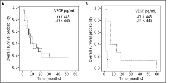
months of survival (p = 0.000) (log-rank test shown in table 5). Kaplan-Meier curves in figure 2 shows the prognostic impact of serum VEGF levels on overall survival by using the cutoff 445 pg/mL.
Figure 2. Prognostic in-fluence of serum VEGF level on overall survival. The sur-vival of patients with serum VEGF levels of < 445 and ≥
445 pg/mL for (A) early HCC disease were 11.0 and 8.0 months, respectively (p = 0.448), (B) advanced HCC disease were 7.0 and 2.0 months, respectively (p = 0.000).
Table 5. Comparison of median survivals of early and advanced HCC disease when stratified by VEGF cutoff 445 pg/mL.
Median survival mo (95% CI) p
VEGF < 445 pg/mL VEGF ≥ 445 pg/mL
HCC patients with early disease 11.0 (7.4-14.6) 8.0 (3.7-12.3) 0.448
HCC patients with advanced disease 7.0 (3.6-10.4) 2.0 (1.4-2.6) 0.000
HCC: hepatocellular carcinoma. VEGF: vascular endothelial growth factor. CI: confidence interval.
Table 4. Comparison of median survivals of stages of four prognostic systems when stratified by VEGF cutoff 445 pg/mL.
Prognostic system Stage Median survival mo (95% CI) p
VEGF < 445 pg/mL VEGF ≥ 445 pg/mL
Okuda I 11.0 (0.1-24.6) 16.0 (5.4-26.6) 0.969
I I 7.0 (2.8-11.2) 8.0 (1.5-14.5) 0.660
III 7.0 (2.1-11.9) 2.0 (1.2-2.7) 0.005
BCLC A 9.0 (5.7-12.2) 16.0 (2.5-29.4) 0.711
B 11.0 (5.4-16.5) 7.0 (0.1-14.2) 0.199
C,D 7.0 (3.6-10.4) 2.0 (1.4-2.5) 0.000
CLIP Early 9.0 (6.0-11.9) 37.0 (12.9-61.0) 0.607
Intermediate 7.0 (5.9-8.0) 7.0 (0.1-14.8) 0.263
Advanced 11.0 (1.1-20.8) 2.0 (0.9-3.0) 0.058
Child-Pugh A 11.0 (7.9-14.0) 16.0 (0.6-31.3) 0.888
B 7.0 (4.7-9.2) 3.0 (0.1-6.0) 0.255
C 14.0 (2.5-25.4) 2.0 (0.4-3.6) 0.094
BCLC: Barcelona Clinic Liver Cancer. CLIP: Cancer of the Liver Italian Program. HCC: hepatocellular carcinoma. VEGF: vascular endothelial growth factor. CI: confidence interval.
A B
1.0
0.8
0.6
0.4
0.2
0.0
0 10 20 30 40 50 60
Time (months) 0 10 Time (months)20 30 40 50 60
Overall survival probability
1.0
0.8
0.6
0.4
0.2
0.0
Overall survival probability
VEGF pg/mL
≥ 445
< 445
≥ 445
Correlation of serum VEGF levels and clinicopathological features
No significant correlation was observed between serum VEGF levels and sex, age, serum AFP level, metastatic disease, presence of varices, etiology of underlying cirrhosis, and extension of tumor.
Serum levels of VEGF in patients with HCC (me-dian 347.09 pg/mL) were significantly higher than those in healthy controls (median 143.35 pg/mL) (p = 0.000), and higher than those in cirrhotic patients without HCC (median 175.55 pg/mL) (p = 0.011). There was also a significant association between high serum VEGF levels and presence of abdominal pain (p = 0.038).
DISCUSSION
In our study, we compared the discriminatory abi-lities of four prognostic staging systems in terms of predicting survival using c-index analysis. As con-cordance probabilities did not reveal any meaningful differences among each other, none of the systems were proved to be more accurate at discriminating survival. The reason for lack of difference may be re-lated to the characteristics of our study population, as we included a heterogeneous group of HCC pa-tients with different underlying etiologies of liver cirrhosis (hepatitis B and C, cryptogenic, alcohol), receiving different forms of treatments (radiofre-quency ablation, chemoembolization, alcohol abla-tion, supportive, surgery). Several other studies comparing HCC staging systems in surgical and nonsurgical series reported varying performances regarding their prognostic stratification and predic-tion abilities.30-35 The characteristics of a study po-pulation might be significantly different from the population which the staging systems were designed originally in terms of tumor extension, treatment strategy, liver function status, genetics, sex, age, geographic area, ethnic group and other demogra-phics. Thus, the relevance of certain parameters and the predictive ability and applicability of staging systems will vary according to the characteristics of the population studied; therefore, it may be challeng-ing to compare them. Certain parameters such as tumor size may only have predictive value in early HCC patients undergoing curative therapies,22,36 whereas some parameters can only be predictive in the setting of advanced HCC patients who benefit from palliative treatments. One other factor complicating the staging process is the incomplete understanding of the highly complex biologic
characteristics of these tumors, thus lack of inte-gration of the intrinsic tumor characteristics in staging systems.37
We performed a detailed analysis to evaluate the prognostic influence of various variables including VEGF as an intrinsic tumor feature and a series of clinical, radiologic and laboratory parameters, con-sidering early and advanced HCC disease separately. For early HCC disease, we identified high serum bili-rubin, a parameter representing liver function as an independent unfavorable prognostic factor for overall survival. BCLC system which was constructed based on the independent prognostic factors derived from the analysis of various studies, has been validated as the best staging system to select early-stage HCC patients who could benefit from curative thera-pies.34,38 In BCLC system portal hypertension and bilirubin levels > 1.5 mg/dL were identified as fac-tors negatively correlating with survival for early HCC patients. For advanced HCC patients, we found serum VEGF level higher than its optimal cutoff of 445 pg/mL as a significant independent predictor of poor survival, while any independent prognostic influence of tumor extension, esophageal varices or bilirubin levels on overall survival were not observed. There was also no significant correlation between serum VEGF levels and tumor extension in our stu-dy. Tumor extension tend to influence survival in our univariate model, but this influence disappeared in our multivariate model. This suggests that the correlation between survival and tumor extension may be mainly indirect, and circulating VEGF levels reflect the tumor angiogenic activity rather than the tumor burden. Furthermore, as reported by other studies, there was no correlation between serum VEGF and AFP levels in our study.18,39,40 Therefore, in advanced HCC disease setting, VEGF is an inde-pendent and the most important prognostic factor and survival seems to be more related to the intrin-sic characteristics of tumor itself rather than liver function parameters for this setting.
non-surgically treated groups, and suggested that choice of therapy was not potentially associated with serum VEGF levels.13 According to this, hetero-geneity of the therapies received in our patient popu-lation would not complicate the generalisability of our results.
In this study, our biomarker cutoff point 445 pg/ mL determined by recursive partitioning method was in agreement with two previous studies based on the same method identifying their cutoff as 450 pg/mL.41,42 Different VEGF cutoffs were identified with different methods in some other studies using the median level or ROC curve method as a cutoff value.16,18,19,44-46 In our study, the serum median level of VEGF was 347.09 pg/mL in HCC patients and was significantly higher than the value in healthy controls and in cirrhotic patients without HCC, but our cutoff value 445 pg/mL best correlated with the survival of our patient population.
When we compared the stages of each scoring sys-tem by pairwise log-rank test, significant survival differences were mostly revealed between early and intermediate-advanced stages. Importantly, we could not show a progressive decrease in survival from the intermediate to the advanced stages in each of all four systems (log-rank Child-Pugh B vs. C p = 0.571; Okuda II vs. III p = 0.103; CLIP intermediate vs. advanced p = 0.537; BCLC B vs. C-D p = 0.174). These proved that the prognostic parameters for intermediate or advanced stages were not well represented in these four conventional prognostic systems and thus, they were inadequate for accurate prognostic stratification. On the other hand, we could stratify advanced stages of four prognostic systems by our VEGF cutoff into risk subgroups with different median survivals; patients with a VEGF level higher than the cutoff value had a worse median survival than VEGF level lower patients (Okuda stage III 2.0 vs. 7.0 mo; BCLC stage C-D 2.0
vs. 7.0 mo; CLIP advanced stage 2.0 vs. 11.0 mo; Child-Pugh stage C 2.0 vs. 14.0 mo). These findings revealed a substantial variation in prognosis among patients within the advanced stages of all four con-ventional prognostic systems, and showed that VEGF could define more homogeneous populations of patients with different outcomes for this group of patients.
Accurate prognostic stratification of advanced HCC population is particularly important, because this population represents the classic patient popu-lation of therapeutic clinical trials. One of the major challenges in designing HCC clinical trials is the he-terogeneity of advanced HCC disease which makes
the results difficult to compare, analyze, and inter-pret. Stratification systems should have an optimal capacity to define homogeneous populations with different outcomes. Thus, tumor prognostic parameters should be well-represented in HCC staging systems, and it seems that this should involve more than the number and size of the tumor or liver function sta-tus, and should reflect the intrinsic biologic charac-teristics of the tumor like angiogenic biomarkers.
Non-invasive means of measuring tumor parame-ters like circulating angiogenic biomarkers is ad-vantageous over evaluation in tumor samples; because it is technically simple, easily accessible and will not induce any bleeding risk in HCC patients with coagulopathy following needle biopsies. Serum VEGF levels have been found to be correlated with VEGF expression in HCC, and can serve as a valid surrogate marker of tumor tissue levels; it seems reasonable to measure serum VEGF levels as a reflection of tumor angiogenic activity in HCC.47
Our study has some limitations. First of all, this is a single-center study. Even though different cen-ters may have their own experience and practice, our patients were followed and treated uniformly based on consensus algorithms widely used by many cen-ters. As a tertiary referral center, although we were able to analyze a broad spectrum of patients with early, intermediate, and advanced tumors, some va-riables had relatively large confidence intervals, which were related to the lack of representative sampling for some subgroups of staging systems. Validation across different geographic populations is especially needed before generalisability of our results due to the global variations related to the complex etiology of HCC, leading to distinct outcomes as revealed between eastern and western patient popu-lations in therapeutic HCC trials.29,48 Due to large variations in serum VEGF levels which made the reliability of cuttoff estimate an issue, additional studies are warranted to optimize the value of VEGF to be used in advanced HCC setting.
by affecting therapeutic decisions may capture bene-fits in advanced HCC outcome. Active research is needed to identify other biomarkers which may allow the stratification of patients not only according to the conventional prognostic measures like liver function or tumor burden, but also more accurately by characterizing intrinsic tumor features.
ABBREVIATIONS
• AFP: α-fetoprotein.
• BCLC: Barcelona Clinic Liver Cancer. • CI: confidence interval.
• CLIP: Cancer of the Liver Italian Program. • HCC: hepatocellular carcinoma.
• VEGF: vascular endothelial growth factor.
ACKNOWLEDGEMENT
This study was supported by Research Founda-tion of Marmara University Faculty of Medicine.
REFERENCES
1. Parkin DM. Global cancer statistics in the year 2000.
Lan-cet Oncol 2001; 2: 533-43.
2. Marrero JA. Hepatocellular carcinoma. Curr Opin
Gas-troenterol 2006; 22: 248-53.
3. Motola-Kuba D, Zamora-Valdes D, Uribe M,
Mendez-San-chez N. Hepatocellular carcinoma. An overview. Ann
He-patol 2006; 5: 16-24.
4. El-Serag HB, Mason AC. Rising incidence of hepatocellular
carcinoma in the United States. N Engl J Med 1999; 340:
745-50.
5. Altekruse SF, McGlynn KA, Reichman ME. Hepatocellular carcinoma incidence, mortality, and survival trends in the
United States from 1975 to 2005. J Clin Oncol 2009; 27:
1485-91.
6. Bosetti C, Levi F, Boffetta P, Lucchini F, Negri E, La Vec-chia C. Trends in mortality from hepatocellular carcinoma
in Europe, 1980-2004. Hepatology 2008; 48: 137.
7. El-Serag HB. Hepatocellular carcinoma: an epidemiologic
view. J Clin Gastroenterol 2002; 35: 72-8.
8. Fattovich G, Stroffolini T, Zagni I, Donato F. Hepatocellu-lar carcinoma in cirrhosis: Incidence and risk factors.
Gastroenterology 2004; 127: 35-50.
9. Jemal A, Bray F, Center MM, Ferlay J, Ward E, Forman
D. Global cancer statistics. CA Cancer J Clin 2011; 61:
69-90.
10. Yang ZF, Poon RT. Vascular Changes in Hepatocellular
Car-cinoma. The Anatomical Record 2008; 291: 721-34.
11. Huang GW, Yang LY, Lu WQ. Expression of hypoxiainduci-ble factor 1alpha and vascular endothelial growth factor in hepatocellular carcinoma: impact on neovascularization
and survival. World J Gastroenterol 2005; 11: 1705-8.
12. Zhang ZL, Liu ZS, Sun Q. Expression of angiopoietins, Tie2 and vascular endothelial growth factor in angiogenesis
and progression of hepatocellular carcinoma. World J
Gas-troenterol 2006; 12: 4241-5.
13. Schoenleber SJ, Kurtz DM, Talwalkar JA, Roberts LR, Gores GJ. Prognostic role of vascular endothelial growth factor in hepatocellular carcinoma: systematic review and
meta-analysis. Br J Cancer 2009; 100: 1385-92.
14. Yao DF, Wu XH, Zhu Y, Shi GS, Dong ZZ, Yao DB, Wu W, et al. Quantitative analysis of vascular endothelial growth factor, microvascular density and their clinicopathologic
features in human hepatocellular carcinoma. Hepatobiliary
Pancreat Dis Int 2005; 4: 220-6.
15. Tseng CS, Lo HW, Chen PH, Chuang WL, Juan CC, Ker CG. Clinical significance of plasma D-dimer levels and serum
VEGF levels in patients with hepatocellular carcinoma.
He-patogastroenterology 2004; 51: 1454-8.
16. Poon RT, Ho JW, Tong CS, Lau C, Ng IO, Fan ST. Prognos-tic significance of serum vascular endothelial growth fac-tor and endostatin in patients with hepatocellular
carcinoma. Br J Surg 2004; 91: 1354-60.
17. Kamel L, Nessim I, Abd-el-Hady A, Ghali A, Ismail A. Assess-ment of the clinical significance of serum vascular endo-thelial growth factor and matrix metalloproteinase-9 in
patients with hepatocellular carcinoma. J Egypt Soc
Para-sitol 2005; 35: 875-90.
18. Chao Y, Li CP, Chau GY, Chen CP, King KL, Lui WY, Yen SH, et al. Prognostic significance of vascular endothelial growth factor, basic fibroblast growth factor, and angio-genin in patients with resectable hepatocellular carcinoma
after surgery. Ann Surg Oncol 2003; 10: 355-62.
19. Poon RT, Lau C, Yu WC, Fan ST, Wong J. High serum levels of vascular endothelial growth factor predict poor response to transarterial chemoembolization in hepatocellular
carci-noma: a prospective study. Oncol Rep 2004; 11: 1077-84.
20. Li X, Feng GS, Zheng CS, Zhuo CK, Liu X. Expression of plasma vascular endothelial growth factor in patients with hepatocellular carcinoma and effect of transcatheter ar-terial chemoembolization therapy on plasma vascular
en-dothelial growth factor level. World J Gastroenterol
2004; 10: 2878-82.
21. Wildi S, Pestalozzi BC, McCormack L, Clavien PA. Critical evaluation of the different staging systems for
hepatoce-llular carcinoma. Br J Surg 2004; 91: 400-8.
22. Okuda K, Ohtsuki T, Obata H, Tomimatsu M, Okazaki N, Hasegawa H, Nakajima Y, et al. Natural history of hepato-cellular carcinoma and prognosis in relation to treatment.
Study of 850 patients. Cancer 1985; 56: 918-28.
23. CLIP Group. A new prognostic system for hepatocellular carcinoma: a retrospective study of 435 patients: the Cancer of the Liver Italian Program (CLIP) investigators.
Hepatology 1998; 28: 751-5.
24. Llovet JM, Bru C, Bruix J. Prognosis of hepatocellular
car-cinoma: the BCLC staging classification. Semin Liver Dis
1999; 19: 329-38.
25. Chen CH, Hu FC, Huang GT, Lee PH, Tsang YM, Cheng AL, Chen DS, et al. Applicability of staging systems for pa-tients with hepatocellular carcinoma is dependent on treatment method-analysis of 2010 Taiwanese patients.
Eur J Cancer 2009; 45: 1630-9.
26. Lu W, Dong J, Huang Z, Guo D, Liu Y, Shi S. Comparison of four current staging systems for Chinese patients with hepatocellular carcinoma undergoing curative resection:
Okuda, CLIP, TNM and CUPI. J Gastroenterol Hepatol
2008; 23: 1874-8.
27. Farinati F, Sergio A, Baldan A, Giacomin A, Di Nolfo MA, Del Poggio P, Benvegnu L, et al. Early and very early hepato-cellular carcinoma: when and how much do staging and choice of treatment really matter? A multi-center study.
28. Bruix J, Sherman M. Management of hepatocellular
carci-noma: an update. Hepatology 2011; 53: 1020-2.
29. Llovet JM, Ricci S, Mazzaferro V, Hilgard P, Gane E, Blanc JF, de Oliveira AC, et al. Sorafenib in advanced
hepatoce-llular carcinoma. N Engl J Med 2008; 359: 378-90.
30. Collette S, Bonnetain F, Paoletti X, Doffoel M, Bouché O, Raoul JL, Rougier P, et al. Prognosis of advanced hepato-cellular carcinoma: Comparison of three staging systems
in two French clinical trials. Ann Oncol 2008; 19: 1117-26.
31. Guglielmi A, Ruzzenente A, Pachera S, Valdegamberi A, San-dri M, D’Onofrio M, Iacono C. Comparison of seven sta-ging systems in cirrhotic patients with hepatocellular carcinoma in a cohort of patients who underwent
radio-frequency ablation with complete response. Am J
Gas-troenterol 2008; 103: 597-604.
32. Kondo K, Chijiiwa K, Nagano M, Hiyoshi M, Kai M, Maehara N, Ohuchida J, et al. Comparison of seven prognostic sta-ging systems in patients who undergo hepatectomy for
hepatocellular carcinoma. Hepatogastroenterology 2007;
54: 1534-8.
33. Grieco A, Pompili M, Caminiti G, Miele L, Covino M, Alfei B, Rapaccini GL, et al. Prognostic factors for survival in pa-tients with early intermediate hepatocellular carcinoma undergoing non-surgical therapy: Comparison of Okuda, CLIP, and BCLC staging systems in a single Italian centre.
Gut 2005; 54: 411-8.
34. Cillo U, Bassanello M, Vitale A, Grigoletto FA, Burra P, Fa-giuoli S, D’Amico F, et al. The critical issue of hepatocellu-lar carcinoma prognostic classification: Which is the best
tool available? J Hepatol 2004; 40: 124-31.
35. Levy I, Sherman M; Liver Cancer Study Group of the Uni-versity of Toronto. Staging of hepatocellular carcinoma: assessment of the CLIP, Okuda, and Child-Pugh staging
systems in a cohort of 257 patients in Toronto. Gut 2002;
50: 881-5.
36. Bruix J, Sherman M, Llovet JM, Beaugrand M, Lencioni R, Burroughs AK, Christensen E, et al. EASL Panel of Experts on HCC. Clinical management of hepatocellular carcinoma. Conclusions of the Barcelona-2000 EASL conference.
Euro-pean Association for the Study of the Liver. J Hepatol
2001; 35: 421–30.
37. Singhal A, Jayaraman M, Dhanasekaran DN, Kohli V. Mole-cular and serum markers in hepatocellular carcinoma:
Pre-dictive tools for prognosis and recurrence. Crit Rev Oncol
Hematol 2012; 82: 116-40.
38. Befeler AS, Di Bisceglie AM. Hepatocellular carcinoma:
diagnosis and treatment. Gastroenterology 2002; 122:
1609-19.
39. Poon RT, Ng IO, Lau C, Zhu LX, Yu WC, Lo CM, Fan ST, et al. Serum vascular endothelial growth factor predicts ve-nous invasion in hepatocellular carcinoma: a prospective
study. Ann Surg 2001; 233: 227-35.
40. Jinno K, Tanimizu M, Hyodo I, Nishikawa Y, Hosokawa Y, Doi T, Endo H, et al. Circulating vascular endothelial growth factor (VEGF) is a possible tumor marker for
me-tastasis in human hepatocellular carcinoma. J
Gastroente-rol 1998; 33: 376-82.
41. Kaseb AO, Hassan MM, Lin E, Xiao L, Kumar V, Pathak P, Lozano R, et al. V-CLIP: Integrating plasma vascular en-dothelial growth factor ýnto a new scoring system to stratify patients with advanced hepatocellular carcinoma
for clinical trials. Cancer 2011; 117: 2478-88.
42. Kaseb AO, Morris JS, Hassan MM, Siddiqui AM, Lin E, Xiao L, Abdalla EK, et al. Clinical and prognostic ýmplications of plasma ýnsulin-like growth factor-1 and vascular endothe-lial growth factor in patients with hepatocellular carcino-ma. J Clin Oncol 2011; 29: 3892-9.
43. Niizeki T, Sumie S, Torimura T, Kurogi J, Kuromatsu R, Iwamoto H, Aino H, et al. Serum vascular endothelial growth factor as a predictor of response and survival in patients with advanced hepatocellular carcinoma
under-going hepatic arterial infusion chemotherapy. J
Gastroen-terol 2012; 47: 686-95.
44. Ho MC, Chen CN, Lee H, Hsieh FJ, Shun CT, Chang CL, Lai YT, et al. Placenta growth factor not vascular endothelial growth factor A or C can predict the early recurrence
af-ter radical resection of hepatocellular carcinoma. Cancer
Lett 2007; 250: 237-49.
45. Poon RTP, Lau C, Pang R, Ng KK, Yuen J, Fan ST. High serum vascular endothelial growth factor levels predict poor prognosis after radiofrequency ablation of hepato-cellular carcinoma: importance of tumor biomarker in
ablative therapies. Ann Surg Oncol 2007; 14: 1835–45.
46. Treiber G, Wex T, Rocken C, Fostitsch P, Malfertheiner P. Impact of biomarkers on disease survival and progression in patients treated with octreotide for advanced
hepatocellu-lar carcinoma. J Cancer Res Clin Oncol 2006;132: 699-708.
47. Poon RT, Lau CP, Cheung ST, Yu WC, Fan ST. Quantitative correlation of serum levels and tumor expression of vascu-lar endothelial growth factor in patients with
hepatocellu-lar carcinoma. Cancer Res 2003; 63: 3121-6.
48. Cheng AL, Kang YK, Chen Z, Tsao CJ, Qin S, Kim JS, Luo R, et al. Efficacy and safety of sorafenib in patients in the Asia-Pacific region with advanced hepatocellular carcino-ma: a phase III randomised, double-blind,