Hepatitis C virus strategies to evade the specific-T cell
response:
a possible mission favoring its persistence
Jorge Fabián Quarleri,* José Raúl Oubiña** * Instituto de Investigaciones Biomédicas en Retrovirus y Sida (INBIRS), Universidad de Buenos Aires-CONICET, Argentina. ** Instituto de Microbiología y Parasitología Médica (IMPAM), Universidad de Buenos Aires-CONICET, Argentina.
A B S T R A C T A B S T R A C T A B S T R A C T A B S T R A C T A B S T R A C T
Hepatitis C virus (HCV) is a small, enveloped RNA virus. The number of HCV-infected individuals worldwide is estimated to be ap-proximately 200 million. The vast majority of HCV infections persist, with up to 80% of all cases leading to chronic hepatitis associa-ted with liver fibrosis, cirrhosis, and hepatocellular carcinoma. The interaction between HCV and the host have a pivotal role in viral fitness, persistence, pathogenicity, and disease progression. The control of HCV infection requires both effective innate and adaptive immune responses. The HCV clearance during acute infection is associated with an early induction of the innate and a delayed initia-tion of the adaptive immune responses. However, in the vast majority of acute HCV infecinitia-tions, these responses are overcome and the virus persistence almost inexorably occurs. Recently, several host- and virus-related mechanisms responsible for the failure of both the innate and the adaptive immune responses have been recognized. Among the latter, the wide range of escape mutations to evade the specific-T-and B-cell responses as well as the T cell anergy and the CD8+ T cell exhaustion together with the interference with its function after prolonged virus exposure hold a pivotal role. Other HCV strategies include the modification or manipulation of molecules playing key roles in the induction of the interferon response and its induced effector proteins. In this review, we at-tempt to gain insights on the main T cell immune evasion strategies used by the virus in order to favor its persistence.
Key words. Key words. Key words. Key words.
Key words. HCV. Immune evasion. CD4+ T cell. CD8+ T cell.
January-February, Vol. 15 No. 1, 2016: 17-26
THE NATURAL
COURSE OF HCV INFECTION
Hepatitis C virus (HCV) infection represents the most common blood-borne viral infection for which a vaccine is still lacking, while an estimated 3% of the world’s popu-lation is infected, representing approximately 200 million individuals.1 The control of the acute infection depends
on both host- and viral-related factors. The former in-cludes both an early induction of innate- and a delayed ini-tiation of adaptive-immune responses. However, in the majority of acutely HCV infected individuals, these re-sponses are insufficient to clear the virus and most of HCV infections persists, with up to 80% (50-90%) of all cases leading to chronic hepatitis, eventually associated with liver fibrosis, cirrhosis and/or hepatocellular carci-noma. The acute phase of infection is usually asymptomat-ic, with rare cases exhibiting fatigue, malaise and jaundice during a few weeks. Viral RNA can be detected 1-3 weeks
after viral entry into the organism, despite a high induction level of the innate immune response.2 Intriguingly, HCV
presence seems to be virtually overlooked by both T cell and B cell adaptive immune responses during the first 8-12 weeks after infection (e.g., antibodies to HCV may be evi-dent by 3 months after infection, in approximately 90% of infected individuals).3 An elevated level of
alanine-ami-notransferase (ALT) within 4-12 weeks after exposure is an indication of liver cell injury. Immune events during this stage of disease are considered to be essential for de-termining the outcome of infection.
INTERFERON LAMBDA (IFN-λλλλλ): AN ESSENTIAL PARTNER OF THE T CELL RESPONSE
Although the impact of both interferon stimulated genes (ISG) and IL-28B (a subclass of IFN-λ, a type III IFN) on the course of HCV natural infection are well
The Official Journal of the Mexican Association of Hepatology, the Latin-American Association for Study of the Liver and
the Canadian Association for the Study of the Liver
Manuscript received: Manuscript received:Manuscript received:
beyond the scope of this concise review, a brief comment about the latter is well deserved, considering its current prognostic usefulness for clinicians and its connection with T cell response. IFN-λ is the most recently group of identified IFN proteins, which includes IFN-λ1, -λ2, -λ3 and -λ4, encoded by IFNL1 (formerly, IL-29),IFNL2 (IL-28A), and IFNL3 (IL28B) and IFNL4 genes, respectively. In humans, IFNL1, IFNL2 and IFNL3 are functional genes, while IFNL4 is a pseudogene in part of the popula-tion due to the single nucleotide polymorphisms (SNP) rs368234815.4
Viruses can be sensed in infected cells through the in-teraction of pathogen-associated molecular patterns (PAMPs) with sensors such as cytosolic helicases (e.g. RIG-1, MDA-5 or LGP-2), membrane-associated Toll-like receptors (TLRs) and Nod-Toll-like receptors (NLRs). After engaging with their respective ligands, these pattern recognition receptors activate IKKε/TANK-binding ki-nase 1 (TBK-1), the IKKα/β/γ complex, as well as MAPK, leading to the activation of transcription factors, such as IFN regulatory factor 3 (IRF-3), IRF-7, NF-κB, and AP-1.
Once activated, these factors translocate into the nucleus and, in turn, activate the transcription of type I IFN. In an autocrine as well as a paracrine way, the IFN-α/β activates the expression of ISGs through the JAK-STAT signaling pathway to establish an antiviral state in universally dis-tributed host cells. Likewise, IFN-λs are believed to acti-vate the same pathway promoting the expression of a set of antiviral proteins in epithelial cells including hepatocytes and in as yet not properly defined hematopoietic cells.4
Indeed, both the T cell response as well as the SNPs in the proximity of the IFNL3 gene appears to be crucial for viral clearance.4,5 The molecular mechanism underlying
the association between SNPs and the outcome of natural clearance of HCV or the response to IFN therapy in pa-tients chronically infected with HCV has been proposed recently by McFarland, et al. They have identified a favora-ble SNP located in the 3’ untranslated region of IFNL3
mRNA that diminishes the binding of HCV-induced mi-croRNAs (miR-208b and miR-499a-5p) during infection. Thus, this SNP dictates transcript stability and influences the outcome of both antiviral treatment and spontaneous
Figure 1. Figure 1. Figure 1. Figure 1.
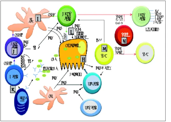
Figure 1. Essential features of the innate and adaptive immune re-sponse during primary HCV infec-tion. A.A.A.A.A. HCV replication in hepatocytes promoting the release of type I and type III IFNs. IFN-α and -λ are also secreted by both plasmocytoid (p) and myeloid (m) dendritic cells (DCs). These cy-tokines activate NK cells for killing HCV-infected hepatocytes. B.B.B.B.B. Küpffer cells (as liver antigen-pre-senting cells) and DCs present HCV-derived epitopes to both CD4+ and CD8+ T cells in the context of HLA class II and HLA class I, respectively. C.C.C.C. CD4+C. helper T cells activate both CD8+ T cells and B cells through production of Th1 (IFN-γ IL-2) and Th2 (IL-4, IL-5, IL-6) cytokines, respectively. D.D.D.D.D. Anti-HCV specific Igs are released from plasma cells during primary infection. E.E.E.E.E. Clearance mechanisms are mediated by CD8+ T cells exerted by cytolytic (perforin and granzymes), or non-cytolytic mechanisms through secretion of IFN-γ and TNF-α. F.F.F.F.F. IL-21 (mainly produced by Th17 cells, but also by NKT cells) sustains CD8+ T cell functions and is able to rescue virus-specific T cells from exhaustion. G.G.G.G.G. Regulatory T cells might decrease primary immune response by secreting the regulatory cytokines TGF-β, IL-10, or by expressing Gal-9 that enhances apoptosis of Tim-3+ CD4+ and CD8+ T cells.
DC
APC BBBBB
HLA-1 CD8T
cell TGFIL-10β
Gal-9
PD-1 Tim-3 CTLA-4 CD39 CD160
IL-4 IL-5 IL-6 IL-10 IL-13
T h 2 T h 2 T h 2 T h 2 T h 2 HLA-II
CD4T cell
C CC C C
IFNγ IL-2
Th 1 Th 1 Th 1 Th 1 Th 1
D DD D D
Igs anti-HCV
Cytolysis
IFNα/β and λ
IFNλ
IFNγ IFNα
pDC
Plasma
cell
NKT cell NIK cell
Th17 Th17
F FF FF
A AA A A
G G G G G
Exhaustion
IL-21
TGFβ Gal-9 IFNγ
TNFα Perferin Granzyme
E EE EE
IFNα
IFNλ
HCV
Hepatocyte
B cell
HLA-1
HLA-II T r e gT r e gT r e gT r e gT r e g
clearance during HCV infection.6 Additionally, a variant
upstream of IFNL3 creating a new interferon gene –re-ferred as the ancestral ‘ΔG’ allele of IFNL4– is associated with impaired clearance of hepatitis C virus. This new genotype results in the expression of four previously un-known proteins, including p179, designated IFN-λ4. This cytokine induces a weak cellular signaling that may reduce the responsiveness of cells to IFN-α, thereby inhibiting efficient HCV clearance.7 In sharp contrast, the TT allele
of such SNP disrupts the open reading frame of IFNL4
(thus, inhibiting its translation) and protects against HCV. These findings suggest that IFN-λ4 is the causative agent of HCV clearance failure. Remarkably, the very recent dis-covery of an additional SNP (rs117648444) acting in com-bination with the ΔG SNP proved also useful to predict the response to anti-HCV treatment.8
The reader is kindly prompted to look at both figures 1 and 2, as well as to table 1 to follow the key concepts de-picted in this concise review.
T CELL RESPONSE TO HCV INFECTION
The outcome of HCV infection is mainly influenced by the coordinated action of heterogeneous cell types. In this regard, numerous reports provide evidence that the
HCV clearance depends on the magnitude, breadth and quality of the virus-specific CD4+ and CD8+ T cell re-sponses to multiple epitopes derived from both structural and non-structural proteins.9 For the first time, and as a
consequence of the delayed induction –but not to a cellu-lar recruitment failure–, such immune responses are de-tected in blood 4 to 8 weeks after infection.10 The primary
effector response is mediated by CD8+ T cells, whose function is sustained by the CD4+ T cells, both of which are responsible for long-lasting control of the HCV.11
Af-ter the onset of the HCV-specific CD8+ T cell response, a direct relationship is established with the HCV clear-ance. A vigorous response related to viral clearance and targeting multiple epitopes is detected in T cells recov-ered either from the liver or from peripheral blood. Such antiviral activity is mainly exerted through IFN-γ -mediat-ed non-cytolytic effector functions and merely to a lower extent through cytolytic effector functions, exerted by per-forin as well as by Fas/FasL interactions. The non-cytolyt-ic antiviral activity is 100- to 1,000-fold more effective than the cytolytic one.12 However, other CD8+ T cell
func-tions with a protective role are also observed, such as the production of IL-17 from a subset of cells able to recog-nize dissimilar epitopes than those targeted by IFN-γ se-creting CD8+ T cells.13
Figure 2. Figure 2. Figure 2. Figure 2.
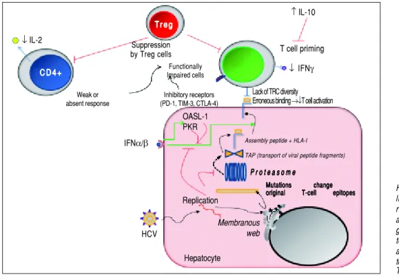
Figure 2. Mechanisms used by HCV to escape from adaptive cel-lular immune response. During viral replication, HCV proteins are gener-ated; then, these molecules are de-graded by the proteasome complex to generate peptide fragments that are translocated to the ER in order to assembly with HLA-I molecules. These complexes are transported to the hepatocyte surface to be presented to CD8+ T cells. Once activated, these cells secrete IFN-γ. The secreted levels of both IL-2 and IFN-γ by CD4+ and CD8+ T cells are respectively dimin-ished. IL-10 is able to suppress naïve HCV-specific CD8+ T cell priming. Red lines indicate strategies to suppress the immune response. Dotted lines show the steps throughout the HCV replication as well as HCV-derived viral epitopes presentation on hepatocyte surface by HLA-I. The green lines denote the stimula-tory effect of type I IFNs on HLA-I expression on cell surface and on both PKR and OASL-1 molecules. For further details, see the main text.
HCV IFNα/β
T r e g T r e g T r e g T r e g T r e g
↑ IL-10
↓ IL-2
C D 4 + C D 4 +C D 4 + C D 4 + C D 4 +
Suppression
by Treg cells T cell priming
Functionally
Impaired cells ↓ IFNγ Weak or
absent response
Inhibitory receptors (PD-1, TIM-3, CTLA-4)
Lack of TRC diversity
Erroneous binding →↓T cell activation
Replication
Hepatocyte
Mutations change Mutations changeMutations change Mutations change Mutations change original T-cell epitopes original T-cell epitopesoriginal T-cell epitopes original T-cell epitopes original T-cell epitopes
P r o t e a s o m e P r o t e a s o m eP r o t e a s o m e P r o t e a s o m e P r o t e a s o m e
Assembly peptide + HLA-I
TAP (transport of viral peptide fragments)
Membranous web OASL-1
From the host, the HCV-specific CD8+ T cell re-sponse is influenced by the HLA class I alleles. HLA class I alleles A*0301, B*2701, B*5701, B*5703 and C*0102 are associated with viral clearance, whereas B8 is linked to vi-ral persistence. The strongest protective effect is observed with HLA-B27.14 Remarkably, HLA class I expression is
up-regulated by both type-I and -II IFNs.
The HCV-specific CD4+ T cells are critical for both limiting immune evasion and priming effector memory CD8+ T cells. Interestingly, among acute HCV patients – and irrespectively of their clinical outcome– primed broad HCV-specific CD4+ T cell responses directed against multiple epitopes are observed in blood, which persist for long periods in patients that spontaneously re-solve the infection. This event almost exclusively takes place during the first year of infection –and usually within the first 6 months–, stressing the fact that the virus tends to gain the battle against the host as time elapses. Moreo-ver, when the infection evolves to persistence, the HCV-specific T cell responses are not sustained and become specifically dysfunctional for HCV.15 In the latter
scenar-io, virus and host cohabit avoiding accelerated liver dam-age. Similarly to the findings described for HLA class I, certain HLA class II alleles influence the outcome of HCV infection as well. The HLA class II alleles most reproduc-ibly associated with viral clearance are DRB1*1101 and DQB1*0301, which are genetically close-linked. Howev-er, HCV-derived CD4+ T cell epitopes showed promis-cuity and can be presented by different HLA II molecules.16 As previously described for CD8+ T cells,
IL-17-producing CD4+ T cells were observed among chronic infected patients, but their role in the outcome is still debated. These T cells displayed a distinct
phenotyp-ic profile, high expression of the homing receptor CD161 and low levels of inhibitory receptors, such as mucin-do-main-containing-molecule-3 (Tim-3) and programmed-death 1 (PD-1).17
ESCAPE MUTATIONS TO EVADE THE SPECIFIC-T CELL RESPONSE
Whether the HCV escape from T cell response is a cause or a consequence of HCV persistence is still under extensive debate. However, several important insights into CD8+ T cell-mediated viral escape have been revealed. The HCV exhibits a proclivity to mutate due to its high rate of replication (up to 1012-14 particles/day or about 106
IU/mL of plasma) associated with a lack of proofreading ability of its polymerase. Therefore, multiple virus vari-ants or quasispecies co-circulate in an individual patient facilitating the selection of CD8+ T-cell escape variants. Hence, their occurrence responds to a complex interplay of the host T cell responses, the viral epitopes evolution under immune selection pressure and the viral fitness costs, although they may be restricted due to the incom-patibility with an abolished viral replicative capacity. Four distinct outcomes may take place, as depicted in the fol-lowing paragraph:
• The viral clearance during acute infection is associated with a sustained CD4+ T cell response with multi-specific CD8+ T cell response that may constrain the development of viral escape mutations. Remarkably, the early CD4+ T cell responses tend to disappear from the blood in those patients who develop HCV persistence.18 This failure of the CD4+ T cell
Table 1. Highlights on HCV strategies for viral evasion from the adaptive cellular immune response.
Viral strategies References
Escape mutations to evade -Proteasomal processing (mutations in the flanking region) 7 the specific-T cell response -Binding to HLA molecules (mutations at the HLA binding anchors)
-TCR recognition (mutations in the centre of the epitope)
Original antigenic sin 27
Clustered accumulated mutations 24
Inhibitory signal to HCV-specific CD8+ T cells 33, 35, 36
T cells primed improperly became anergic 25, 30
T cells exhausted under a prolonged and excessive stimulation 32, 37, 38
CD8+ T cell exhaustion Expression of inhibitory receptors (PD-1, Tim-3, 2B4, KLRG-1, 22 after prolonged virus exposure CTLA-4, and CD160), or their ligands in liver cells
(PDL-1, Gal-9 and other molecules)
Expansion of Tregs 46, 50, 51
CD4+ T cell response failure Decrease of IL-21producing Th17 cells and 22
increase of Gal-9 producing Foxp3+ regulatory CD4+ T cells
response during the acute phase of the HCV infection may be due to a decrease in IL-21-producing Th17 cells and an increase of galectin-9 (Gal-9) producing Foxp3+ regulatory CD4+ T cells.19
• The above mentioned HCV escape mutations do not likely develop when the CD8+ T cell response is weak, associated with an absent or impaired CD4-help and priming. In this scenario, high viral levels and in-tact HCV epitopes are associated with increased levels of T cell inhibitory receptors.
• In contrast, when a discrete failure of the CD4+ T cell response occurs in the presence of a dysfunctional nar-row but vigorous CD8+ T cell response, the appear-ance of escape mutations is favored and additional compensatory mutations may be required for replica-tive fitness improvement. These facts are associated with a decreased inhibitory receptor expression on CD8+ T cells, perhaps accounting for a HCV robust proliferation to wild-type, non-mutated virus.
• Finally, without both a restricting HLA allele and a CD8+immune selective pressure, reversion to the wild-type sequence probably occurs as a consequence of the high fitness cost associated with some escape mutations.9 These HCV mutations may occur at 3
dis-tinct sites regarding T cell epitopes:
a) In a flanking region of these molecules, leading to a proteasomal processing of an abnormal peptide, which may be cleaved within the epitope and hence, without being loaded onto the HLA-I molecule. b) Within the HLA-I anchor site (i.e. P2 and the
C-terminal amino acid which fails to promote CD8+ T cell activation), or
c) Within T cell binding site (i.e. in the center of the epitope, which prevents a proper activation of CD8+ T cells as well or even inducing it as an an-tagonist).9,20
The HLA-I allele background could also play an im-portant role in determining viral escape, since it has been reported a strong association between specific HLA-I alle-les and viral escape within an immunodominant HLA-re-stricted epitope (e.g. HLA-B27). Importantly, the occurrence of clustered accumulated mutations within the epitope is required for efficient CD8+ T-cell escape de-spite the broad cross-recognition of viral variants. Thus, immune escape probably requires a too long elapsed time, providing enough time to the host immune system to clear the virus before escape can take place.21 These strategies
constitute a means to escape HLA-restricted immune re-sponses but considering the HCV fitness costs, such abili-ty is constrained and compensatory mutations may be required to restore HCV replicative capacity.22 When the
mutated epitopes are no longer bound by the HLA-I mol-ecule, no substantial additional diversification is expected. However, those epitopes that still have the capacity to bind to the HLA molecule may trigger the “original anti-genic sin” phenomenon that results in a diminished clear-ance of viral variants and therefore, enhclear-ancement of viral escape. In this regard, two pathways might take place:
• The expansion of wild-type specific CD8+ T cells as a consequence of stimulation by mutated epitopes23 and
• Alternatively, the inhibition of HCV-specific CD8+ T cells responding to HLA-I mutated epitopes after TCR recognition due to inhibitory signaling. Despite its elevated frequency among targeted CD8+ T-cell epitopes, viral escape mutations are not a general con-sequence resulting from CD8+ T-cell pressure. In this regard, a large proportion of intrahepatic virus-specific CD8+ T-cells was detected to target the non-mutated viral antigens among chronically infected patients. Thus, it suggests that factors other than muta-tional escape contribute to the failure of intrahepatic virus-specific CD8+ T-cells.20
Regarding the HLA class II–restricted epitopes escape mutations can also occur, but they are infrequent among chronically infected patients.24 Considering both HLA-I
and -II, it is plausible to infer that additional mechanisms could contribute to CD8+ and CD4+ T-cell failure, such as T-cell dysfunction. In this context, two distin-guishable situations are possible in chronic HCV infec-tion. By the one hand when T cells are improperly primed by signaling through the TCR in the absence of costimulatory or inflammatory signals, they become
aner-gic.25,26 By the other hand, when T cells become exhausted
and primed by antigen, as well as costimulatory and in-flammatory signals, they still develop some effector func-tions. But then, under a prolonged and excessive stimulation, progressive loss of functions occurs.26
CD8+ AND CD4+ T
CELL FUNCTION IMPAIRMENT
Viral persistence is prompted when CD8+ T cell ex-haustion emerges. The prolonged exposure appears to be the mechanism that leads to T cell dysfunction in chronic hepatitis C. Such persistent high level of viremia is firstly accompanied by a good T cell response but then it fails to eliminate the virus with a subsequent gradual decline of CD8+ and CD4+ T cell responses. Two main pathways have been described that mediate this exhaustion:
• The expansion of regulatory T cells (Tregs) that sup-press CD8+ T cell activity.
The former involves changes in cytokines’ levels pro-duction or cell proliferation after antigen-dependent stim-ulation that prime T-cells for activation-induced apoptosis.27 Such T-cell exhaustion is sustained by the
over-expression of inhibitory receptors like Tim-3, PD-1, cytotoxic T-lymphocyte antigen 4 (CTLA-4), CD160 and 2B4 on HCV-specific CD8+ T cells in the blood and liv-er of individuals developing chronic HCV infection. Like-wise, the expression of their respective ligands in the liver may contribute to exhaustion and apoptosis, as occurs when the PD-1 ligand-1 (PDL-1) and the Tim-3 ligand Gal-9 are highly expressed.19 Amazingly, a HLA-I
restrict-ed CD8+ T cell -to-CD4+ T cell cross-differentiation might occur with the same clonotypic T cell receptor. This conversion could produce HLA-class-I-restricted CD4+Foxp3+ Treg cells,28 whose implications still
re-main to be explored in HCV infection. The dysfunctional HCV-specific CD8+ T cells also exhibit a low inter-leukin-7 receptor (CD127) expression, able to regulate CTL reactivity through the balance modulation between Mcl-1(myeloid leukemia cell differentiation protein-1) and Bim (Bcl2-interacting mediator). Bim is a pro-apop-totic molecule blocked by the action of Mcl-1. A low ex vivo Mcl-1 expression and Bim up-regulation after antigen encounter were involved in CD127(low) HCV-specific
CTL hyporeactivity during chronic infection.29,30 While
this manuscript was under review, terminally exhausted CD8+ T cells have been identified as CD39+ cells.31
The T cell exhaustion has been proposed as a mecha-nism underlying the dysfunction of HCV-specific CD4+ and CD8+ T cells during acute infection. However, this proposal is still under intense debate. If true, it should in-dicate that the fate of the HCV infection is already deter-mined at its very early stage. In this regard, at least four groups initially observed contrasting results, since high PD-1 levels on HCV-specific T cells during acute infec-tion were observed to be associated with viral persistence, requiring preservation of cognate antigen during the chronic stage,32 while such high PD-1 level on both
HCV-specific CD8+ and CD4+ T cells during acute HCV infection, appeared to be unrelated to the clinical outcome in other studies.33 Perhaps, a conciliating view
might take into account that T cell exhaustion is a dynam-ic process whdynam-ich exhibits variable stages of impairment, as observed in the experimental lymphocytic choriomenin-gitis virus (LCMV) model,34 and confirmed by elegant ex
vivo experiments on acute HCV patients who either spon-taneously resolved the infection or became persistently in-fected, pointing to crucial and distinct roles of Tim-3 and PD-1 molecules.32 Importantly, the frequency of the
memory precursor marker CD127 on HCV specific T cells has been recently proposed to predict the outcome of the HCV acute infection in experimentally infected chim-panzees, since high levels appeared to be present only in those who cleared the infection.35 The targeting of
multi-ple co-regulatory receptors has very recently opened new avenues for therapeutic approaches.36
Several groups have demonstrated weak or absent HCV-specific CD4+ T cell responses during chronic HCV infection with a failure of IL-2 secretion that may lead to disruption of IFN-γ and cellular proliferation, as opposed to physical deletion or complete functional unre-sponsiveness. Such inhibition of IL-2 secretion -but not of its transcription-has also been recently associated with the interaction of HCV E2 glycoprotein with T cells.37
Af-ter a decreased production of IL-2, the cytotoxicity and production of TNF-α and IFN-γ are impaired sequen-tially. The recent discovery of CD154 molecule (CD40L) as a biomarker of antigen specific activated CD4+T cells might help to overcome some of the drawbacks of func-tional assays and limitations of multimer-based methods used for the detection of this crucial population,38 thus
an-swering a key question: is this a matter of an insufficient number of activated CD4+ T cells and/or a result of func-tional impairment? In this regard, it should be stressed that HCV is able to infect T cells. Interestingly, HCV E2 RNA as well as E2 protein are independently able to sup-press either proximal and distal TCR signaling, respec-tively. Most likely both structures are involved in HCV pathogenesis, contributing to viral persistence.39
Moreo-ver, HCV F protein promotes the PD1 expression on both CD8+ and CD4+ T cells, impairing their functionality.40
The action of Treg cells may also cause exhaustion of HCV-specific CD8+Tcell response. Treg are T lym-phocyte subsets within the CD4+ and CD8+ compart-ments with strong anti-inflammatory functions. Thus, CD4+ Treg and CD8+ Treg inhibit virus-induced im-mune activation.41 Their presence is not privative of
chronic infection, but when a high-level viremia is per-sistent, the suppressive activity in vitro is significantly high-er than that obshigh-erved from cells obtained from patients who spontaneously clear the virus.42 Under immune
pres-sure and escaping host defenses, HCV variants emerge. Such viral HLA-II- epitopes variants are able to induce an-tigen-specific Treg cells to suppress the antiviral T-cell response in an antigen-specific manner, thus attenuating the conventional CD4+ T-cell help required to clear viral infection, and thus promoting its persistence.43
other viruses. However, the in vivo enrichment of the CD4+ Treg cells in the liver might indicate a more spe-cific inhibition on HCV-spespe-cific CD8+ T cell by cell-to-cell contact, thus diminishing their immune-mediated damage. This in vivo suppression is inversely correlated with the PD-1 expression level on Treg cells that pro-motes a lower expansion of intrahepatic Treg cells.44 This
local inhibition of Tregs might be viewed as a true con-trasuppression, suggesting that a delicate balance between suppressor Treg cells (FoxP3+) and contrasuppressor Tregs (PD-1+) might fine-tune this cell population to cut down the harmful responses without switching off those involved in limiting viral spread. In addition, the CD4+Treg cells can indirectly suppress conventional T-cell activity by inhibiting both the DC maturation and the immunostimulatory activity of antigen presenting cell cap-turing, as well as internalizing and degrading CD80 and CD86 molecules in a CTLA-4 mediated way or, alterna-tively, by the CD4-homologous molecule lymphocyte ac-tivation gene-3 (LAG-3 or CD223), that binds MHC class II molecules.45,46
DC are susceptible to HCV infection and viral proteins (core, NS3, NS4 and NS5) exert a marked, inhibitory ef-fect on their capacity to express HLA and cell surface co-stimulatory molecules, synthesize proinflammatory cytokines such as IL-12 and induce allogeneic T cell pro-liferation. Compared to DCs purified from peripheral blood, liver DCs produce significantly higher levels of IL-10 and lower proinflammatory cytokine (i.e., IL-1β, IL-6 and TNF-α) levels. Such HCV-induced impairment on DC function appears to play a key role in the induction and maintenance of HCV-specific Treg cell responses.47-49
Among HCV-infected patients, HCV-specific CD4+ Treg cells are detected in the blood and have the capacity to suppress HCV-specific T cells.50 In liver histological
examinations, the CD4+Treg cells are predominantly lo-calized in piecemeal and lobular necroses, in contact with CD8+T cells, infiltrating the hepatic lymphoid aggre-gates, portal or septal tracts, as well as parenchymatous lobules or nodules. Treg cells are also regulated by the ex-pression of inhibitory signaling pathways. HCV-infected human hepatocytes express high levels of Gal-9 and
TGF-β, and upregulate the Tim-3 expression and the regulatory cytokines TGF-β/IL-10 in conventional CD4+ T cells, converting them into Treg cells.51
As previously mentioned, there are also HCV-specific CD8+ Treg cells that express high levels of IL-10 but its intra-hepatic presence is very scarce.52
During viral infections, IFNs type I (α and β) and III (λ) are produced by infected cells and dendritic cells and IFN type II (-γ) is produced by NK and natural killer T cells as part of the innate immune response, and by anti-gen-specific T cells (both CD4+Th1 and CD8+ cytotoxic
T lymphocytes) representing the adaptive cellular re-sponse. Importantly, increased NK degranulation during the acute phase of HCV infection is correlated with the magnitude of the T cell response.53 The NK cells
func-tions are controlled by inhibitory receptors for HLA I, in-cluding the killer cell immunoglobulin-like receptors (KIR). HLA presented on NK cells is closely associated with the clearance of HCV and clinical recovery. Patients who have the receptor-ligand combination of being ho-mozygous for KIR2DL3 alleles (which encode KIR2DL3) and HLA-C alleles (which encode its ligand HLA-C1) are more likely to recover from HCV infection and a sus-tained virological response after combined therapy.54
In-terestingly, NK cells in chronic HCV infection have been reported to exhibit impaired IFN-γ secretion by several but not all groups.55-58 Several HCV proteins inhibit host
responses when overexpressed in vitro. The HCV core in-hibits signal transducers and activators of transcription 1 (STAT1) activation, the NS3/4A protease blocks IFN reg-ulatory factor 3 (IRF3) and impairs the peptidase activities of the proteasome, and the E2 and NS5A proteins inhibit kinase activity of protein-kinase RNA-dependent (PKR). Strikingly, HCV is able to cleave IFN-λ3 by means of its NS34A protease, pointing to the crucial role of such inhi-bition to favor viral persistence.59
Moreover, NS5A containing apoptotic bodies have been shown to induce monocytes to produce increased levels of IL-10 and decreased amounts of IL-12, leading to a significant downregulation of the activating NKG2D receptor on NK cells via TGF-β.60,61
Paradoxically, IFN-I can also suppress the immune system in ways that promote viral persistence despite a sustained IFN-I signaling, presumably due to ongoing recognition of viral pathogen-associated molecular pat-terns (PAMPs). In this sense, by using the LCMV chronic infection in mice it has been reported that IFN-I has im-munosuppressive capability by triggering PD-1 of activat-ed T cells and increase production of immunosuppressive molecules, including IL-10 and PD-L1.62 Likewise, Lee, et
al. showed that 2’-5’ oligoadenylate synthetase (OAS)-like 1 (OASL1) –a member of ISGs– is a novel translation in-hibitor of IRF7,the IFN-inducible IFN-I master tran-scription factor, and negatively regulates robust IFN-I production during acute viral infections.63 The induction
of IFN-I negative regulators, such as OASL1 acts early during infection inhibiting both efficient viral control and the induction of functional virus-specific T-cell response, favoring viral persistence and CD8+ T-cell exhaustion. These results indicate that OASL1-mediated suppression of IFN-I production is a critical step for permitting chronic viral infection.64 However, HCV proteins per se
HCV replication is able to suppress IFN-induced HLA class I expression after viral-induced phosphorylation of both PKR and the eukaryotic initiation factor 2α (eIF2α)65
while P56 (ISG56) induction attenuates the induction of IFN-stimulated protein expression in HCV-infected cells. If HCV attenuates IFN-induced HLA class I expres-sion, the role of NK cells would need to be closely moni-tored. In this connection, NK cells contribute to antiviral innate immune responses by recognizing virus-infected cells that lack HLA class I or overexpress ligands for acti-vating receptors. Although the diminished expression of IFN-induced HLA class I protein in HCV-infected cells can facilitate immune evasion from HCV-specific CD8+ T cells, it might lead to recognition of HCV-infected cells by NK cells. However, HCV is known to evade the NK cell responses in several clever ways, including its direct suppression by the HCV envelope and down-regulation of NKG2D60 avoiding its activation.
PERSPECTIVES
The knowledge on the adaptive immune response to HCV has increased enormously. It is now well established that the functionality of HCV-specific T cells is regulated by a refined network of an expanding repertoire of co-reg-ulatory receptors, which might be harnessed for immuno-therapeutic interventions. Since the recent targeting of particular pathways during persistent HCV infections has resulted in variable outcomes, and HCV specific CD8+ T cell exhaustion could be more easily reversed during the acute phase of infection, it would seem worthy to evaluate the effect of targeting multiple co-regulatory pathways at earlier stages than currently explored. Since both liver in-jury and HCV control are mediated by the host immune system, a better understanding of the mechanisms that ex-plain the dysfunctional T cell response and the viral strate-gies to evade its effectors is essential to restore such response, thereby possibly limiting the virus infection. Moreover, the viral and host factors that are responsible for the heterogeneity of the strength of the T cell response and the regulatory mechanisms that control HCV-specific immunity will be crucial for the development of effec-tive immune-based therapeutic strategies.
ACKNOWLEDGMENT
We do apologize to those authors whose findings were not explicitly referenced due to the strict limitation on the number of references that can be cited.
CONFLICT-OF-INTEREST STATEMENT
The authors declare not conflict of interest.
REFERENCES
1. Samuel CE. Thematic minireview series: elucidating hepatitis C virus-host interactions at the biochemical level. J Biol Chem 2010; 285: 22723-4.
2. Ireton RC, Gale M, Jr. Pushing to a cure by harnessing innate immunity against hepatitis C virus. Antiviral Res 2014; 108: 156-64.
3. Holz L, Rehermann B. T cell responses in hepatitis C virus in-fection: historical overview and goals for future research.
Antiviral Res 2015; 114: 96-105.
4. Wack A, Terczyñska-Dyla E, Hartmann R. Guarding the fron-tiers: the biology of type III interferons. Nat Immunol 2015; 16: 802-09.
5. Duggal P, Thio CL, Wojcik GL, Goedert JJ, Mangia A, Latan-ich R, Kim AY, et al. Genome-wide association study of spontaneous resolution of hepatitis C virus infection: data from multiple cohorts. Ann Intern Med 2013; 158: 235-45. 6. McFarland AP, Horner SM, Jarret A, Joslyn RC, Bindewald E,
Shapiro BA, Delker DA, et al. The favorable IFNL3 genotype escapes mRNA decay mediated by AU-rich elements and hepatitis C virus-induced microRNAs. Nat Immunol 2014; 15: 72-9.
7. Prokunina-Olsson L, Muchmore B, Tang W, Pfeiffer RM, Park H, Dickensheets H, Hergott D, et al. A variant upstream of IFNL3 (IL28B) creating a new interferon gene IFNL4 is asso-ciated with impaired clearance of hepatitis C virus. Nat Ge-net 2013; 45: 164-71.
8. Terczynska-Dyla E, Bibert S, Duong FH, Krol I, Jorgensen S, Collinet E, Kutalik Z, et al. Reduced IFNλ4 activity is associat-ed with improvassociat-ed HCV clearance and rassociat-educassociat-ed expression of interferon-stimulated genes. Nat Commun 2014; 5: 5699. 9. Rosen HR. Emerging concepts in immunity to hepatitis C
vi-rus infection. J Clin Invest 2013; 123: 4121-30.
10. Shin EC, Park SH, Demino M, Nascimbeni M, Mihalik K, Major M, Veerapu NS, et al. Delayed induction, not impaired recruit-ment, of specific CD8(+) T cells causes the late onset of acute hepatitis C. Gastroenterology 2011; 14: 686-95. 11. Claassen MA, Janssen HL, Boonstra A. Role of T cell
immu-nity in hepatitis C virus infections. Curr Opin Virol 2013; 3: 461-7.
12. Heim MH, Thimme R. Innate and adaptive immune responses in HCV infections. J Hepatol 2014; 61: S14-S25.
13. Grafmueller S, Billerbeck E, Blum HE, Neumann-Haefelin C, Thimme R. Differential antigen specificity of hepatitis C virus-specific interleukin 17- and interferon gamma-producing CD8(+) T cells during chronic infection. J Infect Dis 2012; 205: 1142-6.
14. Kuniholm MH, Kovacs A, Gao X, Xue X, Marti D, Thio CL, Pe-ters MG, et al. Specific human leukocyte antigen class I and II alleles associated with hepatitis C virus viremia. Hepatolo-gy 2010; 51: 1514-22.
15. Klenerman P, Thimme R. T cell responses in hepatitis C: the good, the bad and the unconventional. Gut 2012; 61: 1226-34.
16. Thimme R, Binder M, Bartenschlager R. Failure of innate and adaptive immune responses in controlling hepatitis C virus in-fection. FEMS Microbiol Rev 2012; 36: 663-83.
17. Foster RG, Golden-Mason L, Rutebemberwa A, Rosen HR. Interleukin (IL)-17/IL-22-producing T cells enriched within the liver of patients with chronic hepatitis C viral (HCV) infection.
Dig Dis Sci 2012; 57: 381-9.
19. Kared H, Fabre T, Bedard N, Bruneau J, Shoukry NH. Galec-tin-9 and IL-21 mediate cross-regulation between Th17 and Treg cells during acute hepatitis C. PLoS Pathog 2013; 9: e1003422.
20. Neumann-Haefelin C, Timm J, Spangenberg HC, Wischnio-wski N, Nazarova N, Kersting N, Roggendorf M, et al. Viro-logical and immunoViro-logical determinants of intrahepatic virus-specific CD8+ T-cell failure in chronic hepatitis C virus infection. Hepatology 2008; 47: 1824-36.
21. Dazert E, Neumann-Haefelin C, Bressanelli S, Fitzmaurice K, Kort J, Timm J, McKiernan S, et al. Loss of viral fitness and cross-recognition by CD8+ T cells limit HCV escape from a protective HLA-B27-restricted human immune response. J Clin Invest 2009; 119: 376-86.
22. Ruhl M, Chhatwal P, Strathmann H, Kuntzen T, Bankwitz D, Skibbe K, Walker A, et al. Escape from a dominant HLA-B*15-restricted CD8+ T cell response against hepatitis C vi-rus requires compensatory mutations outside the epitope. J Virol 2012; 86: 991-1000.
23. Tester I, Smyk-Pearson S, Wang P, Wertheimer A, Yao E, Lewinsohn DM, Tavis JE, et al. Immune evasion versus re-covery after acute hepatitis C virus infection from a shared source. J Exp Med 2005; 201: 1725-31.
24. Fleming VM, Harcourt G, Barnes E, Klenerman P. Virological footprint of CD4+ T-cell responses during chronic hepatitis C virus infection. J Gen Virol 2010; 91: 1396-406.
25. Larrea E, Riezu-Boj JI, Aldabe R, Guembe L, Echeverria I, Balasiddaiah A, Gastaminza P, et al. Dysregulation of inter-feron regulatory factors impairs the expression of immunos-timulatory molecules in hepatitis C virus genotype 1-infected hepatocytes. Gut 2014; 63: 665-73.
26. Virgin HW, Wherry EJ, Ahmed R. Redefining chronic viral in-fection. Cell 2009; 138: 30-50.
27. Zhao BB, Zheng SJ, Gong LL, Wang Y, Chen CF, Jin WJ, Zhang D, et al. T lymphocytes from chronic HCV-infected pa-tients are primed for activation-induced apoptosis and expre-ss unique pro-apoptotic gene signature. PLoS One 2013; 8: e77008.
28. Lui JB, Devarajan P, Teplicki SA, Chen Z. Cross-differentia-tion from the CD8 lineage to CD4 T cells in the gut-associated microenvironment with a nonessential role of microbiota.
Cell Rep 2015; 10: 574-85.
29. Seigel B, Bengsch B, Lohmann V, Bartenschlager R, Blum HE, Thimme R. Factors that determine the antiviral efficacy of HCV-specific CD8(+) T cells ex vivo. Gastroenterology
2013; 144: 426-36.
30. Larrubia JR, Lokhande MU, Garcia-Garzon S, Miquel J, Gonzalez-Praetorius A, Parra-Cid T, Sanz-de-Villalobos E. Persistent hepatitis C virus (HCV) infection impairs HCV-spe-cific cytotoxic T cell reactivity through Mcl-1/Bim imbalance due to CD127 down-regulation. J Viral Hepat 2013; 20: 85-94. 31. Gupta PK, Godec J, Wolski D, Adland E, Yates K, Pauken KE, Cosgrove C, et al. CD39 expression identifies terminally ex-hausted CD8+ T Cells. PLoS Pathog 2015; 11: e1005177. 32. McMahan RH, Golden-Mason L, Nishimura MI, McMahon BJ,
Kemper M, Allen TM, Gretch DR, et al. Tim-3 expression on PD-1+ HCV-specific human CTLs is associated with viral persistence, and its blockade restores hepatocyte-directed
in vitro cytotoxicity. J Clin Invest 2010; 120: 4546-57. 33. Kasprowicz V, Schulze Zur Wiesch J, Kuntzen T, Nolan BE,
Longworth S, Berical A, Blum J, et al. High level of PD-1 ex-pression on hepatitis C virus (HCV)-specific CD8+ and CD4+ T cells during acute HCV infection, irrespective of clinical outcome. J Virol 2008; 82: 3154-60.
34. Wherry EJ, Blattman JN, Murali-Krishna K, van der Most R, Ahmed R. Viral persistence alters CD8 T-cell
immunodomi-nance and tissue distribution and results in distinct stages of functional impairment. J Virol 2003; 77: 4911-27.
35. Shin EC, Park SH, Nascimbeni M, Major M, Caggiari L, de Re V, Feinstone SM, et al. The frequency of CD127(+) hepatitis C virus (HCV)-specific T cells but not the expression of ex-haustion markers predicts the outcome of acute HCV infec-tion. J Virol 2013; 87: 4772-7.
36. Owusu Sekyere S, Suneetha PV, Kraft AR, Zhang S, Dietz J, Sarrazin C, Manns MP, et al. A heterogeneous hierarchy of co-regulatory receptors regulates exhaustion of HCV-specific CD8 T cells in patients with chronic hepatitis C. J Hepatol 2015; 62: 31-40.
37. Petrovic D, Stamataki Z, Dempsey E, Golden-Mason L, Free-ley M, Doherty D, Prichard D, et al. Hepatitis C virus targets the T cell secretory machinery as a mechanism of immune evasion. Hepatology 2011; 53: 1846-53.
38. Lokhande MU, Thimme R, Klenerman P, Semmo N. Methodolo-gies for the analysis of HCV-specific CD4 T Cells. Front Im-munol 2015; 6: 57.
39. Bhattarai N, McLinden JH, Xiang J, Kaufman TM, Stapleton JT. Conserved motifs within hepatitis C virus envelope (E2) RNA and protein independently inhibit T cell activation. PLoS Pathog 2015; 11: e1005183.
40. Xiao W, Jiang LF, Deng XZ, Zhu DY, Pei JP, Xu ML, Li BJ, et al. PD-1/PD-L1 signal pathway participates in HCV F protein-induced T cell dysfunction in chronic HCV infection. Immunol Res 2015 [In press].
41. Vignali DA, Collison LW, Workman CJ. How regulatory T ce-lls work. Nat Rev Immunol 2008; 8: 523-32.
42. Smyk-Pearson S, Golden-Mason L, Klarquist J, Burton JR, Jr., Tester IA, Wang CC, Culbertson N, et al. Functional suppres-sion by FoxP3+CD4+CD25(high) regulatory T cells during acute
hepatitis C virus infection. J Infect Dis 2008; 197: 46-57. 43. Cusick MF, Schiller JJ, Gill JC, Eckels DD. Hepatitis C virus
in-duces regulatory T cells by naturally occurring viral variants to suppress T cell responses. Clin Dev Immunol 2011: 806061.
44. Franceschini D, Paroli M, Francavilla V, Videtta M, Morrone S, Labbadia G, Cerino A, et al. PD-L1 negatively regulates CD4+CD25+Foxp3+ Tregs by limiting STAT-5 phosphoryla-tion in patients chronically infected with HCV. J Clin Invest
2009; 119: 551-64.
45. Qureshi OS, Zheng Y, Nakamura K, Attridge K, Manzotti C, Schmidt EM, Baker J, et al. Trans-endocytosis of CD80 and CD86: a molecular basis for the cell-extrinsic function of CTLA-4. Science 2011; 332: 600-3.
46. Liang B, Workman C, Lee J, Chew C, Dale BM, Colonna L, Flores M, et al. Regulatory T cells inhibit dendritic cells by lymphocyte activation gene-3 engagement of MHC class II. J Immunol 2008; 180: 5916-26.
47. Dolganiuc A, Kodys K, Kopasz A, Marshall C, Do T, Romics L Jr, Mandrekar P, et al. Hepatitis C virus core and nonstruc-tural protein 3 proteins induce pro- and anti-inflammatory cytokines and inhibit dendritic cell differentiation. J Immunol
2003; 170: 5615-24.
48. Rodrigue-Gervais IG, Rigsby H, Jouan L, Sauvé D, Sékaly RP, Willems B, Lamarre D. Dendritic cell inhibition is connect-ed to exhaustion of CD8+ T cell polyfunctionality during chronic hepatitis C virus infection. J Immunol 2010; 184: 3134-44.
49. Krishnadas DK, Ahn JS, Han J, Kumar R, Agrawal B. Immu-nomodulation by hepatitis C virus-derived proteins: targeting human dendritic cells by multiple mechanisms. Int Immunol
2010; 22: 491-502.
functional antigen-specific CD25+ FoxP3+ regulatory T cells in hepatitis C virus infection. J Virol 2008; 82: 5043-53. 51. Ji XJ, Ma CJ, Wang JM, Wu XY, Niki T, Hirashima M, Moorman
JP, et al. HCV-infected hepatocytes drive CD4+ CD25+ Foxp3+ regulatory T-cell development through the Tim-3/Gal-9 pathway. Eur J Immunol 2013; 43: 458-67.
52. Sturm N, Thelu MA, Camous X, Dimitrov G, Ramzan M, Dufeu-Duchesne T, Bonorino P, et al. Characterization and role of intra-hepatic regulatory T cells in chronic hepatitis C patho-genesis. J Hepatol 2010; 53: 25-35.
53. Pelletier S, Drouin C, Bedard N, Khakoo SI, Bruneau J, Shoukry NH. Increased degranulation of natural killer cells during acute HCV correlates with the magnitude of virus-specific T cell responses. J Hepatol 2010; 53: 805-16. 54. Knapp S, Warshow U, Hegazy D, Brackenbury L, Guha IN,
Fowell A, Little AM, et al. Consistent beneficial effects of killer cell immunoglobulin-like receptor 2DL3 and group 1 human leukocyte antigen-C following exposure to hepatitis C virus. Hepatology 2010; 51: 1168-75.
55. Kokordelis P, Krämer B, Körner C, Boesecke C, Voigt E, Ingiliz P, Glässner A, et al. An effective interferon-gamma-mediated inhibition of hepatitis C virus replication by natural killer cells is associated with spontaneous clearance of acute hepatitis C in human immunodeficiency virus-positive patients. Hepa-tology 2014; 59: 814-27.
56. Ahlenstiel G, Titerence RH, Koh C, Edlich B, Feld JJ, Rotman Y, Ghany MG, et al. Natural killer cells are polarized toward cytotoxicity in chronic hepatitis C in an interferon-alfa-de-pendent manner. Gastroenterology 2010; 138: 325-35. 57. Oliviero B, Varchetta S, Paudice E, Michelone G, Zaramella
M, Mavilio D, De Filippi F, et al. Natural killer cell functional di-chotomy in chronic hepatitis B and chronic hepatitis C virus infections. Gastroenterology 2009; 137: 1151-6.
58. Wang JM, Cheng YQ, Shi L, Ying RS, Wu XY, Li GY, Moor-man JP, et al. KLRG1 negatively regulates natural killer cell
functions through the Akt pathway in individuals with chron-ic hepatitis C virus infection. J Virol 2013; 87: 11626-36. 59. Ding Q, Huang B, Lu J, Liu YJ, Zhong J. Hepatitis C virus
NS3/4A protease blocks IL-28 production. Eur J Immunol
2012; 42: 2374-82.
60. Sene D, Levasseur F, Abel M, Lambert M, Camous X, Hern-andez C, Pene V, et al. Hepatitis C virus (HCV) evades NKG2D-dependent NK cell responses through NS5A-mediat-ed imbalance of inflammatory cytokines. PLoS Pathog 2010; 6: e1001184.
61. Foy E, Li K, Wang C, Sumpter R, Jr., Ikeda M, Lemon SM, Gale M, Jr. Regulation of interferon regulatory factor-3 by the hepatitis C virus serine protease. Science 2003; 300: 1145-8.
62. Teijaro JR, Ng C, Lee AM, Sullivan BM, Sheehan KC, Welch M, Schreiber RD, et al. Persistent LCMV infection is control-led by blockade of type I interferon signaling. Science 2013; 340: 207-11.
63. Lee MS, Kim B, Oh GT, Kim YJ. OASL1 inhibits translation of the type I interferon-regulating transcription factor IRF7. Nat Immunol 2013; 14: 346-55.
64. Lee MS, Park CH, Jeong YH, Kim YJ, Ha SJ. Negative regula-tion of type I IFN expression by OASL1 permits chronic viral infection and CD8(+) T-cell exhaustion. PLoS Pathog 2013; 9: e1003478.
65. Kang W, Sung PS, Park SH, Yoon S, Chang DY, Kim S, Han KH, et al. Hepatitis C virus attenuates interferon-induced ma-jor histocompatibility complex class I expression and de-creases CD8+ T cell effector functions. Gastroenterology
2014; 146: 1351-60.
Correspondence and reprint request: Jorge Quarleri, Ph.D.
Paraguay 2155, Piso 11 (C1121ABG), Buenos Aires, Argentina. Tel.: 54 11 4508 3689