ATRIAL FIBRILLATION
National clinical guideline for
management in primary and secondary care
for Chronic Conditions
Funded to produce guidelines for the NHS by NICE
Acknowledgements
The Guideline Development Group would like to thank the following people for their valuable input during the development of this guideline:
Mr Steven Barnes, Mrs Susan Clifford, Mr Rob Grant, Dr Bernard Higgins, Ms Jane Ingham, Ms Ester Klaeijsen, Dr Ian Lockhart, Ms Louise Martin, Ms Jill Parnham.
Mission statement
The Royal College of Physicians plays a leading role in the delivery of high quality patient care by setting standards of medical practice and promoting clinical excellence. We provide physicians in the United Kingdom and overseas with education, training and support throughout their careers. As an independent body representing over 20,000 Fellows and Members worldwide, we advise and work with government, the public, patients and other professions to improve health and healthcare.
The National Collaborating Centre for Chronic Conditions
The National Collaborating Centre for Chronic Conditions (NCC-CC) is a collaborative, multi-professional centre undertaking commissions to develop clinical guidance for the NHS in England and Wales. The NCC-CC was established in 2001. It is an independent body, housed within Clinical Standards Department at the Royal College of Physicians of London. The NCC-CC is funded by the National Institute for Health and Clinical Excellence (NICE) to undertake commissions for national clinical guidelines on an annual rolling programme.
Citation for this document
National Collaborating Centre for Chronic Conditions. Atrial fibrillation: national clinical guideline for management in primary and secondary care. London: Royal College of Physicians, 2006.
Copyright
All rights reserved. No part of this publication may be reproduced in any form (including photocopying or storing it in any medium by electronic means and whether or not transiently or incidentally to some other use of this publication) without the written permission of the copyright owner. Applications for the copyright owner’s written permission to reproduce any part of this publication should be addressed to the publisher.
Copyright © 2006 Royal College of Physicians of London
ISBN 1 86016 282 7
ROYAL COLLEGE OF PHYSICIANS 11 St Andrews Place, London NW1 4LE
www.rcplondon.ac.uk
Registered charity No 210508
Typeset by Dan-Set Graphics, Telford, Shropshire
Members of the Guideline Development Group vi
Preface viii
List of abbreviations ix
DEVELOPMENT OF THE GUIDELINE
1 Introduction
1.1 Definition 3
1.2 Classification 3
1.3 Epidemiology 4
1.4 Prognosis 7
1.5 Guideline structure 7
1.6 How to use this guideline 8
2 Methodology
2.1 About the guideline 9
2.2 Guideline development 10
2.3 Disclaimer 15
2.4 Funding 15
3 Key messages of the guideline
3.1 Priorities for implementation 17
3.2 AF care pathway 18
3.3 Treatment strategy decision tree 18
3.4 Audit criteria 19
3.5 Areas for future research 20
THE GUIDELINE
4 Identification and diagnosis
4.1 Presenting symptoms/pulse palpitation 25
4.2 Electrocardiography 27
4.3 Ambulatory ECG recording 29
4.4 Echocardiography 31
5 Cardioversion
5.1 Electrical versus pharmacological cardioversion 35
5.2 Pharmacological cardioversion 37
5.3 Electrical cardioversion with concomitant antiarrhythmic drugs 40 5.4 Transoesophageal echocardiography-guided cardioversion 44
6 Treatment of persistent AF
6.1 Rate control versus rhythm control 49
6.2 Rhythm control for persistent AF 52
6.3 Antithrombotic therapy for persistent AF 55
6.4 Rhythm-control treatment algorithm for persistent AF 58
7 Treatment for permanent AF
7.1 Rate control for permanent AF 59
7.2 Antithrombotic therapy for permanent AF 61
7.3 Rate-control treatment algorithm for permanent AF 66
8 Treatment for paroxysmal AF
8.1 Rhythm control for paroxysmal AF 67
8.2 Treatment strategy for paroxysmal AF 71
8.3 Antithrombotic therapy for paroxysmal AF 73
8.4 Rhythm-control treatment algorithm for paroxysmal AF 75
9 Treatment for acute-onset AF
9.1 Acute AF in haemodynamically unstable patients 77
9.2 Antithrombotic therapy for acute-onset AF 80
9.3 Haemodynamically unstable AF treatment algorithm 82
10 Postoperative AF
10.1 Drug prophylaxis for postoperative AF 83
10.2 Treatment for postoperative AF 88
11 Antithrombotic therapy
11.1 Initiating antithrombotic therapy 91
11.2 Antithrombotic therapy in acute stroke patients 92
11.3 Antithrombotic therapy following a stroke or TIA 94
11.4 Antithrombotic therapy for asymptomatic AF 96
11.5 Risks of long-term anticoagulation 96
11.6 Risk factors for stroke and thromboembolism 100
11.7 Stroke risk stratification algorithm 104
11.8 The cost effectiveness of oral anticoagulation as thromboprophylaxis 105
12 Monitoring and referral
12.1 Anticoagulation self-monitoring 107
12.2 Follow-up post cardioversion 110
APPENDICES
Appendix A: Health economics modelling 121
Appendix B: Stroke risk stratification models 123
Appendix C: Clinical questions and search strategies 125
Appendix D: Drug classification and licensing 129
Appendix E: Glossary of terms 131
Appendix F: Stakeholders 145
Appendix G: Scope 149
Development Group
Dr Michael Rudolf, NCC-CC (Chair)
Consultant Respiratory Physician, Ealing Hospital NHS Trust
Professor Gregory Lip, NCC-CC (Clinical Advisor)
Professor of Cardiovascular Medicine, University Department of Medicine, City Hospital, Birmingham
Mrs Lina Bakhshi, NCC-CC
Information Scientist, Royal College of Physicians of London
Professor John Camm, British Cardiac Society
Consultant Cardiologist, St. George’s Hospital Medical School, London
Dr Mark Davis, Primary Care Cardiovascular Society General Practitioner, Moorfield House Surgery, Leeds
Mr Richard Deacon, Royal College of Nursing
Senior Charge Nurse, Leeds Teaching Hospitals NHS Trust
Dr Richard Dewar, Royal College of Physicians of London
Consultant Physician in General & Elderly Care Medicine, Pontypridd and Rhondda NHS Trust
Dr Martin Fotherby, British Geriatrics Society
Senior Lecturer in Age and Stroke Medicine, University of Leicester
Dr Jane Fisher, NCC-CC
Project Manager, Royal College of Physicians of London
Mrs Bernadette Ford, NCC-CC
Information Scientist, Royal College of Physicians of London
Dr Michael Hughes, NCC-CC
Research Fellow/Project Manager, Royal College of Physicians of London
Professor Lalit Kalra, King’s College London, invited as an expert in stroke medicine to attend GDG meetings 3, 4, 11 and 14
Consultant Stroke Physician, King’s College Hospital NHS Trust, London
Mr Simon Kendall, Society of Cardiothoracic Surgeons, invited as an expert in cardiothoracic surgery to attend GDG meetings 4 and 7
Consultant Cardiothoracic Surgeon, James Cook University Hospital, Middlesbrough
Dr Clifford Mann, British Association for Emergency Medicine
Consultant in Accident and Emergency Medicine, Taunton and Somerset NHS Trust
Dr Duncan McRobbie, Royal Pharmaceutical Society
Mr Leo Nherera, NCC-CC
Health Economist, Royal College of Physicians of London
Dr Stephen Rogers, Royal College of General Practitioners Senior Lecturer in Primary Care, University College, London
Dr Peter Rose, invited as an expert in haematology to attend GDG meetings 3 and 4 Consultant Haematologist, South Warwickshire General Hospitals NHS Trust
Mr Peter Rose, The Stroke Association
Patient/Carer Representative, Information Service Organiser for the East of England, The Stroke Association
Mrs Fiona Sayers, Royal College of Nursing
Nurse Practitioner, Frimley Park Hospital NHS Foundation Trust, Surrey
Mr David Smith, British Cardiac Patients Association
Patient/Carer Representative, Trustee, British Cardiac Patients Association
Dr Neil Sulke, British Cardiac Society
The association of an irregular pulse with morbidity has been recognised since antiquity, and as long ago as 1628 William Harvey observed cardiac irregularity directly in animals. The modern emphasis on electrical demonstration of atrial fibrillation (AF) dates back a mere 100 years to the first publication by William Einthoven of an electrocardiogram showing the abnormality. Even the treatment of this disorder has a long and venerable history: William Withering published An account of the foxglove and some of its medical uses in 1785, and digoxin, the active extract of his remedy, remains in use today.
A patient developing AF in 2006 is faced with a wide array of potential therapies. A number of drugs can be employed to control the rapid heart rate, which is often an intrinsic part of AF; attempts can be made to restore sinus rhythm using drugs or direct current electrical shock; and an increasing number of surgical procedures are described. Despite the wide number of options available, there is an acknowledgement that AF is too frequently treated with the almost automatic prescription of monotherapy with digoxin: this is still a useful drug over 200 years on but the best option for only a minority of patients. This failure to appreciate or implement proper treatment options in such a common condition makes AF an excellent topic for a national clinical guideline.
The guideline covers aspects of diagnosis and the management of AF in a number of different circumstances. It covers paroxysmal, persistent and permanent AF, considers AF developing after surgical procedures, and offers advice on haemodynamically unstable AF. Many of the recommendations relate to control of AF and the important decision of whether to attempt to restore sinus rhythm or concentrate on control of the heart rate. In a linked set of recom-mendations, the importance of considering anticoagulation in all these patients is emphasised. This is sometimes neglected in clinical practice, but anticoagulation is of enormous potential benefit because of its role in stroke prevention, and one of the key recommendations in the guideline is that the risk of thromboembolism should be formally assessed. A simple clinical model that includes advice on appropriate prophylaxis is suggested for this purpose. Other key recommendations cover the use of the electrocardiogram in diagnosis, and the preference in most patients for beta-blockers or rate-limiting calcium antagonists over digoxin for rate control.
The work of producing the guideline has been in the hands of a Guideline Development Group (GDG) comprising a small team from the National Collaborating Centre for Chronic Conditions working together with patients and health professionals with particular interest and experience in the management of AF. They have used the available evidence and their own clinical and personal judgement to produce guidance that is both clinically relevant and methodologically sound.
The GDG has had to evaluate a large amount of evidence during this process, and debate on some of the recommendations has been lively. The members have been driven throughout by the desire to produce a guideline that will be of value throughout the NHS. I am grateful to them for their hard work and for their expertise, and I am confident that they have produced a guideline that deserves to meet that aim.
Dr Bernard Higgins MD FRCP
AF Atrial fibrillation ARR Absolute risk reduction
AV Atrioventricular
AVJ Atrioventricular junction (also called atrioventricular node)
bid Twice daily
BSA Body surface area
CABG Coronary artery bypass graft CHF Congestive heart failure
CI Confidence interval
Class Ic Vaughan-Williams Class Ic antiarrhythmic drug
Class II Vaughan-Williams Class II antiarrhythmic drug Class III Vaughan-Williams Class III antiarrhythmic drug
cm Centimetre
CTR Cardiothoracic ratio
CV Cardioversion
CXR Chest X-ray
DC Direct current
ECG Electrocardiogram
ECV Electrical cardioversion
GDG Guideline Development Group
Hg Mercury
ICD Implantable cardioverter defibrillator
ICER Incremental cost-effectiveness ratio ICH Intracranial haemorrhage
INR International normalised ratio
IV Intravenous
LAA Left atrial appendage LAA-A Left atrial appendage area
LAA-V Left atrial appendage flow velocity
LAD Left atrial diameter (synonymous with left-atrial dimension)
LA Left atrium
LBBB Left bundle branch block
LMWH Low molecular weight heparin
LV Left ventricular
LVEDD Left ventricular end diastolic diameter LVESD Left ventricular end systolic diameter
LVFS Left ventricular fractional shortening
LVH Left ventricular hypertrophy
M Metre
mg Milligram
MI Myocardial infarction
mm Millimetre
mmol Joule
N Number of study participants
NCC-CC National Collaborating Centre for Chronic Conditions NICE National Institute for Health and Clinical Excellence
NNT Numbers needed to treat
NPV Negative predictive value
NR Not reported
NS Not statistically significant
NSAID Non-steroidal anti-inflammatory drug NSF National service framework
NYHA New York Heart Association (functional classification)
OR Odds ratio
p Probability of a result happening by chance rather than because of a genuine effect
PCV Pharmacological cardioversion PPV Positive predictive value PVI Pulmonary vein isolation
QALY Quality-adjusted life-years
qid Four times daily
RA Right atrium
RAA Right-atrial appendage RBBB Right bundle branch block
RCT Randomised controlled trial
RR Relative risk
sec Second
SVT Supraventricular tachycardia
TDP Torsades des pointes
TEE Thromboembolic event
TIA Transient ischaemic attack
tid Three times daily
TOE Transoesophageal echocardiogram
TTE Transthoracic echocardiogram
UK United Kingdom
WMI Wall motion index
1.1
Definition
Atrial fibrillation (AF) is an atrial tachyarrhythmia characterised by predominantly uncoordinated atrial activation with consequent deterioration of atrial mechanical function. Another closely related atrial arrhythmia is atrial flutter, and this will also be discussed in the guideline insofar as its treatment coincides with that of AF itself.
On the electrocardiogram, AF is described by the absence of consistent P waves; instead there are rapid oscillations or fibrillatory waves that vary in size, shape and timing and are generally associated with an irregular ventricular response when atrioventricular (AV) conduction is intact. The patient may experience AF as palpitations, chest pain, dizziness, or in extreme cases loss of consciousness. In many cases, however, it may occur asymptomatically.
The ventricular response in AF depends on many things, including AV nodal properties, the level of vagal and sympathetic tone and drugs that affect AV nodal conduction such as beta-blockers, non-dihydropyridine calcium-channel blockers (calcium antagonists) and digitalis glycosides. However, regular relative risk (RR) intervals on the electrocardiogram (ECG) may occur, for example, in the presence of heart block associated with conduction disease or drug therapy. In patients with permanent ventricular pacing, the diagnosis may require temporary pacemaker inhibition in order to visualise AF activity. A rapid, irregular, sustained, wide QRS complex tachycardia could suggest AF with conduction via an accessory pathway.
1.2
Classification
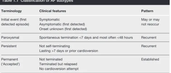
Recent guidelines suggest classification of AF based on the temporal pattern of the arrhythmia (see Table 1.1).
Terminology Clinical features Pattern
Initial event (first Symptomatic May or may
detected episode) Asymptomatic (first detected) not reoccur
Onset unknown (first detected)
Paroxysmal Spontaneous termination <7 days and most often <48 hours Recurrent
Persistent Not self-terminating Recurrent
Lasting >7 days or prior cardioversion
Permanent Not terminated Established
(‘Accepted’) Terminated but relapsed No cardioversion attempt
Table reprinted with permission from Levy S, Camm AJ, Saksena S et al. International consensus on nomenclature and classification of atrial fibrillation. Europace 2003;5:119–22.1
AF is considered recurrent when a patient experiences two or more episodes. These episodes may be paroxysmal if they terminate spontaneously, defined by consensus as 7 days, or persistent if the arrhythmia requires electrical or pharmacological cardioversion for termination. Successful termination of AF does not alter the classification of persistent AF in these patients.
Long-standing AF (defined as over a year) that is not successfully terminated by cardioversion, or when cardioversion is not pursued, is classified as permanent.
Paroxysmal AF, in which the frequency of paroxysms is low, may degenerate into either paroxysmal AF with more frequent paroxysms, or a sustained form of AF. Similarly, persistent AF may degenerate into permanent AF. Despite its name, the reversion of permanent AF to normal sinus rhythm is also possible, particularly in those cases where the AF is caused by an underlying disease process which is successfully treated (eg thyroid disease), or where a specialist procedure is performed that modifies the electrophysiological properties of the heart.
Without treatment, AF can sometimes result in a degree of haemodynamic instability which can represent a critical condition that requires immediate intervention to alleviate symptoms of breathlessness, chest pain and loss of consciousness, and restore haemodynamic stability.
1.3
Epidemiology
1.3.1
Prevalence
AF is the commonest sustained cardiac arrhythmia. Much of the epidemiology of AF is derived from data from predominantly white populations, and information on AF in non-white populations is scarce. Hospital practice data may give a biased view of the clinical epidemiology of AF, since only one-third of patients with AF may actually have been admitted to hospital.2
The prevalence of AF roughly doubles with each advancing decade of age, from 0.5% at age 50–59 years to almost 9% at age 80–89 years.3Conversely, AF is very uncommon in infants and children, unless concomitant structural or congenital heart disease is present.
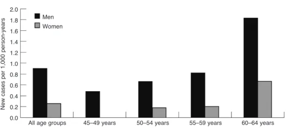
In the UK, the Renfrew–Paisley study4found that of an original cohort of men and women aged 45–64 years (N=15,406) there were 100 (0.65%; 95% CI 0.53 to 0.79%) documented cases of AF. The prevalence of AF increased with age and more cases were detected in men (53 of 7,052) than women (47 of 8,354)4as shown in Figure 1.1. In the West Birmingham AF project, the prevalence of AF was 2.4% in two general practices2 and further extension of this project showed that the prevalence of AF among Indo-Asians aged over 50 years in the general practice population was 0.6%.5The Newcastle survey screened 4,843 people aged 65 years or more in general practices and found a prevalence of AF of 4.7%.6Among UK hospital admissions, AF is present in 3–6% of acute medical admissions.7,8
1.3.2
Risk factors
There are many risk factors for developing AF. As discussed above, there is an increasing prevalence and incidence of AF with increasing age. In the Framingham study9the develop-ment of AF was associated with increasing age (odds ratio (OR) 2.1 for men and 2.2 for women, p <0.0001), diabetes (OR 1.4 for men and 1.6 for women), hypertension (OR 1.5 for men and 1.4 for women), and valve disease (OR 1.8 for men and 3.4 for women). It is also commonly associated with, and complicated by, congestive heart failure and strokes.
AF is often caused by coexisting medical conditions. These causes can be cardiac and non-cardiac (see Table 1.2).
Figure 1.1 Prevalence of AF in the Renfrew–Paisley project.4*
All age groups 45–49 years 50–54 years 55–59 years 60–64 years Men
Women 16
14
12
10
8
6
4
2
0
Number of cases per 1,000 person-years
Figure 1.2 Incidence of AF in the Renfrew–Paisley project.4*Figures 1.1 and 1.2 were reproduced with permission of BMJ Publishing Ltd from Stewart S, Hart CL, Hole DJ et al. Population prevalence, incidence, and predictors of atrial fibrillation in the Renfrew/Paisley study. Heart (British Cardiac Society)
2001;86:516–21.
All age groups 45–49 years 50–54 years 55–59 years 60–64 years 2.0
1.8
1.6
1.4
1.2
1.0
0.8
0.6
0.4
0.2
0.0
New cases per 1,000 person-years
AF is also common after surgery, especially cardiothoracic operations such as thoracotomy and coronary artery bypass graft. Overall, the presence of the AF after surgery not only results in prolongation of hospital stay but may also increase risk of heart failure, stroke, or thromboembolism, and greater hospital costs. The incidence of postoperative AF depends on many risk factors apart from the type of procedure, such as age and the patient’s preoperative physiology and electrolyte balance.
Many dietary and lifestyle factors have also been associated with AF. These include excessive alcohol or caffeine consumption and emotional or physical stress. In the case of alcohol, AF may develop as a consequence of an excessive intake of alcohol over a relatively short period – a so-called ‘holiday heart’. In one series of younger patients (aged <65 years) with new onset AF, alcohol caused or contributed to the arrhythmia in 63% of cases.10
Lone AF is defined as AF without overt structural heart disease, and defined by a normal clinical history and examination, ECG, chest X-ray and, more recently, the echocardiogram. There are implications of labelling patients with this diagnosis, as this group is often considered to be at ‘low risk’, although recent data have been inconclusive.
A diagnosis of lone AF is only considered a diagnosis of exclusion if there is:
no history of cardiovascular disease or hypertension
no abnormal cardiac signs on physical examination
a normal chest X-ray and, apart from the presence of AF, a normal ECG (ie no indication
of prior myocardial infarction or left ventricular hypertrophy)
normal atria, valves and left ventricular size and function by echocardiography. Cardiac causes of AF
Common cardiac causes:
ischaemic heart disease rheumatic heart disease hypertension
sick sinus syndrome pre-excitation syndromes
(eg Wolff–Parkinson–White).
Less common cardiac causes:
cardiomyopathy or heart muscle disease pericardial disease (including effusion and
constrictive pericarditis) atrial septal defect atrial myxoma.
Non-cardiac causes of AF
Acute infections, especially pneumonia
Electrolyte depletion
Lung carcinoma
Other intrathoracic pathology (eg pleural effusion)
Pulmonary embolism
Thyrotoxicosis
1.4
Prognosis
The adverse effects of AF are the result of haemodynamic changes related to the rapid and/or irregular heart rhythm, and thromboembolic complications related to a prothrombotic state associated with the arrhythmia. AF is associated with an odds ratio for death of 1.5 for men and 1.9 in women, which does not vary by age, but most of the excess of mortality attributed to AF occurs early after diagnosis of AF.11
Onset of AF can result in a reduction in cardiac output of up to 10–20% regardless of ventricular rate. The presence of fast ventricular rates can push an already compromised ventricle into heart failure. An uncontrolled AF rate may even precipitate critical cardiac ischaemia.
AF is associated with a prothrombotic state – intra-atrial blood stasis, structural heart disease or blood vessel abnormalities and abnormal platelets and haemostasis – leading to a predisposition to thrombus formation (thrombogenesis).12 This prothrombotic state predisposes to stroke and thromboembolism in AF, with an approximately five-fold greater risk than that of people without AF.13In stroke patients, concurrent AF is associated with greater disability, longer in-hospital patient stay and lower rate of discharge to own home. The incidence of strokes attributable to AF increases from 1.5% at age 50–59 years to 23.5% at age 80–89 years.14
In terms of the direct effects on patients’ quality of life, AF can also result in reduced exercise tolerance, as well as impairment in cognitive function.
1.5
Guideline structure
Chapter 2 details the methodology used in the construction of this guideline. This includes the protocols used for the searching, selection and appraisal of evidence, and the grading of recommendations according to the strength of their supporting evidence.
The remaining chapters deal with specific issues in the diagnosis and treatment of AF. For ease of reference, wherever possible different forms of AF have been considered in distinct chapters, as follows:
persistent AF (Chapter 6)
permanent AF (Chapter 7)
paroxysmal AF (Chapter 8)
acute-onset AF (Chapter 9)
postoperative AF (Chapter 10).
Most of these chapters include an easy-to-use flowchart showing the recommendations relevant to the particular form of AF.
1.6
How to use this guideline
The purpose of this guideline is to support clinical judgement, not to replace it. This
means the treating clinician should:
– take into consideration any contraindications in deciding whether or not to administer any treatment recommended by this guideline
– consider the appropriateness of any recommended treatment for a particular patient in terms of the patient’s relevant clinical and non-clinical characteristics.
Wherever possible, before administering or changing any treatment the treating clinician
should follow good practice in terms of:
– discussing with the patient why the treatment is being offered and what health outcomes are anticipated
– highlighting any possible adverse events or side effects that have been associated with the treatment
– obtaining explicit consent for the treatment.
For those recommendations involving pharmacological treatment, the most recent
edition of the British National Formulary (www.bnf.org.uk) should be followed for the determination of:
– indications – drug dosage
– method and route of administration – contraindications
– supervision and monitoring – product characteristics.
Exceptions to the above are cases where guidance is provided within the recommendation itself.
The guideline will normally only make drug recommendations that fall within licensed
2.1
About the guideline
2.1.1
Aim
With this document the National Collaborating Centre for Chronic Conditions (NCC-CC) has aimed to provide a user-friendly, clinical, evidence-based guideline for the National Health Service (NHS) in England and Wales that:
offers best clinical advice for AF
is based on best published evidence and expert consensus
takes into account patient choice and informed decision-making
defines the major components of NHS care provision for AF
indicates areas suitable for clinical audit
details areas of uncertainty or controversy requiring further research
provides a choice of guideline versions for differing audiences.
2.1.2
Scope
The guideline was developed in accordance with a scope which detailed the remit of the guideline originating from the Department of Health and specified those aspects of AF to be included and excluded. Prior to the commencement of the guideline development, the scope was subjected to stakeholder consultation in accordance with processes established by the National Institute for Health and Clinical Excellence (NICE).15The full scope is shown in Appendix G.
This guideline sets out best practice for the diagnosis and treatment of AF in both primary and secondary care. It also considers which patients benefit from referral for specialist investigations or procedures and offers guidance in this respect.
Most of the recommendations made in this guideline apply to both AF and the closely related arrhythmia atrial flutter (in those cases where the two conditions are indistinguishable). Exceptions are recommendations concerning diagnosis and opportunistic case-detection, which apply only to AF.
Although the guideline covers the majority of AF cases, it does not consider those patients for whom there is a need for highly specialised clinical input. In particular, it does not consider paediatric cases of AF, or gestational AF.
With the exception of the use of prophylactic drugs to prevent postoperative AF and the use of typical symptoms in guiding opportunistic case-detection, the guideline will not consider issues of public health screening or interventions.
2.1.3
Audience
The guideline is intended for use by the following people or organisations:
all healthcare professionals
people with AF and their carers
patient support groups
commissioning organisations
service providers.
2.1.4
Patient involvement
The NCC-CC was keen to ensure that the views and preferences of people with AF and their carers informed all stages of the guideline. This was achieved by:
having a person with AF and a representative of a user organisation on the Guideline
Development Group (GDG)
consulting the Patient Information Unit (PIU) housed within NICE during the
pre-development (scoping) and final validation stages of the guideline.
2.1.5
Guideline limitations
Limitations to the guideline are as follows:
Clinical guidelines usually do not cover issues of service delivery, organisation or
provision (unless specified in the remit from the Department of Health).
NICE is primarily concerned with health services and so recommendations are not
provided for social services and the voluntary sector. However, the guideline may address important issues in how NHS clinicians interface with these other sectors.
Generally the guideline does not cover rare, complex, complicated or unusual conditions.
2.2
Guideline development
2.2.1
Background
The development of this evidence-based clinical guideline draws upon the methods described by the NICE Guideline development methods manual16(www.nice.org.uk/page.aspx?o=201982) and the methodology pack17specifically developed by the National Collaborating Centre for Chronic Conditions (NCC-CC) for each chronic condition guideline (www.rcplondon.ac.uk/ college/ceeu/ncccc_index.htm). The developers’ roles and remit are summarised below.
s National Collaborating Centre for Chronic Conditions
The NCC-CC was set up in 2001 and is housed within the Royal College of Physicians. The NCC-CC undertakes commissions received from the National Institute for Health and Clinical Excellence (NICE).
s NCC-CC technical team
The technical team met approximately two weeks before each GDG meeting and comprised:
the GDG group leader
the GDG clinical advisor
an information scientist
a research fellow
a health economist
a project manager
administrative personnel.
s Guideline Development Group16
The GDG met 14 times between July 2004 and December 2005 and comprised a multidis-ciplinary team of professionals, service users (people with AF or carers), and user organisation representatives who were supported by the technical team.
The GDG membership details, including patient representation and professional groups, are listed at the front of this guideline (see pages vi–vii).
Members of the GDG declared any interests in accordance with the NICE technical manual.16A register is available from the NCC-CC for inspection upon request ([email protected]).
s Sign-off workshop17
At the end of the guideline development process the GDG met to review and agree the guideline recommendations.
2.2.2
The guideline development process
There are ten basic steps in the process of producing a guideline.
s Step 1: Developing evidence-based questions
The technical team drafted a series of clinical questions that covered the guideline scope. The GDG and Project Executive refined and approved these questions, shown in Appendix C.
s Step 2: Systematically searching for the evidence
The information scientist developed a search strategy for each question. Key words for the search were identified by the GDG. In addition, the health economist searched for additional papers to inform detailed health economic work (eg modelling). Papers that were published or accepted for publication in peer-reviewed journals were considered as evidence by the GDG. Conference paper abstracts and non-English language papers were excluded from the searches.
relevant to the question. Exclusion lists were generated for each question together with the rationale for the exclusion. The exclusion lists were presented to the GDG. Full papers were obtained where relevant. See Appendix C for literature search details.
s Step 3: Critically appraising the evidence
The research fellow or health economist, as appropriate, critically appraised the full papers. In general no formal contact was made with authors but there were ad hoc occasions when this was required in order to clarify specific details. Critical appraisal checklists were compiled for each full paper. One research fellow undertook the critical appraisal and data extraction. The evidence was considered carefully by the GDG for accuracy and completeness.
All procedures are fully compliant with:
NICE methodology as detailed in the Guideline development methods – information for
National Collaborating Centres and guideline developers manual.16
NCC-CC quality assurance document and systematic review chart, available at
www.rcplondon.ac.uk/college/ceeu/ncccc_index.htm
s Step 4: Incorporating health economic evidence
There were constraints in the health economic resources and so the following approach was agreed for this guideline. Health economics was incorporated alongside the clinical questions.
Searches in relevant databases were done by the information scientist using economic
filters on the related clinical questions.
No study design criteria were imposed a priori, ie the searches were not limited to
randomised control trials (RCTs) or formal economic evaluations.
Titles and abstracts identified in the economic searches were reviewed by the health
economist and full papers were obtained once they met the inclusion/exclusion criteria.
The full papers were critically appraised by the health economist and the relevant data
were presented to the GDG.
Cost-effectiveness evidence from the UK was preferred, but all relevant evidence was considered, including non-UK studies.
The GDG identified areas for additional economic work. Five key areas were identified and three were given priority. The GDG agreed on the model structures. The health economist performed supplemental literature searches using key search terms in Medline and an Internet search engine to obtain additional information for modelling. None of the identified priority areas were modelled for various reasons (see Appendix A for details).
s Step 5: Distilling and synthesising the evidence and developing recommendations
s Step 6: Grading the evidence statements and recommendations
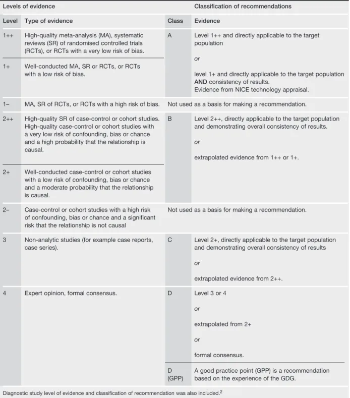
The criteria for grading evidence and classifying recommendations are shown in Table 2.1.
Evidence tables are available at
www.rcplondon.ac.uk/pubs/online_home.htm
Table 2.1 Criteria for grading evidence and recommendations. Note that diagnostic study levels of evidence and classification of recommendations were also included.16
Levels of evidence Classification of recommendations
Level Type of evidence Class Evidence
1++ High-quality meta-analysis (MA), systematic A Level 1++ and directly applicable to the target reviews (SR) of randomised controlled trials population
(RCTs), or RCTs with a very low risk of bias.
or
1+ Well-conducted MA, SR or RCTs, or RCTs
with a low risk of bias. level 1+ and directly applicable to the target population
ANDconsistency of results.
Evidence from NICE technology appraisal.
1– MA, SR of RCTs, or RCTs with a high risk of bias. Not used as a basis for making a recommendation.
2++ High-quality SR of case-control or cohort studies. B Level 2++, directly applicable to the target population High-quality case-control or cohort studies with and demonstrating overall consistency of results. a very low risk of confounding, bias or chance
and a high probability that the relationship is or
causal.
extrapolated evidence from 1++ or 1+.
2+ Well-conducted case-control or cohort studies with a low risk of confounding, bias or chance and a moderate probability that the relationship is causal.
2– Case-control or cohort studies with a high risk Not used as a basis for making a recommendation. of confounding, bias or chance and a significant
risk that the relationship is not causal
3 Non-analytic studies (for example case reports, C Level 2+, directly applicable to the target population
case series). and demonstrating overall consistency of results
or
extrapolated evidence from 2++.
4 Expert opinion, formal consensus. D Level 3 or 4
or
extrapolated from 2+
or
formal consensus.
D A good practice point (GPP) is a recommendation (GPP) based on the experience of the GDG.
s Step 7: Agreeing the recommendations
The sign-off workshop employed formal consensus techniques to:
ensure that the recommendations reflected the evidence base
approve recommendations based on lesser evidence or extrapolations from other
situations
reach consensus recommendations where the evidence was inadequate
debate areas of disagreement and finalise recommendations.
The sign-off workshop also reached agreement on the following:
five priorities for implementation
five key research recommendations
algorithms.
In prioritising key recommendations for implementation, the sign-off workshop also took into account the following criteria:
high clinical impact
high impact on reducing variation
more efficient use of NHS resources
allowing the patient to reach critical points in the care pathway more quickly.
The audit criteria provide suggestions of areas for audit in line with the key recommendations for implementation.16
s Step 8: Structure of the guideline
The guideline is divided into sections for ease of reading. For most sections the layout is similar and is described below:
The clinical introduction sets a succinct background and describes the current clinical
context.
The methodological introduction describes any issues or limitations that were apparent
when reading the evidence base.
Evidence statements provide a synthesis of the evidence base and usually describe what the
evidence showed in relation to the outcomes of interest.
Health economics presents, where appropriate, an overview of the cost effectiveness of the
evidence base.
From evidence to recommendations highlights the debate of the GDG. This section sets out
the GDG decision-making rationale providing a clear and explicit audit trail from the evidence to the evolution of the recommendations.
The recommendations section provides stand-alone, action-orientated recommendations.
Evidence tables are not published as part of the full guideline but are available online at
s Step 9: Writing the guideline
The first draft version of the guideline was drawn up by the technical team in accordance with the decision of the GDG. The guideline was then submitted for two formal rounds of public and stakeholder consultation prior to publication.16The registered stakeholders for this guideline are detailed in Appendix F. Editorial responsibility for the full guideline rests with the GDG.
Table 2.2 describes the versions of the guideline that are available.
s Step 10: Updating the guideline
Literature searches were repeated for all of the evidence-based questions at the end of the GDG development process allowing any relevant papers published up until 30 June 2006 to be considered. Future guideline updates will consider evidence published after this cut-off date.
Two years after publication of the guideline, NICE will commission a National Collaborating Centre to determine whether the evidence base has progressed significantly to alter the guideline recommendations and warrant an early update. If not, the guideline will be updated approximately 4 years after publication.16
2.3
Disclaimer
Healthcare providers need to use clinical judgement, knowledge and expertise when deciding whether it is appropriate to apply guidelines. The recommendations cited here are a guide and may not be appropriate for use in all situations. The decision to adopt any of the recom-mendations cited here must be made by the practitioner in light of individual patient circumstances, the wishes of the patient, clinical expertise and resources.
The NCC-CC disclaims any responsibility for damages arising out of the use or non-use of these guidelines and the literature used in support of these guidelines.
2.4
Funding
The National Collaborating Centre for Chronic Conditions was commissioned by the National Institute for Health and Clinical Excellence to undertake the work on this guideline.
Full version Details the recommendations. The supporting evidence base and the expert considerations of the GDG. Available at
www.rcplondon.ac.uk/pubs/online_home.htm
NICE version Documents the recommendations without any supporting evidence. Available at www.nice.org.uk/page.aspx?o=guidelines.completed
Quick reference guide An abridged version.
Available at www.nice.org.uk/page.aspx?o=guidelines.completed
Information for the public A lay version of the guideline recommendations.
Available at www.nice.org.uk/page.aspx?o=guidelines.completed
3.1
Priorities for implementation
The following five recommendations have been identified by the GDG as priorities for implementation:
1. An electrocardiogram (ECG) should be performed in all patients, whether symptomatic or not, in whom AF is suspected because an irregular pulse has been detected.
2. As some patients with persistent AF will satisfy criteria for either an initial rate-control or rhythm-control strategy (for example, aged over 65 but also symptomatic):
the indications for each option should not be regarded as mutually exclusive and the
potential advantages and disadvantages of each strategy should be explained to patients before agreeing which to adopt
any comorbidities that might indicate one approach rather than the other should be
taken into account
irrespective of whether a rate-control or a rhythm-control strategy is adopted in
patients with persistent AF, appropriate antithrombotic therapy should be used.
3. In patients with permanent AF, who need treatment for rate control:
beta-blockers or rate-limiting calcium antagonists should be the preferred initial
monotherapy in all patients
digoxin should only be considered as monotherapy in predominately sedentary
patients.
4. In patients with newly diagnosed AF for whom antithrombotic therapy is indicated (see section 11.6), such treatment should be initiated with minimal delay after the appropriate management of comorbidities.
5. The stroke risk stratification algorithm (Figure 11.1) should be used in patients with AF to assess their risk of stroke and thromboembolism, and appropriate thromboprophylaxis given.
Each of these recommendations highlights areas of current clinical practice that the GDG believe would particularly benefit from guidance. Compliance with each of these key priority areas may be audited according to the corresponding audit criteria below.
3.2
AF care pathway
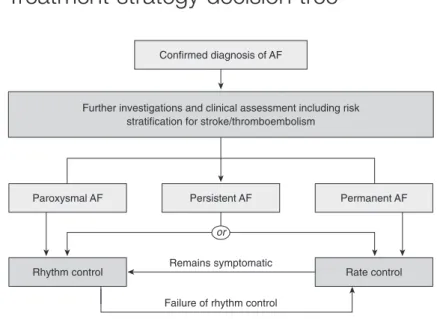
3.3
Treatment strategy decision tree
Figure 3.1 AF care pathway
Yes Continued AF
Sinus rhythm No symptoms – opportunistic
case – finding leads to clinical suspicion of AF
ECG to confirm diagnosis
Further investigations and clinical assessment (including risk stratification)
Further management* in community and/or
secondary care
Continued AF or sinus rhythm at
follow-up?
Need for further follow-up?
Develop management plan
Assess need for further follow-up? Further follow-up** Regular review Further referral Follow-up Emergency referral if appropriate Symptomatic
presentation and clinical suspicion of AF
See treatment strategy decision tree (Figure 3.3) or *Further management to include rate- or rhythm-control treatment strategy (see Figures 7.1 and 8.1) and appropriate antithrombotic therapy based on stroke risk stratification model (see Figure 11.1).
**Further follow-up for coexisting conditions and assessment for ongoing anticoagulation.
Figure 3.2 Treatment strategy decision tree
Rate control or
Confirmed diagnosis of AF
Paroxysmal AF Persistent AF
Remains symptomatic
Failure of rhythm control
Permanent AF Further investigations and clinical assessment including risk
stratification for stroke/thromboembolism
3.4
Audit criteria
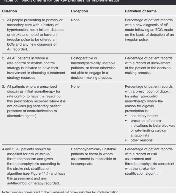
Table 3.1 lists the audit criteria identified to evaluate the impact of the implementation of the five key priority areas detailed above on clinical practice and health outcomes.
Criterion Exception Definition of terms
1. All people presenting to primary or None. Percentage of patient records
secondary care with a history of with a new diagnosis of AF
hypertension, heart failure, diabetes made following an ECG made
or stroke and noted to have an on the basis of detection of an
irregular pulse to be offered an irregular pulse.
ECG and any new diagnosis of AF recorded.
2. All AF patients in whom a Postoperative or Percentage of patient records
rate-control or rhythm-control haemodynamically unstable with a record of involvement strategy is initiated to have their patients, or those otherwise of the patient in the decision-involvement in choosing a treatment not able to engage in a making process.
strategy recorded. decision-making process.
3. All patients who are prescribed None. Percentage of patient records
digoxin as initial monotherapy for with a prescription of digoxin
rate control to have the reason for for initial rate-control
this prescription recorded where it is monotherapy where the
not obvious (eg sedentary patient, reason for digoxin
presence of contraindication to prescription is:
alternative agents). • sedentary patient
• presence of contra-indications to beta-blockers or rate-limiting calcium antagonists
• other reasons.
4 and 5. All patients should be Haemodynamically unstable Percentage of patient records assessed for risk of stroke/ patients or those in whom with a record of risk
thromboembolism and given assessment is impossible or assessment and
thromboprophylaxis according to inappropriate. thromboprophylaxis consistent
the stroke risk stratification with the stroke risk
algorithm (see Figure 11.1) and have stratification algorithm.
this assessment and any antithrombotic therapy recorded.
Note: numbers correspond to the numbered list of key priorities for implementation.
3.5
Areas for future research
The GDG has identified the following five questions as key areas for further research:
s Cardioversion
Although cardioversion is a core treatment for many patients with AF, there is little evidence that compares the different modes (electrical and pharmacological), particularly in terms of cost effectiveness. Further, the studies that have considered the efficacy of preloading with antiarrhythmic drugs prior to electrical cardioversion have not reported long-term efficacy in maintaining sinus rhythm, or the cost effectiveness of this strategy.
In patients scheduled for elective cardioversion what is the optimal form of cardioversion,
in terms of the pre-cardioversion use of antiarrhythmic drugs, the mode of cardioversion (electrical or pharmacological), the cost effectiveness of each procedure and the impact on quality of life?
s Echocardiography
Echocardiography allows cardiac abnormalities such as left ventricular impairment to be diagnosed earlier than would be possible from signs and symptoms alone. However, no study has addressed the issue of whether performing routine echocardiography on all newly diagnosed AF patients would be more cost effective in diagnosing and treating heart disease earlier, than performing echocardiography only on those patients in whom there is a clinical suspicion of undiagnosed heart disease.
What is the cost effectiveness of performing a routine echocardiographic examination in
all newly diagnosed AF patients, compared to only selective examination based on clinical criteria?
s Anticoagulation with antiplatelet therapy
In the general AF population, the evidence suggests that combined therapeutic anticoagulation with antiplatelet therapy does not reduce the incidence of stroke or thromboembolism compared with therapeutic anticoagulation alone, and it may increase the incidence of bleeding. However, it is unclear whether there are certain subgroups of patients with AF for whom the therapeutic effects of combination therapy may be greater than either monotherapy. In particular, it is unclear whether combination therapy is justified in those AF patients who have stent implantation or a history of myocardial infarction (MI).
Is there any additional benefit, in terms of overall vascular events, from combined
anticoagulation with antiplatelet therapy for any subgroups of patients with AF such as those with prior MI or stent implantation?
s Pill-in-the-pocket treatment
compared with either the emergency department administration of treatment or continuous antiarrhythmic drug therapy.
What is the clinical and cost effectiveness of pill-in-the-pocket treatment for those with
paroxysmal AF compared with hospital-based administration or continuous antiarrhythmic therapy?
s Anticoagulation in paroxysmal AF
The frequency of paroxysms in patients with paroxysmal AF varies widely between patients. It remains unclear, however, whether the risk of stroke or thromboembolism varies between those with only infrequent paroxysms and those with more frequent paroxysms. It is also unclear whether, if the risk of stroke or thromboembolism is reduced in those with infrequent paroxysms, the use of anticoagulation is justified in such a low-risk group.
What is the optimal anticoagulation strategy for those patients with paroxysmal AF who
4.1
Presenting symptoms/pulse palpitation
AF can present in the setting of a wide variety of cardiac and non-cardiac conditions, it is often asymptomatic and can present with vague non-specific symptoms. Too often, AF is only detected after the patient presents with serious complications of AF, such as a stroke, thromboembolism or heart failure. The initial diagnosis of AF depends on associating symptoms such as breathlessness, dyspnoea, palpitations, syncope/dizziness or chest discomfort with AF.
Most of the data on presentation of AF patients have been based on white Caucasian populations, and limited data are available in relation to ethnicity and AF.18Furthermore, there may be important differences between hospital-based cohorts compared with community or population-based studies, as many do not present to hospital care, and if they do, it is often in the context of associated comorbidity such as ischaemic heart disease or heart failure. Indeed, many patients with AF in general practice remain asymptomatic. However, as AF commonly occurs in association with risk factors, such as hypertension, diabetes and ischaemic heart disease, opportunistic assessment of such patients for the presence of AF may be prudent, especially since such patients are frequently seen for check-ups in primary care.
While general population screening is beyond the scope of this guideline, targeted/opportunistic screening of symptomatic patients or those with risk factors may allow identification of AF patients. One recent study19,20aims to determine the baseline prevalence and the incidence of AF based on a variety of screening strategies and in doing so to evaluate the incremental cost effectiveness of different screening strategies, including targeted or whole population screening, compared with routine clinical practice, for detection of AF in people aged 65 and over. This study20– whose publication date fell outside of the date limits of the systematic literature search – reported that the baseline prevalence of AF in subjects older than 65 was 7.2%, with a higher prevalence in men (7.8%) and among patients aged 75 or older (10.3%), and indicated that the only strategy that improved on routine practice was opportunistic screening.
4.1.1
Methodological introduction
The results of nine studies are included in this report. Seven studies were critically appraised.7,8,21–25Of these, none reported the frequency of presenting symptoms in primary care in a UK (or other) population. Patients presenting to secondary care generally present with more severe symptoms. The studies:
did not use a consistent terminology to classify AF symptoms. ‘Dizziness’ was also
referred to as ‘near syncope’21and ‘chest pain’ was also referred to as ‘chest discomfort’22 were single-centre studies
had a variable proportion of patients presenting with de novo AF versus those with a
previous history of AF.
4.1.2
Evidence statements
Dyspnoea, chest pain and palpitations were found to be the most common presenting symptoms in emergency admissions with newly diagnosed or previously diagnosed AF in the UK7,8or USA.26–28
Similar results were found in patients with chronic23 (more than 7 days since onset of symptoms) and lone AF.21
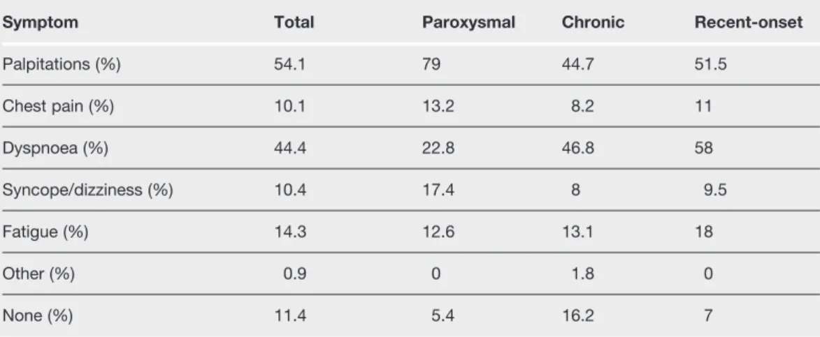
In one study24(N=756) dyspnoea was the most commonly reported symptom in chronic and recent-onset AF (46.8%); palpitations were the most commonly reported symptom in paroxysmal AF (79.0%).28
In two studies, stroke was reported as a presenting symptom of AF at rates of 5.1% and 3.2% respectively22,25and occurred at a rate of 12.7% in a study population combining both new-onset and previously diagnosed AF.8,28
Dizziness Dyspnoea Chest pain Palpitations /syncope
Study N % % % %
Zarifis et al8 245 47.1 19.9 16.2 16.2
Lip et al7 170 51.8 34.1 25.9 18.8
Michael et al26 289 7 10 78 3
Burton et al27 266 12 24 40 9
Table 4.1 Presenting symptoms associated with emergency AF admissions
Symptom Total Paroxysmal Chronic Recent-onset
Palpitations (%) 54.1 79 44.7 51.5
Chest pain (%) 10.1 13.2 8.2 11
Dyspnoea (%) 44.4 22.8 46.8 58
Syncope/dizziness (%) 10.4 17.4 8 9.5
Fatigue (%) 14.3 12.6 13.1 18
Other (%) 0.9 0 1.8 0
None (%) 11.4 5.4 16.2 7
4.1.3
From evidence to recommendations
Those with undiagnosed AF can receive treatment sooner if an opportunistic case finding is undertaken using manual pulse palpation in those presenting with symptoms commonly associated with AF. It was therefore considered good practice to check the blood pressure and pulse (manually) in all patients who present with breathlessness, dyspnoea, palpitations, syncope/dizziness or chest discomfort.
Many patients presenting with stroke are also found to be in AF, indicating a missed opportunity to diagnose the pre-existing AF and administer appropriate antithrombotic therapy.
RECOMMENDATION
R1 In patients presenting with any of the following:
breathlessness/dyspnoea
palpitations
syncope/dizziness
chest discomfort
stroke/TIA
manual pulse palpation should be performed to assess for the presence of an irregular
pulse that may indicate underlying AF. C
4.2
Electrocardiography
As with many chronic disorders, AF may be symptomatic or asymptomatic, and episodes of either can occur in the same patient.
Most symptomatic patients with AF present with symptoms related to the arrhythmia. However, such patients can have a wide variety of other cardio-respiratory presenting symptoms and clinical features7,24(see section 4.1).
Many patients with AF are asymptomatic and are picked up in general practice. One study2 found that a third of AF patients had not had hospital contact for symptoms related to AF. Asymptomatic AF can be discovered incidentally during clinical examination by cardiac auscultation, 12-lead ECG recording, or 24-hour Holter recording that may have been performed for unrelated reasons.
The patient may also have presented with associated medical problems, such as heart failure, stroke or thromboembolism, and coincidental AF is detected. The duration of AF may be unknown in such patients, and whether AF was the cause or effect of the acute problem (eg stroke or heart failure) may be uncertain.
4.2.1
Methodological introduction
The two studies performed in UK primary care29,30evaluated the finding of an irregular pulse as a screening test for AF. Both studies included populations of over 65-year-olds and confirmed the diagnosis of AF by ECG.
4.2.2
Evidence statements
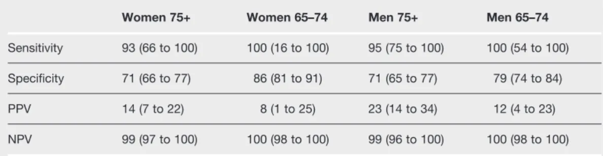
In one study,29the diagnostic accuracy of pulse palpation was compared between different age and gender groups in a primary care population aged 65 or over, and is summarised in Table 4.3. (II)
One study30measured the diagnostic accuracy of three different methods of nurse-based screening for AF based on the presence of either continuous or intermittent pulse irregularities over a minimum of 20 seconds in a population aged over 65. The results are as shown in Table 4.4. (II)
Women 75+ Women 65–74 Men 75+ Men 65–74
Sensitivity 93 (66 to 100) 100 (16 to 100) 95 (75 to 100) 100 (54 to 100)
Specificity 71 (66 to 77) 86 (81 to 91) 71 (65 to 77) 79 (74 to 84)
PPV 14 (7 to 22) 8 (1 to 25) 23 (14 to 34) 12 (4 to 23)
NPV 99 (97 to 100) 100 (98 to 100) 99 (96 to 100) 100 (98 to 100)
All values are percentages with 95% confidence intervals.
Table 4.3 Diagnostic accuracy of pulse palpitation between different age and gender groups
Method 1 Method 2 Method 3
Sensitivity 91 (82 to 97) 72 (59 to 82) 54 (41 to 66)
Specificity 74 (72 to 77) 94 (93 to 96) 98 (97 to 99)
PPV 19 (15 to 23) 44 (35 to 54) 61 (47 to 73)
NPV 99 (98 to 100) 98 (97 to 99) 97 (96 to 98)
All values are percentages with 95% confidence intervals. Method 1: diagnostic accuracy based on the detection of any pulse irregularity; method 2: diagnostic accuracy based on the detection of frequent or continuous irregularities; method 3: diagnostic accuracy based on the detection of only continuous irregularities.
4.2.3
From evidence to recommendations
An irregular pulse was found to be sensitive to the presence of AF.29 The positive predictive value was greater in those over 75 years old, as the prevalence of AF is known to be higher in this population. The negative predictive value of a regular pulse (>96%) was also emphasised. The results of a second study30suggested it would be prudent to consider any pulse irregularity as requiring further investigation to determine whether AF is present.
One study19,20whose publication date fell outside of the date limits of the systematic literature search confirmed the above results in an elderly UK population (over 65 years old). The study also showed that opportunistic case-detection for AF is a more cost-effective strategy than systematic screening and is associated with fewer ischaemic strokes and a greater proportion of diagnosed AF cases.
The evidence did not consider clinical indicators other than an irregular pulse and it was agreed that where there were other clinical indicators suggestive of AF, an ECG should still be performed. Nonetheless, the majority of patients presenting with AF will have an irregular pulse that may occur in the absence of any symptoms, and it is unlikely that AF will be present if the pulse is normal.
The diagnosis of AF does not require a 12-lead ECG recording. In the case of atrial flutter, however, a 12-lead ECG may be necessary, and may also occur in the presence of a regular pulse. The recommendation made below therefore applies only to AF case detection.
RECOMMENDATION
R2 An electrocardiogram (ECG) should be performed in all patients, whether symptomatic or not, in whom AF is suspected because an irregular pulse has been detected. B(DS)
4.3
Ambulatory ECG recording
Many patients with intermittent AF have asymptomatic paroxysms. In one study31 it was estimated that only 1 in 12 paroxysms are symptomatic. Nonetheless, these patients remain at risk of complications associated with AF.
In patients with daily paroxysms, clinical practice is to perform a 24-hour Holter monitor, but this is less useful in patients who get paroxysms at intervals of more than 24 hours. In the latter category of patients, event ECGs (including transtelephonic monitors (‘cardiomemos’) and some implanted systems) are commonly used to detect/diagnose AF.
4.3.1
Methodological introduction
Event-ECG was defined as any electrocardiographic recording device which recorded only particular events, identified either automatically by a software program to detect arrhythmic episodes or by the onset of symptoms (when the patient manually switches on the device for the duration of the symptomatic episode), or a combination of the two. As with ambulatory-ECGs, event-ECGs record cardiac electrical activity while the patient is able to move around relatively freely without hindrance.
Studies were included if the sample population was reported to be patients with either suspected AF or suspected atrial arrhythmia. No studies compared the diagnostic accuracy of event-ECG devices with ambulatory-ECG devices over the same duration.
4.3.2
Evidence statements
One cross-over study32 of patients suspected of atrial arrhythmia based on palpitations compared a patient-triggered event recorder over a mean period of 70 hours with a 48-hour Holter monitor. The event recorder detected proportionately more symptomatic episodes than the Holter monitor (67% of recorded episodes associated with symptoms versus 35% respectively; p<0.001) (1b). Similarly, the event recorder yielded more arrhythmia diagnoses (19% versus 0% respectively; p<0.005). (1b)
In one study33which compared 24-hour Holter monitoring with automatic and patient triggered event recording (each over 30 days), the automatically-triggered event recorder had a higher diagnostic yield than the patient-triggered event recorder, which in turn had a higher diagnostic yield for diagnoses of AF than the Holter monitor (24%, 13% and 5% respectively). The automatically triggered event recorder was also more effective than the patient-triggered event recorder in detecting asymptomatic episodes of AF (52 events versus 1 event respectively).34
In one study35of 139 patients admitted with symptoms of acute stroke or transient ischaemic attack (TIA) who were ECG-negative for AF/flutter, seven (5%) were picked up in a second round of monitoring using a 24-hour Holter monitor. A further five (6%) patients were diagnosed with AF/flutter in a third round of monitoring using a 7-day event recorder (with both patient and automated triggering).34
4.3.3
From evidence to recommendations
No studies were found to compare the positive diagnostic yield per unit time between an ambulatory-ECG diagnostic tool and an event-ECG tool where the recordings were interpreted in a comparable manner.
One study35found that the use of event-ECG detected cases of AF remained undetected by both non-ambulatory and ambulatory-ECG. In addition, the study found that the use of ambulatory-ECG detected cases of AF remained undetected by non-ambulatory-ECG.
RECOMMENDATION
R3 In patients with suspected paroxysmal AF undetected by standard ECG recording:
a 24-hour ambulatory ECG monitor should be used in those with suspected
asymptomatic episodes or symptomatic episodes less than 24 hours apart
an event recorder ECG should be used in those with symptomatic episodes more
than 24 hours apart. B(DS)
4.4
Echocardiography
Although most cardiologists will perform a transthoracic echocardiogram (TTE) on patients with AF referred to cardiology clinics,36 echocardiography is not undertaken on all patients seen in primary or non-specialist secondary care.2,7,8,37
Regarding the use of echocardiography to identify stroke risk factors, although most stroke risk stratification criteria (see Appendix A) lay emphasis on clinical risk factors, there is a perception that TTE is mandatory to decide on antithrombotic therapy. In one study38echocardiography revealed cardiac abnormalities in many AF patients, although most had other clinical risk factors for thromboembolism and often echocardiography did not alter the management decision.
In clinical practice echocardiography has also been used to assess the risk of recurrent AF post cardioversion, as well as to assess the risk of developing postoperative AF. Finally, transoesophageal echocardiography (TOE) has been used to guide cardioversion (TOE-guided cardioversion (see section 5.4)), but this is a specialist investigation. TOE can also be used by specialists to assess the risk of stroke and thromboembolism.39
4.4.1
Methodological introduction
The results of 29 studies were included in this report. Studies were considered for inclusion if echocardiographic (TTE or TOE) variables were stratified into normal and abnormal ranges and tested, alongside clinical variables, as independent risk factors for clinically defined outcomes.
The clinical outcomes considered were:
AF pathophysiology
the recurrence of AF following successful cardioversion
stroke or thromboembolism
vascular death.
The presence of intracardiac thrombus was not considered as an echocardiographic measure of structural or functional heart disease. Rather, it was considered as a consequence of the disease.
4.4.2
Evidence statements
s AF pathophysiology