R E S E A R C H
Open Access
Usefulness of carotid ultrasonography in
the diagnosis of coronary artery disease in
patients undergoing exercise
echocardiography
Raúl Franco-Gutiérrez
1*, Alberto José Pérez-Pérez
1, Virginia Franco-Gutiérrez
2, Ana María Testa-Fernández
1,
Rafael Carlos Vidal-Pérez
1, Manuel Lorenzo López-Reboiro
3, Víctor Manuel Puebla-Rojo
1, Melisa Santás-Álvarez
1,
María Generosa Crespo-Leiro
4,5,6and Carlos González-Juanatey
1Abstract
Background:Relationship between carotid and coronary artery disease (CAD) in patients undergoing invasive and
non-invasive test is unclear. The aim of the study is to evaluate whether carotid disease is associated with CAD in patients submitted to exercise echocardiography (EE) and if it improves the EE ability to predict CAD.
Methods:We retrospectively studied 156 subjects without previous vascular disease who underwent EE, carotid ultrasonography and coronary angiography between 2002 and 2013. Positive EE was defined as exercise induced wall motion abnormalities, carotid disease according to Manheim and American Society of Echocardiography Consensus and significant CAD as stenosis≥50%.
Results:Eighty-nine (57.1%) subjects had significant CAD. Factors associated with CAD in multivariate analysis were fasting plasma glucose (odds ratio [OR] 1.02,p= 0.031), pre-test probability of CAD > 65% (OR 3.71,p< 0.001), positive EE (OR 10.51,p< 0.001) and carotid plaque (CP) presence (OR 2.95,p= 0.013). There was neither statistical significant difference in area under the curve after addition of CP to EE results (0.77 versus 0.81,p= 0.525) nor sensitivity, specificity, predictive values or efficiency. CP presence reclassified as very high-risk according to Systematic COronary Risk Evaluation 13 patients (34.2%) with negative EE and 22 (33.3%) without CAD.
Conclusion:CP is associated with CAD in patients undergoing EE, however its addition to EE does not improve CAD prediction, probably due to insufficient statistical power. CP reclassified one third of patients to very high-risk category despite negative EE or CAD absence, these subjects benefit from aggressive primary prevention interventions.
Keywords:Stress echocardiography, Exercise test, Carotid artery disease, Coronary artery disease, Area under curve
Background
Ischaemic heart disease is a major problem due to its prevalence, health cost and mortality [1–3]. Stress echo-cardiography is a well-validated tool for diagnosis and risk stratification in patients with new onset chest pain, but it has some limitations that can impair its diagnostic capacity such as the dependence of pre-test probabilities
(PTP) of coronary artery disease (CAD), the need to achieve submaximal heart rate, the presence of subopti-mal echocardiographic windows, the inability to detect non limiting flow coronary stenosis or pathologies that can produce wall motion abnormalities during exercise [2–4].
Carotid disease, defined as increased carotid intima-media thickness (CIMT) or the presence of ath-erosclerotic plaques (CP), has been associated with myo-cardial infarction, stroke and death [5–7]. Post-mortem studies have also demonstrated a correlation between ca-rotid and CAD [8]. These findings encouraged
* Correspondence:[email protected];
1Department of Cardiology, Hospital Universitario Lucus Augusti (HULA),
Avenida doctor Ulises Romero n° 1, 27003 Lugo, Spain Full list of author information is available at the end of the article
investigators to evaluate the possibility of using carotid disease in the diagnosis of CAD of patients undergoing invasive and non-invasive tests, however the studies published so far have shown inconsistent results [9–19]. In that sense a meta-analysis of 34 studies focused on the relation of CIMT with coronary atherosclerosis, 30 showed a positive but modest relationship with correl-ation positive coefficients between 0.12 and 0.51 with only one study being above 0.5 and some studies showed no relationship at all [19].
Our group has broad experience in the ultrasono-graphic assessment of carotid arteries, having demon-strated its usefulness as a marker of subclinical atherosclerosis in subjects with autoimmune diseases [20]. The studies mentioned before [5–8], along with our findings, led to the systematic use of carotid ultra-sound in subjects with suspected CAD undergoing exer-cise echocardiography (EE) at our cardiovascular imaging laboratory since 2002. This approach has been endorsed by the European Society of Cardiology (ESC) stable CAD guidelines as a IIa level C recommendation [2].
A clinical study was designed to evaluate if carotid dis-ease is associated with significant CAD in patients with suspected ischaemic heart disease undergoing treadmill exercise stress echocardiography at our institution and if it improves the EE ability to predict significant CAD.
Methods
Study population
Between Jan. 1st 2002 and Dec. 31st 2013 4024 consecu-tive Caucasian subjects older than 18 years with sus-pected CAD underwent EE and carotid ultrasonography at our institution. Of them, 390 patients (9.7%) were also submitted to a coronary angiography. 234 patients (60%) were excluded: 29 (7.4%) due to prior stroke, transient ischaemic attack or peripheral artery disease and 205 due to prior CAD (52.6%) defined as previous myocar-dial infarction [21], coronary revascularization or angio-graphic documentation of any coronary stenosis ≥50%. All patients signed informed consent before testing. The study was approved by the Regional Ethics Committee.
Demographic, clinical, baseline echocardiography, ca-rotid ultrasonography and stress testing data were col-lected. PTP of CAD and Systematic COronary Risk Evaluation (SCORE) were assessed according to current ESC guidelines [1,2].
Treadmill exercise stress echocardiography
Treadmill exercise was the stress modality chosen using a Philips Sonos 5500 ultrasound machine between 2002 and 2005 and a Philips iE33 after 2005 (Philips Medical Systems).
Heart rate, blood pressure and 12-lead electrocardio-gram were obtained at baseline and at each exercise
stage. EE was finished in case of physical exhaustion, dis-abling chest pain, significant arrhythmia and severe hypertensive or hypotensive response. Apical long-axis, apical 4- and 2-chamber and parasternal long- and short-axis views were obtained at rest, peak and immedi-ately after exercise. Echocardiographic analysis was per-formed using a 17-segment model of the left ventricle to evaluate regional wall motion. Each segment was graded on a 4-point scale depending on its motion. Wall motion score index was calculated as the sum of the scores di-vided by the number of segments at rest and at peak exercise.
Ischaemic electrocardiographic abnormalities were defined as development of ST-segment deviation 80 msec after J point ≥1 mm. Echocardiographic ischaemia was defined as exercise induced new or worsening wall motion abnormalities, except worsening from akinesia to dyskinesia and isolated hypokinesia of the inferobasal segment. Extensive ischemia was defined as ischaemia involving ≥3 myocardial segments and multivessel ischemia as ischemia involving≥2 different coronary ter-ritories [4].
Carotid ultrasonography
Carotid scans were performed immediately after stress testing in the same EE ultrasound equipment using a high-resolution, B-mode ultrasound system with a linear array (3–11 MHz) transducer. Measurement of the CIMT and CP definition were done following the ARIC protocol study [5] and expert consensus [22–25]. Semi-automated edge detection software was used (QLAB; Philips 110 Medical Systems, Andover, MA, USA).
Age- and sex-specific CIMT percentile values were ob-tained from previously published data in our country [26].
Both EE and carotid ultrasonography stored images were analysed by two imaging expert cardiologists blinded to angiography results. In case of disagreement a third expert was consulted.
Coronary angiography
The physician in charge of the patient carried out a coronary angiography considering the results of the EE and other conditions such as persistence of symptoms despite optimal medical treatment, patients’ preferences and/or other clinical criteria. Coronary angiography was performed using standard technique. Significant angio-graphic disease was defined as stenosis ≥50% by visual assessment in any major epicardial arteries or in their branches.
Statistical analysis
Categorical variables were reported as percentages and comparison between groups were based on chi-square or Fisher’s exact tests. Continuous variables were re-ported as mean (standard deviation) or median [inter-quartile range] when their distribution departed from normal and differences were assessed via the unpaired t test or the Mann-Whitney U test where appropriate. Binary and continuous quantitative variables were com-pared using logistic binary regression. To create predict-ive models for the presence of significant CAD, backward stepwise binary logistic regression was used with an entry set at 0.2 significance level and a retention set of 0.1. Apvalue of < 0.05 was considered statistically significant. ! DT V2009.06.26® macro for SPSS Statistics (Autonomous University of Barcelona) and IBM SPSS Statistics for Windows, Version 20.0. (Armonk, NY) was used to calculate sensitivity, specificity, positive (PPV) and negative predictive values (NPV), positive (PLR) and negative likelihood ratios (NLR) and efficiency of EE alone and combined with carotid ultrasonography. Area under the curve (AUC) was calculated by means of a re-ceiver operating characteristic curve analysis; compari-son between AUC was done by the DeLong method.
Results
One hundred fifthy six patients were enrolled in the study. Mean age was 66.1 ± 10.4 years and 102 (65.4%) were men. There were no major complications during or after the tests.
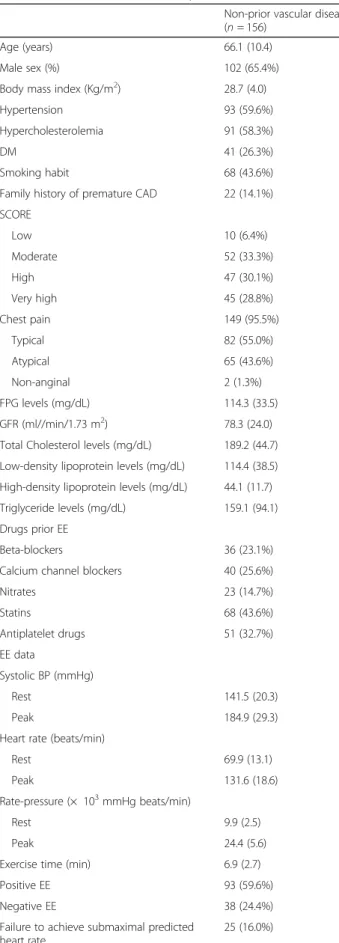
Baseline characteristics are summarized in Table1.
Prediction of CAD
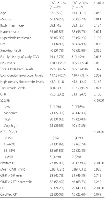
Mean time between non-invasive tests and coronary angiography was 4.2 (3.2) months. Of the 156 patients 89 (57.1%) had significant CAD. This subgroup was older (p= 0.045), with male predominance (p= 0.011), had more frequently diabetes mellitus (DM), smoking habit (p= 0.023) and higher levels of fasting plasma glucose (FPG) (p= 0.003). Higher SCORE, PTP of CAD as well as positive EE and CP presence (all of themp< 0.001) were also significantly more frequent in patients with CAD.
In multivariate analysis FPG (p= 0.031), PTP > 65% (p< 0.001), positive EE (p< 0.001) and CP (p= 0.013) were predictors of significant CAD.
Comparisons of subgroups with and without signifi-cant CAD and multivariate analysis are represented in Tables2and3respectively.
Regarding the subgroup of 21 (13.6%) subjects with resting wall motion abnormalities 4 (19%) had global left ventricular hypokinesia. Of the 21 patients 17 (81.0%) developed worsening wall motion abnormalities during EE and all of them showed significant CAD in the angi-ography, 2 (9.5%) were defined as negative EE and did
not have significant CAD and 2 (9.5%) could not achieve submaximal predicted heart rate, both without significant CAD in the angiography.
AUC, sensitivity, specificity, predictive values, PLR and NLR and efficiency
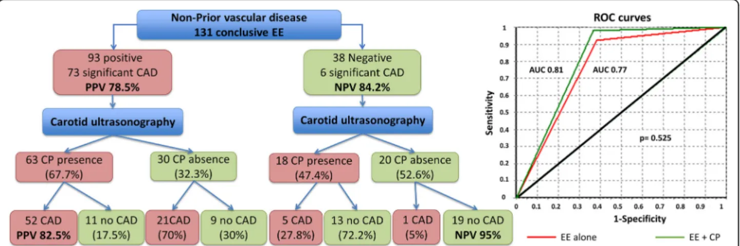
AUC of EE alone was 0.77 (95% confidence interval [CI] 0.68–0.86), whereas AUC combining CP findings was 0.81 (95%CI 0.70–0.92) (p= 0.525). Results are summa-rized in Fig.1and Table4.
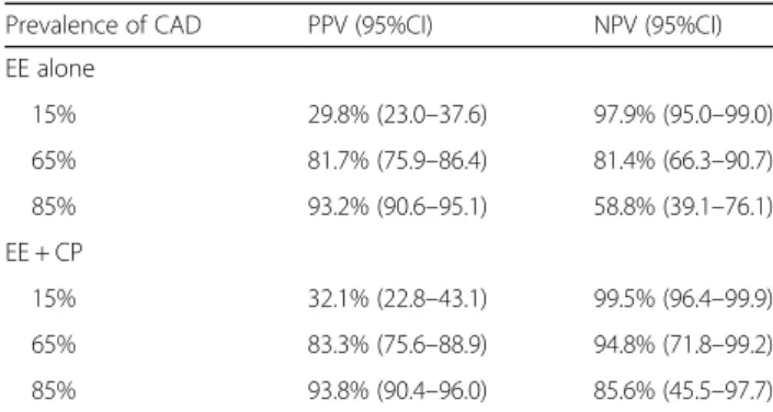
Sensitivity, specificity, predictive values, PLR and NLR and efficiency of EE alone and EE combined with CP are also summarized in Table 4. Table 5 shows predictive values according to established intermediate PTP.
SCORE reclassification according to carotid ultrasound According to European guidelines on cardiovascular disease prevention [1] 10 subjects (6.4%) had low-risk at the time of EE, 52 (33.3%) had moderate-risk, 47 (30.1%) had high-risk, 45 (28.8%) had very high-risk and 2 patients (1.3%) could not be classified. When carotid ultrasonography findings were applied 59 patients (37.8%) were reclassified as very high-risk according to CP presence. Focusing in the 62 patients with low or moderate SCORE risk, 28 (45.2%) had CP.
Of the 38 patients with negative EE 5 subjects (13.2%), 16 (42.1%), 10 (26.3%) and 7 (18.4%) had low, moderate, high and very high-risk respectively. Considering CP presence 13 patients (34.2%) were reclassified as very high-risk. Regarding the 21 patients with low or moder-ate SCORE risk and negative EE, 7 (33.3%) had CP being thereby considered as very high-risk.
Finally, of the 67 patients without CAD, 9 subjects (13.4%) had low-risk, 28 (41.8%) had moderate-risk, 19 (28.4%) had high-risk, 10 (14.9%) had very high-risk and 1 (1.5%) could not be classified. Considering CP results, 22 patients (33.3%) were classified as very high-risk des-pite normal angiography. Of the 37 patients without sig-nificant CAD initially classified as low or moderate SCORE risk 12 (32.4%) presented CP.
Discussion
This study correlates carotid disease with CAD in a real life cohort of patients without prior vascular disease undergoing EE. However, its addition to stress test does not improve CAD prediction by angiography. It is neces-sary to highlight the fact that nearly one third of patients with negative EE and without CAD are reclassified to high-risk group according to carotid ultrasonography findings.
162 (68.6%) subjects had stress test performed (the type was not described in their study) with a low PPV (36%) and also 95%CI were not reported. Kanwar et al. [14] re-ported a study on 50 symptomatic patients without prior CAD who underwent coronary angiography after stress testing. CP, especially those with heterogeneous compos-ition, irregular surface or calcification, was a predictor of significant CAD showing a NPV of 100% in patients with negative/equivocal stress test and CP absence. In contrast to our study, 28% were non-Caucasians and they used different modalities of stress imaging test with a high incidence (64%) of equivocal results. Coskun et al. [15] identified hypertension and CIMT ≥1 mm as pre-dictors of significant CAD in patients without previous CAD or stroke, scheduled for coronary angiography after a positive stress test. Similarly to Akosah et al. [13], the PPV of the stress test was lower compared to our results (61%). Finally, Ahmadvazir et al. [16] identified PTP, positive stress test and presence of CP as predictors of significant CAD in 591 patients with suspected CAD undergoing stress echocardiography. As in previous studies, the NPV combining stress test and carotid ultra-sonography was high (80%) and, in agreement with our findings, nearly one third of the patients were reclassi-fied for risk score according to CP results. However, only 35% of their patients were Caucasian, exercise as stress method was only used in 62% and only 83 (14%) under-went coronary angiography and, similar to the other
Table 1Baseline characteristics of patients
Non-prior vascular disease (n= 156)
Age (years) 66.1 (10.4)
Male sex (%) 102 (65.4%)
Body mass index (Kg/m2) 28.7 (4.0)
Hypertension 93 (59.6%)
Hypercholesterolemia 91 (58.3%)
DM 41 (26.3%)
Smoking habit 68 (43.6%)
Family history of premature CAD 22 (14.1%)
SCORE
Low 10 (6.4%)
Moderate 52 (33.3%)
High 47 (30.1%)
Very high 45 (28.8%)
Chest pain 149 (95.5%)
Typical 82 (55.0%)
Atypical 65 (43.6%)
Non-anginal 2 (1.3%)
FPG levels (mg/dL) 114.3 (33.5)
GFR (ml//min/1.73 m2) 78.3 (24.0)
Total Cholesterol levels (mg/dL) 189.2 (44.7)
Low-density lipoprotein levels (mg/dL) 114.4 (38.5)
High-density lipoprotein levels (mg/dL) 44.1 (11.7)
Triglyceride levels (mg/dL) 159.1 (94.1)
Drugs prior EE
Beta-blockers 36 (23.1%)
Calcium channel blockers 40 (25.6%)
Nitrates 23 (14.7%)
Statins 68 (43.6%)
Antiplatelet drugs 51 (32.7%)
EE data
Systolic BP (mmHg)
Rest 141.5 (20.3)
Peak 184.9 (29.3)
Heart rate (beats/min)
Rest 69.9 (13.1)
Peak 131.6 (18.6)
Rate-pressure (× 103mmHg beats/min)
Rest 9.9 (2.5)
Peak 24.4 (5.6)
Exercise time (min) 6.9 (2.7)
Positive EE 93 (59.6%)
Negative EE 38 (24.4%)
Failure to achieve submaximal predicted heart rate
25 (16.0%)
Table 1Baseline characteristics of patients(Continued)
Non-prior vascular disease (n= 156)
Metabolic equivalents 7.5 (2.6)
Left ventricular ejection fraction (%)
Rest 62.5 (7.1)
Peak 64.3 (12.4)
Resting wall motion abnormality 21 (13.6%)
Wall motion score index
Rest 1.04 (0.17)
Peak 1.22 (0.28)
Carotid ultrasound data
Mean CIMT (mm) 0.88 (0.19)
Mean CIMT percentile Spanish population
≤25th 18 (11.5%)
25th - 75th 40 (25.6%)
≥75th 98 (62.8%)
CP 95 (60.9%)
Calcified CP 47 (30.5%)
BPBlood pressure,CADcoronary artery disease,CIMTcarotid intima-media thickness,CPcarotid plaque,DMdiabetes mellitus,EEexercise
echocardiography,FPGfasting plasma glucose,GFRglomerular filtration rate,
studies [13–15], CI or comparison between AUC were not reported. In contrast with previous results, Sachpe-kidis [17] did not find any statistical association between carotid and CAD (defined as positive dobutamine stress test) in 130 patients, 43% of them with previous CAD. However, the study population was small with only 38.5% yielding positive results, prior CAD could have hampered its findings and there was no comparison with angiography.
Atherosclerosis is a systemic disease and it is likely that patients with carotid disease also have CAD. This fact, as previously mentioned, was demonstrated in post-mortem studies [8] and in Bots’ meta-analysis [19]. The highly variability of the association, with a correl-ation range between −0.04 - 0.51 in the aforementioned meta-analysis, could be due to methodological differ-ences in carotid ultrasound assessment and/or variability in atherosclerosis development between the vascular ter-ritories [19]. According to European and American guidelines on the management of stable CAD [2,3] PTP of CAD must be established and then a non-invasive test must be performed for diagnostic or prognostic pur-poses depending on the degree of PTP. Both agree that a history of cerebrovascular or peripheral artery disease increases the likelihood of CAD [2,3].
In our study most of the patients (96.2%) had inter-mediate PTP and, most importantly, none of them had previous vascular or CAD. Predictors positively associated with significant CAD were positive EE (OR = 10.51), PTP > 65% (OR = 3.71), CP (OR = 2.95) and FPG levels (OR = 1.02). It is interesting to men-tion that other important risk factors associated with CAD such as hypertension, hypercholesterolemia, cholesterol levels or smoking habit [1–3] were not significantly associated with CAD in our study, this fact can be explained due to insufficient statistical power and due to treatment effect, for example 42 patients (47.2%) with significant CAD were taking sta-tins at the time of EE performance while only 26 (38.8%) of subjects without CAD were taking them, also 56 (62.9%) subjects with significant CAD where on antihypertensive drugs compared to only 35 (52.2%) of patients without CAD. FPG not DM was associated with CAD, the reason may be because the development of macrovascular disease occurs with in-sulin resistance, prior to DM diagnosis [27]; high or very high-risk SCORE was not also associated with CAD, probably because it is not designed to estimate it, just the risk of a fatal atherosclerotic event [1]. Al-though CP is the third in order in multivariable ana-lysis after positive EE and PTP of CAD > 65%, it increases by nearly 3 the likelihood of having signifi-cant CAD so carotid ultrasound could be useful in case of intermediate PTP, where diagnosis must be
Table 2Clinical, demographic, exercise and carotid ultrasound data in the subgroup of patients with and without CAD
CAD≥50% (n= 89)
CAD < 50% (n= 67)
pvalue
Age 67.6 (9.2) 64.1 (11.6) 0.045
Male sex 66 (74.2%) 36 (53.7%) 0.011
Body mass index 29.1 (4.2) 28.1 (3.7) 0.134
Hypertension 55 (61.8%) 38 (56.7%) 0.621
Hypercholesterolemia 56 (62.9%) 35 (52.2%) 0.193
DM 31 (34.8%) 10 (14.9%) 0.006
Smoking habit 46 (51.7%) 18 (32.8%) 0.023
Family history of early CAD 14 (15.7%) 8 (11.9%) 0.643
FPG levels 120.7 (38.7) 105.7 (22.4) 0.03
Total Cholesterol levels 192.0 (47.5) 185.5 (40.8) 0.379
Low-density lipoprotein levels 117.2 (40.7) 110.7 (36.1) 0.308
High-density lipoprotein levels 43.0 (11.3) 45.6 (12.1) 0.168
Triglyceride levels 160.6 (91.1) 157.2 (98.7) 0.824
GFR 75.6 (23.2) 81.1 (24.7) 0.105
SCORE < 0.001
Low 1 (1.1%) 9 (13.6%)
Moderate 24 (27.3%) 28 (42.4%)
High 28 (31.8%) 19 (28.8%)
Very high 35 (39.8%) 10 (15.2%)
PTP of CAD < 0.001
< 15% 0 (0%) 3 (4.5%)
15–65% 31 (34.8%) 42 (62.7%)
65–85% 55 (61.8%) 22 (32.8%)
> 85% 3 (3.4%) 0 (0%)
Positive EE 73 (82.0%) 20 (29.9%) < 0.001
Mean CIMT (mm) 0.88 (0.21) 0.89 (0.18) 0.926
CIMT > 0.9 mm 38 (42.7%) 31 (46.3%) 0.745
CIMT > 75th
percentile 52 (58.4%) 46 (68.7%) 0.242
CP 66 (74.2%) 29 (43.3%) < 0.001
Calcified CP 32 (36.0%) 15 (22.4%) 0.079
PTPPre-test probability. Rest of abbreviations as in Table1
Table 3Multivariate significant CAD analysis
Variable B p
value
OR 95% CI
Lower Higher
Constant −4.83 < 0.001 0.01
Smoking habit 0.84 0.057 2.31 0.98 5.46
FPG 0.02 0.031 1.02 1.00 1.04
PTP of CAD > 65% 1.31 0.003 3.71 1.57 8.79
Positive EE 2.35 < 0.001 10.51 4.38 25.20
CP 1.08 0.013 2.95 1.25 6.93
confirmed, or in equivocal EE. Moreover, and similar to Ahmadvazir et al. [16], CP presence reclassified around one third of patients to a high-risk category despite a negative EE or a normal coronary angiog-raphy. This is a very remarkable finding because these subjects benefit from aggressive primary preventive therapies [1] and, although ESC guidelines on cardio-vascular disease prevention in clinical practice estab-lish atherosclerotic plaque detection by carotid artery scanning in cardiovascular risk assessment as a IIb class level of evidence B recommendation [1], consid-ering previously mentioned studies [7, 16] it might be changed to a IIa recommendation. Finally, although CP is associated with significant CAD its addition to EE did not improve AUC (p= 0.525), predictive values, efficiency and likelihood ratios due to CI overlap. These facts can be explained by insufficient statistical power, however it is important to mention the markedly but statistically non-significant increase in both NPV, especially in the moderate and high PTP of CAD groups, and in the NLR. These findings, although non-significant, are consistent to Kanwar et al. [14] and Ahmadvazir et al. [16] studies where CI were not reported. In this sense we considered our study only as hypothesis generating and increasing sample could cor-roborate it. Although there is a study addressing the utility of carotid ultrasonography for selecting patients who do not require coronary angiography before heart valve sur-gery [28], in our study 25.8% of patients with significant
CAD did not have CP and 43.3% of patients without significant CAD have CP in the carotid ultrasonog-raphy. For that reason we consider non-invasive stress test as the first line test in symptomatic patients with intermediate PTP and carotid ultrasonography as an additional tool for decision making. Unlike Kanwar et al. [14] we did not specifically analysed CP morph-ology, nevertheless we did not find significant associ-ation between calcified CP and significant CAD, this fact can be related to insufficient sample size.
Our study has some limitations. First of all, it is a retrospective single institution study with a low recruit-ment rate and therefore it is hampered by the use of dif-ferent equipments and methods of image storage. One alternative could be a multicentre prospective study. Secondly, not all subjects with exercise and carotid tests were submitted to angiography. As a consequence, there are few patients with a negative EE (24.4%) in the sample and prevalence of CAD could be higher in our group than in the global population. Ideally, all subjects sched-uled for EE and carotid ultrasonography should undergo angiography. However, it seems unethical to submit to an invasive, ionizing radiation exposing and expensive procedure asymptomatic people after optimal lifestyle and pharmacological management without bad-prognosis EE. Other important limitation is that the coronary artery stenosis percentage was assessed visually and not by using more accurate tools such as intravascular ultrasound or optical coherence tomography or by physiological
Fig. 1Relationship between EE and CP and ROC curve representation. ROC: Receiver Operating Characteristic. Rest of abbreviation as in Tables1,
2,3and4
Table 4Sensitivity, specificity, predictive values, AUC and likelihood ratios for CAD diagnosis
Conclusive EE (N= 131)
Sensitivity (95% CI) Specificity (95%CI) PPV (95% CI) NPV (95% CI) Efficiency (95% CI) AUC (95% CI) PLR NLR
EE 92.4% (84.4–96.5) 61.5% (48.0–73.5) 78.5% (69.1–85.6) 84.2% (69.6–92.6) 80.2% (72.5–86.1) 0.77 (0.68–0.86) 2.40 0.12
EE + CP 98.1% (90.1–99.7) 63.3% (45.5–78.1) 82.5% (71.4–90.0) 95.0% (76.4–99.1) 85.5% (76.4–91.5) 0.81 (0.70–0.92) 2.68 0.03
assessment of CAD stenosis in the cardiac catheterization laboratory (fractional flow reserve). This is a consequence of a retrospective study design, when some techniques were not available at the time of the angiography perform-ance and it also reflects the usual clinical practice where intermediate stenosis are treated in case of a positive stress test and the methods mentioned before are used according to interventional cardiologist criteria, if negative or no stress test available. Comparison between carotid ultrasound and intracoronary imaging techniques in case of normal angiography could have helped to estab-lish a better correlation between carotid and coronary artery disease, however the aim of the study was to find an association between carotid disease and significant and possibly flow limiting epicardial coronary stenosis causing chest pain. It is also important to keep in mind that this is a real life cohort study and using intravascu-lar ultrasound or optical coherence tomography in people without intermediate CAD increases the cost and the duration of the procedure. Finally, there are 13.6% of patients with resting wall motion abnormal-ities, but we must consider that there are several condi-tions other than ischemic heart disease, such as cardiac sarcoidosis, myocarditis or cardiomyopathies that can also cause them.
Conclusions
In conclusion, our study shows that carotid disease, in particular the presence of CP, is associated with significant CAD in patients submitted to EE. Its addition to EE does not improve sensitivity, specificity, predictive values, likeli-hood ratios, efficiency and AUC for significant CAD diag-nosis; probably due to insufficient statistical power. However, CP reclassified one third of patients to very high-risk SCORE category despite a negative EE or CAD absence and these subjects benefit from aggressive pri-mary prevention interventions.
Abbreviations
AUC:Area under the curve; BP: Blood pressure; CAD: Coronary artery disease; CI: Confidence interval; CIMT: Carotid intima-media thickness; CP: Carotid
plaque; DM: Diabetes mellitus; EE: Exercise echocardiography; ESC: European Society of Cardiology; FPG: Fasting plasma glucose; GFR: Glomerular filtration rate; NLR: Negative likelihood ratio; NPV: Negative predictive value; OR: Odds ratio; PLR: Positive likelihood ratio; PPV: Positive predictive value; PTP: Pre-test probability; ROC: Receiver Operating Characteristic; SCORE: European Systematic COronary Risk Evaluation.
Acknowledgements
The authors want to thank Miss Leonor Ortega Fernández for her assistance in data collection.
Funding
This research was supported by the“Fundación Ramón Domínguez para la Investigación, el Desarrollo y la Innovación biosanitaria”, a non-profit foundation created by the merger of two Hospital Foundations: [Grant code ECOES].
Availability of data and materials
The datasets used and/or analyzed during the current study are available from the corresponding author on reasonable request.
Author’s contributions
RFG (first author, corresponding author) and CGJ conceived and designed the research, RFG performed statistical analysis, CGJ handled funding and supervision, RFG, VFG, LOF and MLLR acquired the data, RFG, VFG and AJPP drafted the manuscript, all authors made critical revision of the manuscript for key intellectual content, and final approval of the manuscript submitted was done by CGJ and RFG. All authors read and approved the final manuscript.
Ethics approval and consent to participate
The study has been approved by the Regional Ethics committee:“Comité Territorial de Ética de Investigación de Santiago-Lugo”with the committee’s reference number 2015/270, and have therefore been performed in accordance with the ethical standards laid down in the 1964 Declaration of Helsinki and its later amendments or comparable ethical standards.
Consent for publication
Not applicable.
Competing interests
The authors declare that they have no competing interests.
Publisher’s Note
Springer Nature remains neutral with regard to jurisdictional claims in published maps and institutional affiliations.
Author details
1Department of Cardiology, Hospital Universitario Lucus Augusti (HULA),
Avenida doctor Ulises Romero n° 1, 27003 Lugo, Spain.2Department of Otolaryngology, Hospital Universitario Marqués de Valdecilla, Avenida Valdecilla n° 25, Santander 39008, Spain.3Department of Internal Medicine, Hospital Universitario Lucus Augusti (HULA), Avenida doctor Ulises Romero n° 1, Lugo 27003, Spain.4Department of Cardiology, Complejo Hospitalario Universitario A Coruña (CHUAC), As Xubias de Arriba n° 84, A Coruña 15006, Spain.5Intitituto de Investigación Biomédica A Coruña (INIBIC), Xubias de Arriba n° 84, A Coruña 15006, Spain.6Universidad de La Coruña (UDC), Calle de la Maestranza n° 9, A Coruña 15001, Spain.
Received: 30 July 2018 Accepted: 12 September 2018
References
1. Piepoli MF, Hoes AW, Agewall S, Albus C, Brotons C, Catapano AL, et al. 2016 European Guidelines on cardiovascular disease prevention in clinical practice: The Sixth Joint Task Force of the European Society of Cardiology and Other Societies on Cardiovascular Disease Prevention in Clinical Practice (constituted by representatives of 10 societies and by invited experts): Developed with the special contribution of the European Association for Cardiovascular Prevention & Rehabilitation (EACPR). Eur Heart J. 2016;37:2315–81.
Table 5Predictive values for significant CAD prediction depending on its prevalence
Prevalence of CAD PPV (95%CI) NPV (95%CI)
EE alone
2. Montalescot G, Sechtem U, Achenbach S, Andreotti F, Arden C, Budaj A, et al. 2013 ESC guidelines on the management of stable coronary artery disease: the task force on the management of stable coronary artery disease of the European Society of Cardiology. Eur Heart J. 2013;34:2949–3003. 3. Fihn SD, Blankenship JC, Alexander KP, Bittl JA, Byrne JG, Fletcher BJ, et al.
2014 ACC/AHA/AATS/PCNA/SCAI/STS focused update of the guideline for the diagnosis and management of patients with stable ischemic heart disease: a report of the American College of Cardiology/American Heart Association Task Force on Practice Guidelines, and the American Association for Thoracic Surgery, Preventive Cardiovascular Nurses Association, Society for Cardiovascular Angiography and Interventions, and Society of Thoracic Surgeons. J Am Coll Cardiol. 2014;64:1929–49.
4. Pellikka PA, Nagueh SF, Elhendy AA, Kuehl CA, Sawada SG. American Society of Echocardiography recommendations for performance, interpretation, and application of stress echocardiography. J Am Soc Echocardiogr. 2007;20:1021–41. 5. Chambless LE, Heiss G, Folsom AR, Rosamond W, Szklo M, Sharrett AR, et al.
Association of coronary heart disease incidence with carotid arterial wall thickness and major risk factors: the atherosclerosis risk in communities (ARIC) study, 1987-1993. Am J Epidemiol. 1997;146:483–94.
6. Lorenz MW, Markus HS, Bots ML, Rosvall M, Sitzer M. Prediction of clinical cardiovascular events with carotid intima-media thickness: a systematic review and meta-analysis. Circulation. 2007;115:459–67.
7. Inaba Y, Chen JA, Bergmann SR. Carotid plaque, compared with carotid intima-media thickness, more accurately predicts coronary artery disease events: a meta-analysis. Atherosclerosis. 2012;220:128–33.
8. Iwakiri T, Yano Y, Sato Y, Hatakeyama K, Marutsuka K, Fujimoto S, et al. Usefulness of carotid intima-media thickness measurement as an indicator of generalized atherosclerosis: findings from autopsy analysis.
Atherosclerosis. 2012;225:359–62.
9. Heuten H, Claeys M, Goovaerts I, Ennekens G, Bosmans J, Vrints C. Can measurement of the intima-media thickness of the carotid artery improve the diagnostic value of exercise stress tests? Acta Cardiol. 2006;61:501–5. 10. Schroeder B, Francis G, Leipsic J, Heilbron B, John Mancini GB, Taylor CM.
Early atherosclerosis detection in asymptomatic patients: a comparison of carotid ultrasound, coronary artery calcium score, and coronary computed tomography angiography. Can J Cardiol. 2013;29:1687–94.
11. Teragawa H, Kato M, Kurokawa J, Yamagata T, Matsuura H, Chayama K. Usefulness of flow-mediated dilation of the brachial artery and/or the intima-media thickness of the carotid artery in predicting coronary narrowing in patients suspected of having coronary artery disease. Am J Cardiol. 2001;88:1147–51.
12. Nowak J, Nilsson T, Sylven C, Jogestrand T. Potential of carotid ultrasonography in the diagnosis of coronary artery disease: a comparison with exercise test and variance ECG. Stroke. 1998;29:439–46.
13. Akosah KO, McHugh VL, Barnhart SI, Schaper AM, Mathiason MA, Perlock PA, et al. Carotid ultrasound for risk clarification in young to middle-aged adults undergoing elective coronary angiography. Am J Hypertens. 2006;19:1256–61. 14. Kanwar M, Rosman HS, Fozo PK, Fahmy S, Vikraman N, Gardin JM, et al.
Usefulness of carotid ultrasound to improve the ability of stress testing to predict coronary artery disease. Am J Cardiol. 2007;99:1196–200. 15. Coskun U, Yildiz A, Esen OB, Baskurt M, Cakar MA, Kilickesmez KO, et al.
Relationship between carotid intima-media thickness and coronary angiographic findings: a prospective study. Cardiovasc Ultrasound. 2009;7:59.
16. Ahmadvazir S, Zacharias K, Shah BN, Pabla JS, Senior R. Role of simultaneous carotid ultrasound in patients undergoing stress echocardiography for assessment of chest pain with no previous history of coronary artery disease. Am Heart J. 2014;168:229–36.
17. Sachpekidis V, Bhan A, Paul M, Gianstefani S, Smith L, Reiken J, et al. The additive value of three-dimensional derived left atrial volume and carotid imaging in dobutamine stress echocardiography. Eur J Echocardiogr. 2011; 12:46–53.
18. Djaberi R, Schuijf JD, de Koning EJ, Rabelink TJ, Smit JW, Kroft LJ, et al. Usefulness of carotid intima-media thickness in patients with diabetes mellitus as a predictor of coronary artery disease. Am J Cardiol. 2009;104:1041–6. 19. Bots ML, Baldassarre D, Simon A, de Groot E, O'Leary DH, Riley W, et al.
Carotid intima-media thickness and coronary atherosclerosis: weak or strong relations? Eur Heart J. 2007;28:398–406.
20. Gonzalez-Gay MA, Gonzalez-Juanatey C, Pineiro A, Garcia-Porrua C, Testa A, Llorca J. High-grade C-reactive protein elevation correlates with accelerated atherogenesis in patients with rheumatoid arthritis. J Rheumatol. 2005;32: 1219–23.
21. Thygesen K, Alpert JS, Jaffe AS, Simoons ML, Chaitman BR, White HD, et al. Third universal definition of myocardial infarction. Circulation. 2012;126: 2020–35.
22. Stein JH, Korcarz CE, Hurst RT, Lonn E, Kendall CB, Mohler ER, et al. Use of carotid ultrasound to identify subclinical vascular disease and evaluate cardiovascular disease risk: a consensus statement from the American Society of Echocardiography carotid intima-media thickness task force. Endorsed by the Society for Vascular Medicine. J Am Soc Echocardiogr. 2008;21:93–111 quiz 189-190.
23. Touboul PJ, Hennerici MG, Meairs S, Adams H, Amarenco P, Desvarieux M, et al. Mannheim intima-media thickness consensus. Cerebrovasc Dis. 2004; 18:346–9.
24. Touboul PJ, Hennerici MG, Meairs S, Adams H, Amarenco P, Bornstein N, et al. Mannheim carotid intima-media thickness consensus (2004-2006). An update on behalf of the advisory board of the 3rd and 4th watching the risk symposium, 13th and 15th European stroke conferences, Mannheim, Germany, 2004, and Brussels, Belgium, 2006. Cerebrovasc Dis. 2007;23:75–80. 25. Touboul PJ, Hennerici MG, Meairs S, Adams H, Amarenco P, Bornstein N, et
al. Mannheim carotid intima-media thickness and plaque consensus (2004-2006-2011). An update on behalf of the advisory board of the 3rd, 4th and 5th watching the risk symposia, at the 13th, 15th and 20th European stroke conferences, Mannheim, Germany, 2004, Brussels, Belgium, 2006, and Hamburg, Germany, 2011. Cerebrovasc Dis. 2012;34:290–6.
26. Grau M, Subirana I, Agis D, Ramos R, Basagana X, Marti R, et al. Carotid intima-media thickness in the Spanish population: reference ranges and association with cardiovascular risk factors. Rev Esp Cardiol (Engl Ed). 2012; 65:1086–93.
27. Low Wang CC, Hess CN, Hiatt WR, Goldfine AB. Atherosclerotic cardiovascular disease and heart failure in type 2 diabetes–mechanisms, management, and clinical considerations. Circulation. 2016;133:2459–502.