TítuloExercise training in patients with ventricular assist devices: a review of the evidence and practical advice A position paper from the Committee on Exercise Physiology and Training and the Committee of Advanced Heart Failure of the Heart Failure As
18
0
0
Texto completo
(2) Introduction Around 5–25% of heart failure (HF) patients reach an end‐stage condition, despite the use of optimal medical therapy.1,2 At this stage three options are currently indicated: ventricular assist device (VAD) implantation, heart transplantation (HT), or palliative care. 3,4 In the modern setting of an increasing HF population and because of the scarcity of heart donors, the VAD option is emerging as a strategy for bridge to HT or as a destination therapy (DT) for those ineligible for HT 3: a small number of VAD patients may have sufficient recovery of myocardial function (bridge to recovery) to allow device to be explanted.4 Although functional capacity usually improves compared to the pre‐implantation status, VAD recipients still experience an impaired exercise capacity.5,6 Exercise training (ET) is highly recommended in HF because of its beneficial effects on functional capacity and prognosis.3,7-10 More recently, it has been proposed also in VAD recipients. 11,12 This latter strategy, however, has not been uniformly implemented, as shown by the European Exercise Training Survey,13 a fact that has been attributed to lack of knowledge, low prioritization, variability in official recommendations, heterogeneity of the surgical intervention (simple or shared device implantation, combined valve surgery, linked with ventricular ablation), indication (INTERMACS ranking), or simply because the patients were considered too severely frail (such as very elderly HF patients). Based on the current consistent, but still limited evidence supporting the safety and the benefit of early mobilisation (EM) and ET in the VAD population, the Heart Failure Association (HFA) of the European Society of Cardiology has developed this document with the aim of promoting the implementation of exercise in VAD recipients in clinical practice. First, the current knowledge on the origin of limitations in exercise capacity and the available evidence concerning the benefit of ET in VAD recipients are reviewed, thereafter advice on the optimal modalities to implement ET in clinical practice are presented and, finally, the gaps in knowledge are discussed.. Exercise capacity in ventricular assist device recipients The transition from rest to exercise induces cardiovascular adjustments to allow adequate tissue perfusion as well as increased peripheral oxygen extraction. The normal physiologic response to exercise is characterised by increased heart rate (HR) and cardiac contraction force at a given left ventricular (LV) pressure, leading to higher cardiac output (CO), through activation of the sympathetic nervous system. During maximal exercise, CO normally increases four‐ to six‐fold, shifting the Frank–Starling curve to the left and upward, associated with peripheral vasodilatation. 14 Although age, sex, fitness, inherited factors and the presence of congenital cardiovascular abnormalities may have some influence, the ability to augment CO in response to the higher metabolic demand is one of the key factors regulating the cardiovascular response to exercise.14 This situation is markedly different in individuals with heart disease, when coexistent demographic factors seem to be less critical.14 Usually, HF patients are limited in their exercise capacity by maladaptive changes in the cardiovascular, musculoskeletal, and respiratory systems. 15,16 In particular in response to stress, advanced HF patients are unable to augment CO adequately due to impaired myocardial contractility, with consequent multi‐organ under‐perfusion, hypoxia and muscular inefficiency.17 Exercise limitation and deconditioning favour a feedback negative loop.3 Cardiopulmonary exercise testing (CPET) is considered the gold standard tool in assessing the physiologic response to exercise,3,18 and in identifying individuals in need of advanced therapies (e.g. HT or VAD implantation).18 However, pre‐implantation VAD patients may be too ill to perform CPET 4,5 and thus comparisons of peak oxygen consumption (VO2) values post‐implantation are mostly lacking19,20 or show conflicting results.21-23 In fact, there is evidence that, over longer implant periods (i.e. 2 years), recipients can show an enhanced exercise performance,24,25 either in terms of CPET parameters or 6 min walking test (6MWT) distances.6,19,25-28 In contrast, some patients may still exhibit significant impairment in exercise capacity6 due to a variety of causes:.
(3) Device characteristics (e.g. inability to increase CO during exercise due to the absence of ramp function, unloading speed, the presence of the operating console and the drive line). Cardiac abnormalities (e.g. native LV contribution, right ventricular dysfunction, chronotropic incompetence). Co‐morbidities (e.g. impaired pulmonary function, skeletal myopathy, endothelial dysfunction, anaemia). Patient's characteristics (i.e. age, gender, disease aetiology and duration of disease, length of hospitalisation, physical deconditioning, and frailty). Left ventricular unloading is important during VAD support, 5 and as a result, device speed is adjusted accordingly. The pump flow is determined by the pump motor activity, the rotational speed, and the VAD characteristics;29 however, only approximate estimation of CO is possible because of the unknown volume of orthograde blood flow across the aortic valve, which is affected by residual LV myocardial function and pre‐ and afterload conditions. Rotational speed affects flow and exercise tolerance as well,30,31 and speed increase affects exercise capacity and peak VO2.32,33 After VAD implantation, the contribution of native ventricular contractility is complex: at rest, the VAD provides most of the CO, whereas, during exercise, a variable contribution of the native heart to CO has been described depending on right and left ventricular contractile reserve interplay. 6,27,30 The role of HR seems to be less important, because it does not affect LVAD flows, 34 although chronotropic incompetence, at least during the early phase of the post‐implant period, has been witnessed.35 Right ventricular (RV) dysfunction may significantly limit maximal CO during exercise, 33,36,37 but tricuspid annular plane systolic excursion, a known marker of RV function, and RV diastolic dimension did not correlate with symptom‐limited exercise capacity.34 Possibly, RV longitudinal strain, a less load‐ dependent index of RV function, might represent a much more pathophysiologically relevant contributor to exercise capacity in a VAD patient, from the right side of the heart. Finally the contribution of pulmonary function and peripheral factors remain unclear, 35-37 but a key role of the peripheral circulation has been proposed. There is evidence that the improvement in leg blood flow accounts for most, if not all, of the increase in CO observed post‐implantation of a continuous‐flow VAD.35 Also an increased venous return associated with reduced peripheral resistance and increased cardiac contractility has been advocated.36 Exercise training may enhance the benefit on peripheral haemodynamic factors induced by VAD implantation. In conclusion, exercise adaptation of the VAD recipient is complex to understand: most studies have been focused on one limiting factor rather describing the integrated exercise response, therefore a complete description of exercise adjustment has not yet fully been worked out. There is recent evidence of the multiple effects of VAD during exercise, such as improvements in central (CO) and peripheral haemodynamics (muscle blood flow and oxygenation) but with a detrimental effect on pulmonary function (lung diffusion deterioration with increased obstructive apnoea, likely due to an increase of intra‐ thoracic fluids).38.
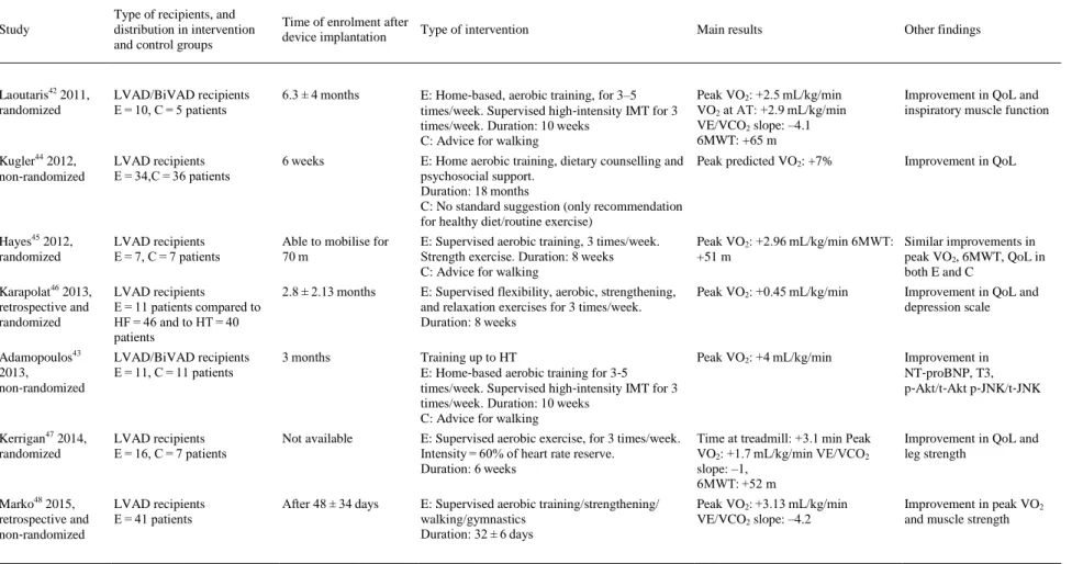
(4) Review of the evidence in favour of exercise testing in ventricular assist device patients Limited but promising data are available concerning the safety and efficacy of EM (7–10 days post‐ implant) and ET in VAD recipients (Table 1).39-48 In 2011, Laoutaris et al.42 provided the first evidence of the feasibility and efficacy of ET in patients with either left ventricular (LVAD) or biventricular (BiVAD) assist devices participating in a 10‐week exercise program, 6.3 ± 4 months post‐implantation. ET improved functional capacity (peak VO2, 6MWT), exertional ventilatory response (VE/VCO2 slope), and quality of life (QoL). Subsequently, Adamopoulos et al.43 extended these findings, showing that long‐ term ET also decreased N‐terminal pro B‐type natriuretic peptide (NT‐proBNP) and triggered myocardial growth factors involved in evolution signalling pathways, in both LVAD and BiVAD patients. A multi‐ model long‐term (18 months) ET intervention increased the percentage of predicted peak VO 2 in LVAD recipients,44 while a shorter (8‐week) ET, started early after implantation and on a small population (14 patients), provided no benefit with respect to the control group. 45 In a retrospective analysis, Karapolat et al.46 observed that an 8‐week ET programme improved peak VO2, pulmonary function and QoL similarly in LVAD recipients, HF or HT patients. Kerrigan et al.47 in a 2:1 randomisation trial comparing usual care vs. ET (which included 18 aerobic exercise sessions at 60–80% of HR reserve), showed that ET improved exercise capacity (peak VO2 by 10%, treadmill time by 3.1 min, 6MWT distance by 52.3 m), QoL (Kansas City Cardiomyopathy Questionnaire score by 14.4 points), and leg strength (17%). More recently, Marko et al.48 confirmed the improvement in peak VO2 and muscle strength in patients with LVAD after ET..
(5) Table 1. Main studies on exercise training in cardiac rehabilitation of patients with ventricular assist devices. Study. Type of recipients, and distribution in intervention and control groups. Laoutaris42 2011, randomized. LVAD/BiVAD recipients E = 10, C = 5 patients. 6.3 ± 4 months. Kugler44 2012, non‐randomized. LVAD recipients E = 34,C = 36 patients. Hayes45 2012, randomized. Time of enrolment after Type of intervention device implantation. Main results. Other findings. E: Home‐based, aerobic training, for 3–5 times/week. Supervised high‐intensity IMT for 3 times/week. Duration: 10 weeks C: Advice for walking. Peak VO2: +2.5 mL/kg/min VO2 at AT: +2.9 mL/kg/min VE/VCO2 slope: –4.1 6MWT: +65 m. Improvement in QoL and inspiratory muscle function. 6 weeks. E: Home aerobic training, dietary counselling and psychosocial support. Duration: 18 months C: No standard suggestion (only recommendation for healthy diet/routine exercise). Peak predicted VO2: +7%. Improvement in QoL. LVAD recipients E = 7, C = 7 patients. Able to mobilise for 70 m. E: Supervised aerobic training, 3 times/week. Strength exercise. Duration: 8 weeks C: Advice for walking. Peak VO2: +2.96 mL/kg/min 6MWT: Similar improvements in +51 m peak VO2, 6MWT, QoL in both E and C. Karapolat46 2013, retrospective and randomized. LVAD recipients E = 11 patients compared to HF = 46 and to HT = 40 patients. 2.8 ± 2.13 months. E: Supervised flexibility, aerobic, strengthening, and relaxation exercises for 3 times/week. Duration: 8 weeks. Peak VO2: +0.45 mL/kg/min. Improvement in QoL and depression scale. Adamopoulos43 2013, non‐randomized. LVAD/BiVAD recipients E = 11, C = 11 patients. 3 months. Training up to HT E: Home‐based aerobic training for 3‐5 times/week. Supervised high‐intensity IMT for 3 times/week. Duration: 10 weeks C: Advice for walking. Peak VO2: +4 mL/kg/min. Improvement in NT‐proBNP, T3, p‐Akt/t‐Akt p‐JNK/t‐JNK. Kerrigan47 2014, randomized. LVAD recipients E = 16, C = 7 patients. Not available. E: Supervised aerobic exercise, for 3 times/week. Intensity = 60% of heart rate reserve. Duration: 6 weeks. Time at treadmill: +3.1 min Peak VO2: +1.7 mL/kg/min VE/VCO2 slope: –1, 6MWT: +52 m. Improvement in QoL and leg strength. Marko48 2015, retrospective and non‐randomized. LVAD recipients E = 41 patients. After 48 ± 34 days. E: Supervised aerobic training/strengthening/ walking/gymnastics Duration: 32 ± 6 days. Peak VO2: +3.13 mL/kg/min VE/VCO2 slope: –4.2. Improvement in peak VO2 and muscle strength. 6MWT, 6‐minute walking test; AI, after intervention; AT, aerobic threshold; BiVAD, biventricular assist device; C, control group; E, exercise group; HF, heart failure; HT, heart transplantation; IMT, inspiratory muscle training; LVAD, left ventricular assist device; NT‐proBNP, N‐terminal pro B‐type natriuretic peptide; QoL, quality of life; VE/VCO2 slope, ventilation vs. carbon dioxide response to exercise; VO2, oxygen consumption.
(6) In conclusion, although the small study populations limit the evidence regarding the role of ET in VAD recipients, all data support the feasibility, safety, and potential for benefit. 40. How to implement exercise Based on the available data, the HFA Committees hereby presents practical advice on the modality of exercise implementation in VAD patients. However the reader should bear in mind that the following are only general recommendations: the implementation in clinical practice is conditioned by local expertise, individual recipient factor (e.g. timing of referral, type of intervention delivered, multidisciplinary approach), characteristics of the VAD recipients (e.g. combined vs. single surgical interventions, indications for implantation, underlying clinical condition, co‐morbidities), and available national recommendations and facilities. Preliminary step – clinical assessment and health professionals' education Medical professionals may be hesitant to start mobilisation because of the presence of the device in a still debilitated patient, and specific skills and expertise are required. Thus, health care providers should be familiar not only with exercise physiology and the different exercise modalities but also with device functioning,44 in order to face promptly all potential complications. Tables 2-6 provide information on what health care providers/exercise therapists should know before, during and after EM and ET..
(7) Table 2. Instruction to reduce the risk of adverse events when exercising ventricular assist device patients. Table 3. Preliminary evaluation and precautions during early mobilisation in ventricular assist device recipients. 1. 2. 3. 4. 5. 6.. 1.. Individualised assessment and prescription. Pre‐screening with risk stratification. Prolonged graduated warm‐up and cool‐down. Low‐to‐moderate intensity exercise training. Avoiding breath holding and Valsalva manoeuvre. Avoiding any trauma, as ventricular assist device recipients are anticoagulated and (some, not all) treated with antiplatelet drugs. 7. Adaptation for co‐morbidities. 8. Monitoring and supervision. 9. Keeping the feet moving during active recovery, if appropriate. 10. Observation of patients for 15 min post‐cessation of exercise.. Assessment a. Recent and past medical history, and level of exercise capacity previous to disease state. b. Mental status and cognitive ability. c. Vital signs and risk of cardiovascular instability (haemodynamic, arrhythmic, clinical). d. Clinical assessment (persistence of VAD‐related and HF symptoms, even medications have been prescribed). e. Medications, i.e. need for continuous or intermittent infusions (inotropic drugs), ventilator settings or oxygen requirements. f. Screen range of motion, coordination, balance, strength, endurance, functional capacity (bed mobility, transfers, gait, daily living activities). g. Baseline haemochromocytometric, ionic and renal functional assessment. We suggest to start exercise when haemoglobin < 9 g/dL, sodium < 130 mEq/L, potassium < 3.8 mEq/L and/or creatininemia <1.9 mg/dL.. 2. 3. 4.. 5.. 6.. Follow sternotomy (6 weeks post‐surgery screening of wound) and skin integrity. Patients should always wear a driveline stabilization belt during exercise. The patient should have his/her travel bag nearby at all times. It should include a back‐up controller, battery clips and spare batteries. Make early mobilization and exercise sessions comfortable. Organize an appropriate place to put monitor, console‐controller and batteries (visible for patient and health care professionals). Discuss this topic with everybody implicated in the exercise programme. The VAD equipment location should not impede emergency procedures..
(8) Table 4. How to set up an early mobilisation programme in ventricular assist device recipients. Table 5. Criteria for exercise contraindications in ventricular assist device recipients. Consider. 1.. 1. 2. 3. 4. 5.. 6. 7. 8.. Positioning. Bed mobility activities. Sitting on edge of bed, in association with exercises. Transfers from bed to stretcher‐chair, chair or commode. Gait, with pre‐gait activities: weight shifting, stepping in place and sideways. Gait training is allowed with rolling walker. Breathlessness management and recovery strategies. Attempt to achieve a target of 11 to 14 out of 20 of the Rate of Perceived Exertion scale. Patient's native heart rate should not exceed 120 b.p.m. during exercise, unless under physician's supervision: heart rate is not always detectable during early mobilisation/exercise, and its monitoring depends on device.. 2.. 3. 4. 5.. Symptoms and signs compatible with exercise intolerance including light headedness, severe intolerable dyspnoea, chest pain or discomfort, tachycardia and exaggerated blood pressure response. Symptomatic hypotension (fainting, dizziness, or diaphoresis, as extreme fatigue or claudication and new onset of neurological changes). Supine resting heart rate > 100 b.p.m. Oxygen saturation < 90% (caveat: oxymetry readings might be difficult to obtain due to low pulsatility). VAD complications during or after exercise sessions: a.. b. Promote Low‐to‐moderate intensity dynamic large muscle group work (e.g. walking, stationary cycling), or involving upper body muscles. ‘Walk & talk’ approach is suggested.. c.. d.. Limit Knee lifts. Resistance training (low weight/high repetitions) and with seated exercise (reduced venous return). Avoid Excessive muscle fatigue. Abrupt postural changes and stooped activities. Rowing machine. Initially, biking due to increased risk of infection near ventricular assist device percutaneous line exit site.. e.. 6. 7. 8.. Alarm activation curves, numbers and alarms should be displayed on the VAD monitor: trends are useful to track pump function and patient perfusion. Significant drop in LVAD flow, or suction alarm are criteria for interrupting the session. Complex and frequent ventricular arrhythmia on exertion (caveat: may be asymptomatic). Infection, mainly at the driveline site (infection control procedures should be followed at all times, e.g. cleaning of equipment, hand washing, disposal of sharps). Evidence of bleeding as VAD recipients are anticoagulated or treated with antiplatelet drugs (not all): these drugs are essential for device working, but they can also enhance exercise‐ related bleedings and haematomas. Thrombus (usually evidenced by an increase in the number of watts/energy necessary for device working).. Request of VAD recipient to stop. Increase > 1.8 kg in body mass over the previous 1 to 3 days. Implantable cardioverter‐defibrillator intervention (anti‐tachycardia pacing and shocks)..
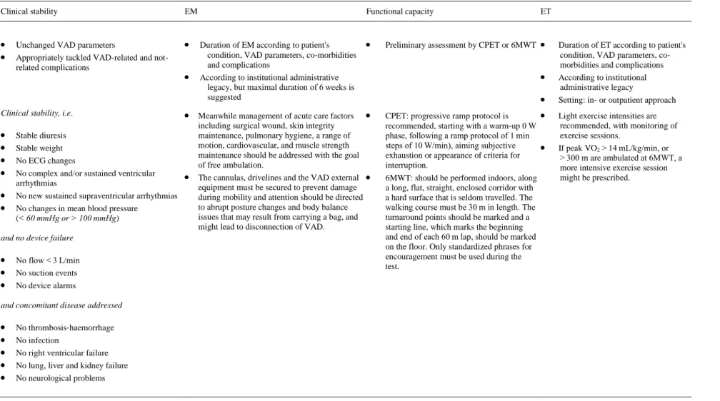
(9) Table 6. Summary of clinical parameters to be considered when exercising in ventricular assist device patients Clinical stability. EM. Unchanged VAD parameters Appropriately tackled VAD‐related and not‐. Duration of EM according to patient's. related complications. Clinical stability, i.e.. . Stable diuresis Stable weight No ECG changes No complex and/or sustained ventricular arrhythmias No new sustained supraventricular arrhythmias No changes in mean blood pressure (< 60 mmHg or > 100 mmHg). . including surgical wound, skin integrity maintenance, pulmonary hygiene, a range of motion, cardiovascular, and muscle strength maintenance should be addressed with the goal of free ambulation. The cannulas, drivelines and the VAD external equipment must be secured to prevent damage during mobility and attention should be directed to abrupt posture changes and body balance issues that may result from carrying a bag, and might lead to disconnection of VAD.. and no device failure. No flow < 3 L/min No suction events No device alarms and concomitant disease addressed. . ET. . . Preliminary assessment by CPET or 6MWT. condition, VAD parameters, co‐morbidities and complications According to institutional administrative legacy, but maximal duration of 6 weeks is suggested. Meanwhile management of acute care factors. . Functional capacity. No thrombosis‐haemorrhage No infection No right ventricular failure No lung, liver and kidney failure No neurological problems. LVAD, left ventricular assist device; VAD, ventricular assist device.. . . CPET: progressive ramp protocol is recommended, starting with a warm‐up 0 W phase, following a ramp protocol of 1 min steps of 10 W/min), aiming subjective exhaustion or appearance of criteria for interruption. 6MWT: should be performed indoors, along a long, flat, straight, enclosed corridor with a hard surface that is seldom travelled. The walking course must be 30 m in length. The turnaround points should be marked and a starting line, which marks the beginning and end of each 60 m lap, should be marked on the floor. Only standardized phrases for encouragement must be used during the test.. . Duration of ET according to patient's condition, VAD parameters, co‐ morbidities and complications According to institutional administrative legacy Setting: in‐ or outpatient approach Light exercise intensities are recommended, with monitoring of exercise sessions. If peak VO2 > 14 mL/kg/min, or > 300 m are ambulated at 6MWT, a more intensive exercise session might be prescribed..
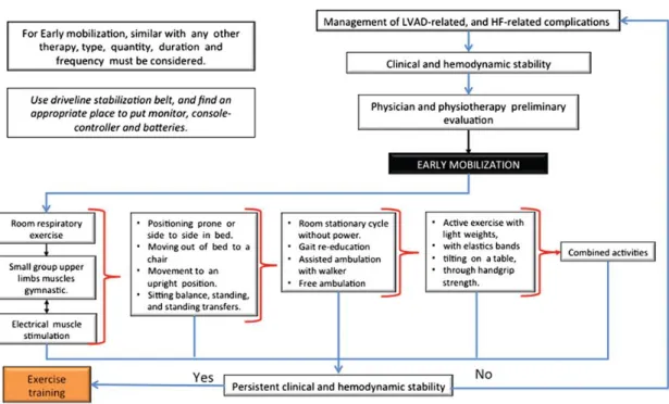
(10) A full patient medical history and clinical and functional evaluations are prerequisites along with HR monitoring for the detection and treatment of arrhythmias. Vital signs, self‐reported symptom scores, and VAD function should be monitored, in particular including the mean arterial pressure in patients on non‐ pulsatile VAD support because hypertension would affect the VAD capacity to pump blood forward; hypotension and VAD blood flow alterations might be related to under‐filling of the left ventricle secondary to high pump speed, RV failure, arrhythmias, etc. The VAD team should be consulted if the mean arterial pressure is below 70 mmHg or higher than 90 mmHg, especially when accompanied by VAD alarm activation. It is also important that the patient is well informed, reassured and feels safe and secure. An exercise physiologist or physical therapist should be responsible for securing the cannulas, drivelines and the VAD external equipment, to prevent damage during mobility. 49,50 Once the recipient is confident with transfers from bed, and shows the ability to carry and to manage the VAD, batteries, and controller, 49 EM can start. The external controller and batteries of recent generations of VADs are highly portable and do not significantly interfere with exercise activities, however, some attention should be directed to avoid abrupt postural changes and body balance issues that may result from carrying a bag, weighing from 2 to 2.5 kg. Complications such as disconnection from the VAD external power supply have been described.51,52 Table 2 provides instruction to reduce the risk of adverse events when exercising VAD patients, while Table 3 lists the preliminary evaluation and precautions during EM. Early mobilisation In every patient, as well as in VAD recipients, EM is defined as initiating physical exercise within the early illness phase, i.e. the first step for initiation of exercise therapy, and it constitutes the basic standard modality for ET implementation during the post‐acute phase (Table 4). This preliminary phase is important but not standardised,53,54 as it is conditioned by the patient's status, facilities and timing of referral. According to the patient's needs, EM should be adapted, and every day treatment changes should be considered: supervision from family members and/or nursing staff is warmly requested, to monitor VAD and clinical parameters. This phase is important to rule out contraindications to exercise49 (Table 5) and should start only when troublesome accounts after VAD implantation are mostly over. 51,52 Early mobilisation prevents the complications of muscle deconditioning and cachexia and, through a broad range of activities, facilitates independence.54 EM favours ambulation and includes functional strengthening, muscle endurance and aerobic training, as for all other HF patients. 55-58 Changes in gait are possible as a result of premature fatigue, appearance of new symptoms or unexpected VAD/clinical parameters changes. Possible falls and some complications such as disconnection from the VAD external power supply, due to the fact that the driveline has a relatively short distance from the skin to the controller, have been described.51 The duration of EM is individualised according to progresses and facilities.19,51 As regards the timing to start, limited available data suggest the safety of a 6‐week interval after implantation,59,60 but in our expert opinion EM should be considered as soon as the patient's haemodynamic and clinical status is stable (including surgical wound, skin integrity maintenance, and pulmonary hygiene), and VAD functioning and troubleshooting have been correctly directed. 49,51,59 An algorithm for EM for VAD‐supported patients and the transition to ET is here proposed, based on expert opinion and patient's aptitudes/clinical state (Figure 1)..
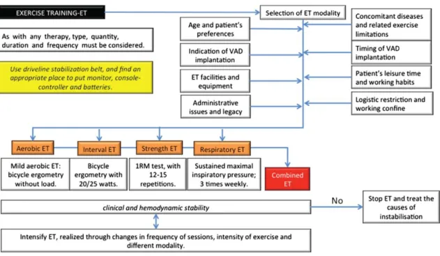
(11) Figure 1. Ventricular assist device recipients and early mobilisation. HF, heart failure; LVAD, left ventricular assist device.. Exercise training No guidelines describing the specific ET setting, modality and duration for VAD‐supported patients are available, but, as described in Table 1, only limited evidence of implementation of light exercise intensities is available. A proper evaluation of symptoms and clinical signs, and functional capacity may help in identifying the most appropriate settings. Although it is reasonable to assume that longer ET interventions could improve physical fitness and QoL, the length and the long‐term adherence to these interventions has not yet been described. Monitoring of exercise sessions is, however, crucial, at least initially, including supervision of the patient, clinical adaptation, and VAD functioning. 49,59 To optimise exercise workload prescription, a symptom‐limited CPET (or 6MWT) is advisable according to administrative constraints and local availability,60,61 in order to aim at a peak workload below the pre‐determined ventilatory anaerobic threshold. If the patient demonstrates a peak VO 2 >14 mL/kg/min or 6MWT distance >300 m, a more intensive exercise test can be considered. Figure 2 provides an algorithm for ET in VAD patients..
(12) Figure 2. Ventricular assist device (VAD) recipients and exercise training. ET, exercise training.. Additionally, caution is recommended to avoid excessive sweating and dehydration, as well as rapid changes of posture from supine to upright positions, which could reduce venous return and negatively impact VAD function40,49,51,59: patients should be urged to drink regularly. Each single ET session starts with a warm up phase and is followed by cool down phase and includes conditioning and endurance exercises: some exercise activities exert torsion on the driveline and, therefore, must be avoided. In summary, based on the available experience in both HF8,9 and VAD patients,18,20-24 both dynamic and resistance exercise are indicated: treadmill (increase ramp, not speed), static bike, hamstring curls in standing position, leg press, bicep curls, core stability, respiratory muscle training, or arm ergometry. Contraindicated forms of exercise include running, rowing machine, cross trainer, abdominals exercises, bilateral arms above the head with weights or abduction with weights or swimming. When to stop exercise The exercise programme should be stopped if: 1. 2. 3.. New symptoms or signs are elicited (i.e. fainting, headache, shortness of breath, chest pain or thoracic pressure, fever, supraventricular or ventricular arrhythmias). VAD alarms or related problems occur. Unexpected changes are detected in VAD parameters, i.e. flow, speed and watt operation.. Thus, monitoring of new signs and symptoms and VAD activity (alarms and related problems) during EM and ET sessions are needed (Table 5). Of note, arrhythmias may appear during EM and ET: ventricular arrhythmias are frequent in VAD supported patients, 62 due to a variety of causes.63,64 Sometimes, ventricular arrhythmias persist over time: arrhythmias do not seem to be a major concern in recipients, since they provide mostly only modest haemodynamic deterioration, but they should be.
(13) carefully evaluated for, if sustained, they might cause device dysfunction, through a detrimental effect on the right ventricle, and they might also promote VAD‐related symptoms. Before the prescription of specific anti‐arrhythmic drugs, some features should be considered: optimization of pharmacological therapy (fluid infusion and/or reduction of daily dose of diuretics), device setting change (if appropriate), and postural changes during exercise sessions (i.e. different sitting position during bicycle ergometer). Of note, initially ET should be discontinued. When arrhythmias have been controlled at rest, exercise can be resumed, albeit at lower frequency and intensity, and all exercise activities should be strictly supervised and ECG monitored. This is a cautionary attitude, not yet supported by scientific evidence. During exercise, atrial fibrillation might occur: atrial fibrillation may worsen symptoms and lead to deterioration of the patient's clinical status, because of loss of atrioventricular synchrony and impaired ventricular filling; thus, electrical cardioversion should be considered.62,63 Different factors should be considered when planning an exercise programme in VAD recipients,40,49,51,58,59,65 as summarised in Table 6.. Gaps in knowledge 1.. 2. 3. 4.. 5.. 6.. 7.. 8.. 9.. The majority of studies so far have included mainly aerobic ET, and data focusing on different components of exercise physiology in VAD recipients are scarce. Thus, the potential benefits of long‐term alternative ET programmes, such as resistance training, balance training and electrical muscle stimulation, should be still investigated. Prolonged periods of ET are needed to observe significant effects in myocardial bioenergetics. This effect might be associated with ventricular unloading and better organ perfusion provided by the VAD. The vast majority of studies included only LVAD recipients, which highlights the need to investigate the benefit of ET in BiVAD patients. Studies comparing exercise capacity pre‐ and post‐VAD implantation are lacking and might add understanding on the role of ET in these patients. Based on individual response and adaptation, EM usually starts early, a few days after the intervention, while ET is considered to continue indefinitely. However, the optimal timing and duration of each single ET session are not yet known. At the beginning, ECG and clinical monitoring are vital: for how long VAD recipients should be monitored is as yet unknown. Intuitively, more complicated VAD recipients need more prolonged supervision. The most effective way to make a patient confident and feeling safe, and the role of the caregiver have rarely been addressed. The potential beneficial contribution of patient education or of a dedicated website needs to be investigated.66,67 The important role of CPET in the exact prescription of ET is advocated: unfortunately, CPET is poorly implemented, and the interpretation is unclear in VAD patients. Where evidence is scant, anecdotal actions predominate, and therefore, for cautionary reasons, low intensity of aerobic training is here recommended.51,66,67 Arterial blood pressure is frequently indeterminable during ET or EM sessions; this is a limiting factor in monitoring EM and ET in VAD recipients. If detectable, measurement of blood pressure before and after exercise is useful, as an excessive rise in blood pressure may induce adverse events, including cerebral haemorrhage, stroke and pump thrombosis. Unfortunately, the blood pressure warning level is not known yet; new symptoms due to exertional maladaptations and alert device‐ related problems (i.e. excessive work) should be considered as alarming signs. Ideally, ET may increase the possibility for a VAD implantation episode to lead a bridge‐to‐ recovery situation, favouring the possibility of weaning, throughout the occurrence of metabolic changes in the failing myocardium and anabolic effects, together with the positive role an adjuvant pharmacotherapy.68-70 The activation of thyroid hormone signalling has been suggested to act as a biological driver for the up‐regulation of physiological growth signalling pathways as indicated by the training‐induced activation of the pro‐survival signalling Akt and inactivation of the anti‐ hypertrophic JNK in cardiomyocytes, leading to physiological growth even in the failing.
(14) 10.. 11.. 12.. 13.. myocardium. Anabolic pharmacotherapy (such as β2‐adrenergic receptor agonist clenbuterol) has been used to facilitate myocardial recovery. VAD unloading therapy and ET might work together for the improvement of exercise capacity. Vignati et al.32 showed that LVAD per se might improve exertional profile in recipients, independently of any ET approach. Only a randomised, non‐exercising control group would be able to differentiate the improvements due to ET from those of device implantation. The factors involved in determining exercise capacity in the post‐VAD patient need to be investigated in more detail: it would be an advantage to identify the most important determinants in an individual patient and focusing therapeutic strategies on them. For instance, the approach to the sarcopenic patient may differ from that to the patient whose main limitation is RV dysfunction. When ET should be started after implantation? Although ET should be commenced as soon as possible, warning and cautionary criteria are still to be established, and, up‐to‐now, personal experience guides the timing of ET in VAD recipients. Target HR of ET sessions need to be defined, with an individualised approach being recommended.. Conclusions Despite LV unloading, impairments persist in VAD patients, with functional capacity frequently below 50% of predicted peak VO2. ET may provide additional benefit. Despite some encouraging small trials, clinical evidence remains limited. Actions should be taken to expand our understanding fn the potential role of ET therapy in VAD recipients to promote its wider implementation in clinical practice.. Acknowledgements We wish to thank M. Frigerio (Milan, Italy) and S. Nalbantgil (Izmir, Turkey) for their thoughtful and constructive comments.. Conflict of interest: none declared.. References 1. 2.. 3.. 4.. Lund LH, Matthews J, Aaronson K. Patient selection for left ventricular assist devices. Eur J Heart Fail 2010;12:434–443. Lloyd‐Jones D, Adams RJ, Brown TM, Carnethon M, Dai S, De Simone G, Ferguson TB, Ford E, Furie K, Gillespie C, Go A, Greenlund K, Haase N, Hailpern S, Ho PM, Howard V, Kissela B, Kittner S, Lackland D, Lisabeth L, Marelli A, MM MD, Meigs J, Mozaffarian D, Mussolino M, Nichol G, Roger VL, Rosamond W, Sacco R, Sorlie P, Roger VL, Thom T, Wasserthiel‐Smoller S, Wong ND, Wylie‐ Rosett J, American Heart Association Statistics Committee and Stroke Statistics Subcommittee. Heart disease and stroke statistics 2010 update: a report from the American Heart Association. Circulation 2010;121:e46–e215. Ponikowski P, Voors AA, Anker SD, Bueno H, Cleland JG, Coats AJ, Falk V, González‐Juanatey JR, Harjola VP, Jankowska EA, Jessup M, Linde C, Nihoyannopoulos P, Parissis JT, Pieske B, Riley JP, Rosano GM, Ruilope LM, Ruschitzka F, Rutten FH, van der Meer P. 2016 ESC Guidelines for the diagnosis and treatment of acute and chronic heart failure: The Task Force for the diagnosis and treatment of acute and chronic heart failure of the European Society of Cardiology (ESC). Developed with the special contribution of the Heart Failure Association (HFA) of the ESC. Eur J Heart Fail 2016;18:891–975. Mc Ilvennan CK, Magid KH, Ambardekar AV, Thompson JS, Matlock DD, Alen LA. Clinical outcomes after continuous‐flow left ventricular assist device: a systematic review. Circ Heart Fail 2014;7:1003–1013..
(15) 5.. 6. 7.. 8. 9.. 10.. 11.. 12.. 13.. 14.. 15.. 16.. 17. 18.. 19.. 20. 21.. Feldman D, Pamboukian SV, Teuteberg JJ, Birks E, Lietz K, Moore SA, Morgan JA, Arabia F, Bauman ME, Buchholz HW, Deng M, Dickstein ML, El‐Banayosy A, Elliot T, Goldstein DJ, Grady KL, Jones K, Hryniewicz K, John R, Kaan A, Kusne S, Loebe M, Massicotte MP, Moazami N, Mohacsi P, Mooney M, Nelson T, Pagani F, Perry W, Potapov EV, Eduardo Rame J, Russell SD, Sorensen EN, Sun B, Strueber M, Mangi AA, Petty MG, Rogers J; International Society for Heart, Lung Transplantation. The 2013 International Society for Heart and Lung Transplantation Guidelines for mechanical circulatory support: executive summary. J Heart Lung Transplant 2013;32:157–187. Jung MH, Gustafsson F. Exercise in heart failure patients supported with a left ventricular assist device. J Heart Lung Transplant 2015;34:489–496. Pina IL, Apstein CS, Balady GJ, Belardinelli R, Chaitman BR, Duscha BD, Fletcher BJ, Fleg JL, Myers JN, Sullivan MJ. Exercise and heart failure: a statement from the American Heart Association Committee on exercise, rehabilitation, and prevention. Circulation 2003;107:1210–1225. Piepoli MF, Davos C, Francis DP, Coats AJ, ExTraMATCH Collaborative. Exercise training meta‐ analysis of trials in patients with chronic heart failure (ExTraMATCH). BMJ 2004;328:189. O'Connor CM, Whellan DJ, Lee KL, Keteyian SJ, Cooper LS, Ellis SJ, Leifer ES, Kraus WE, Kitzman DW, Blumenthal JA, Rendall DS, Miller NH, Fleg JL, Schulman KA, McKelvie RS, Zannad F, Piña IL; HF‐ACTION Investigators. Efficacy and safety of exercise training in patients with chronic heart failure: HF‐ACTION randomized controlled trial. JAMA 2009;301:1439–1450. Piepoli MF, Binno S, Corrà U, Seferovic P, Conraads V, Jaarsma T, Schmid JP, Filippatos G, Ponikowski PP. Committee on Exercise Physiology & Training of the Heart Failure Association of the ESC. ExtraHF survey: the first European survey on implementation of exercise training in heart failure patients. Eur J Heart Fail 2015;17:631–638. Corrà U, Pistono M, Mezzani A, Gnemmi M, Tarro Genta F, Caruso R, Giannuzzi P. Cardiovascular prevention and rehabilitation for patients with ventricular assist device from exercise therapy to long‐ term therapy. Part I: Exercise therapy. Monaldi Arch Chest Dis 2011;76:27–32. Corrà U, Pistono M, Piepoli MF, Giannuzzi P. Ventricular assist device patients on the horizon of cardiovascular prevention and rehabilitation. Can we convert challenges into opportunities? Eur J Prev Rehabil 2011;19:490–493. Ben Gal T, Piepoli MF, Corrà U, Conraads V, Adamopoulos S, Agostoni P, Piotrowicz E, Schmid JP, Seferovic PM, Ponikowski P, Filippatos G, Jaarsma T; Committee on Exercise Physiology & Training of Heart Failure Association and endorsed by Cardiac Rehabilitation Section of European Association for Cardiovascular Rehabilitation and Prevention of ESC. Exercise programs for LVAD supported patients: a snapshot from the ESC affiliated countries. Int J Cardiol 2015;201:215–219. Thompson PD, Franklin B, Balady GJ, Blair SN, Corrado D, Estes IIIM, Fulton JE, Gordon NF, Haskell WL, Link MS, Maron BJ, Mittleman MA, Pelliccia A, Wenger NK, Willich SN, Costa F. Exercise and acute cardiovascular events placing the risks into perspective: a scientific statement from the American Heart Association Council on Nutrition, Physical Activity, and Metabolism and the Council on Clinical Cardiology. Circulation 2007;115:2358–2368. Piepoli MF, Guazzi M, Boriani G, Cicoira M, Corrà U, Dalla Libera L, Emdin M, Mele D, Passino C, Vescovo G, Vigorito C, Villani GQ, Agostoni P. Exercise intolerance in chronic heart failure: mechanisms and therapies. Part I. Eur J Cardiovasc Prev Rehabil 2010;17:637–642. Piepoli MF, Guazzi M, Boriani G, Cicoira M, Corrà U, Dalla Libera L, Emdin M, Mele D, Passino C, Vescovo G, Vigorito C, Villani G, Agostoni P. Exercise intolerance in chronic heart failure: mechanisms and therapies. Part II. Eur J Cardiovasc Prev Rehabil 2010;17:643–648. Levine BD, Cornwell WK 3rd, Drazner MH. Factors influencing the rate of flow through continuous‐ flow left ventricular assist devices at rest and with exercise. JACC Heart Fail 2014;2:331–334. Corrà U, Agostoni PG, Anker SD, AJS C, Crespo Leiro MG, de Boer RA, Harjola VP, Hill L, Lainscak M, Lund LH, Metra M, Ponikowski P, Riley J, Seferović PM, Piepoli MF. Role of cardiopulmonary exercise testing in clinical stratification in heart failure. A position paper from the Committee on Exercise Physiology and Training of the Heart Failure Association of the European Society of Cardiology. Eur J Heart Fail 2018;20:3–15. Scheiderer R, Belden C, Schwab D, Haney C, Paz J. Exercise guidelines for inpatients following ventricular assist device placement: a systematic review of the literature. Cardiopulm Phys Ther J 2013;24:35–42. Adler E, Enciso JS. Functional improvement after ventricular assist device implantation. Is ventricular recovery more common than we thought? J Am Coll Cardiol 2013;61:1995–1997. de Jonge N, Kirkels H, Lahpor JR, Klöpping C, Hulzebos EJ, de la Rivière AB, Robles de Medina EO. Exercise performance in patients with end‐stage heart failure after implantation of a left ventricular assist device and after heart transplantation: an outlook for permanent assisting. J Am Coll Cardiol 2001;37:1794–1799..
(16) 22.. 23.. 24. 25. 26.. 27. 28.. 29. 30.. 31.. 32.. 33.. 34. 35.. 36.. 37.. 38.. 39.. 40.. 41.. Kugler C, Malehsa D, Tegtbur U, Guetzlaff E, Meyer AL, Bara C, Haverich A, Strueber M. Health‐ related quality of life and exercise tolerance in recipients of heart transplants and left ventricular assist devices: a prospective, comparative study. J Heart Lung Transplant 2011;30:204–210. Dunlay SM, Allison TG, Pereria NL. Changes in cardiopulmonary exercise testing parameters following continuous flow left ventricular assist device implantation and heart transplantation. J Card Fail 2014;20:548–554. Marko C, Elili E, Lackner T, Zimpfer D, Schima H, Moscato F. Exercise performance during the first two years after left ventricular assist device implantation. ASAIO J 2017;63:408–413. Abshire M, Dennison Himmelfarb CR, Russell SD. Functional status in left ventricular assist device supported patients: a literature review. J Card Fail 2014;20:973–983. Jung MH, Hansen PB, Sander K, Olsen PS, Rossing K, Boesgaard S, Russell SD, Gustafsson F. Effect of increasing pump speed during exercise on peak oxygen uptake in heart failure patients supported with a continuous‐flow left ventricular assist device. A double‐blind randomized study. Eur J Heart Fail 2014;16:403–408. Loyaga‐Rendon RY, Plaisance EP, Arena R, Shah K. Exercise physiology, testing, and training in patients supported by a left ventricular assist device. J Heart Lung Transplant 2015;34:1005–1016. Schmidt T, Bjarnason‐Wehrens B, Bartsch P, Deniz E, Schmitto J, Schulte‐Eistrup S, Willemsen D, Reiss N. Exercise capacity and functional performance in heart failure patients supported by a left ventricular assist device at discharge from inpatient rehabilitation. Artif Organs 2018;42:22–30. Nakata K, Akiyama K, Sankai K, Shiono M, Orime Y, Saito Y, Hata M, Sezai A, Minami T, Negishi N. Estimation of cardiac function with rotary blood pump. Ann Thorac Cardiovasc Surg 2007;13:240–246. Riess N, Schimdt T, Workowski A, Willemsen D, Schmitto JD, Haverich A, Bjarnason‐Wehrens B. Physical capacity in LVAD patients: hemodynamic principles, diagnostic tools and training control. Int J Artif Organs 2016;39:451–459. Jung MT, Gustafsson F, Houston B, Russel SD. Ramp study hemodynamics, functional capacity, and outcome in heart failure patients with continuous‐flow left ventricular assist devices. ASAIO J 2016;62:442–446. Vignati C, Apostolo A, Cattadori G, Farina S, Del Torto A, Scuri S, Gerosa G, Bottio T, Tarzia V, Bejko J, Sisillo E, Nicoli F, Sciomer S, Alamanni F, Paolillo S, Agostoni P. LVAD pump speed increase is associated with increased peak exercise cardiac output and VO2, postponed anaerobic threshold and improved ventilatory efficiency. Int J Cardiol 2017;230:28–32. Mezzani A, Pistono M, Corrà U, Giordano A, Gnemmi M, Imparato A, Centofanti P, Rinaldi M, Colombo S, Canal E, Giannuzzi P. Systemic perfusion at peak incremental exercise in left ventricular assist device recipients: partitioning pump and native left ventricle relative contribution. IJC Heart & Vessels 2014;4:40–45. Levine BD, Cornwell WK 3rd, Drazner MH. Factors influencing the rate of flow through continuous‐ flow left ventricular assist devices at rest and with exercise. JACC Heart Fail 2014;2:331–334. Dimopoulos S, Diakos N, Tseliou E, Tasoulis A, Mpouchla A, Manetos C, Katsaros L, Drakos S, Terrovitis J, Nanas S. Chronotropic incompetence and abnormal heart rate recovery early after left ventricular assist device implantation. Pacing Clin Electrophysiol 2011;34:1607–1614. Brassard P, Jensen AS, Nordborg N, Gustafsson F, Møller JE, Hassager C, Boesgaard S, Hansen PB, Olsen PS, Sander K, Secher NH, Madsen PL. Central and peripheral blow flow during exercise with continuous‐flow left ventricular assist device: constant versus increasing pump speed: a pilot study. Circ Heart Fail 2011;4:554–560. Jakovljevic DG, Birks EJ, George RS, Trenell MI, Seferovic PM, Yacoub MH, Brodie DA. Relationship between peak cardiac pumping capability and selected exercise‐derived prognostic indicators in patients treated with left ventricular assist devices. Eur J Heart Fail 2011;13:992–999. Apostolo A, Paolillo S, Contini M, Vignati C, Tarzia V, Campodonico J, Mapelli M, Massetti M, Bejko J, Righini F, Bottio T, Bonini N, Salvioni E, Gugliandolo P, Parati G, Lombardi C, Gerosa G, Salvi L, Alamanni F, Agostoni P. Comprehensive effects of left ventricular assist device speed changes on alveolar gas exchange, sleep ventilatory pattern, and exercise performance. J Heart Lung Transplant 2018;37:1361–1371. Ganga HV, Leung A, Jantz J, Choudhary G, Stabile L Levine DJ, Sharma SC, Wu WC. Supervised exercise training versus usual care in ambulatory patients with left ventricular assist devices: a systematic review. PLoS One 2017;12:e0174323. Mahfood Haddad T, Saurav A, Smer A, Azzouz MS, Akinapelli A, Williams MA, Alla VM. Cardiac rehabilitation in patients with left ventricular assist device: a systematic review and meta‐analysis. J Cardiopulm Rehabil Prev 2017;37:390–396. Alsara O, Reeves RK, Pyfferoen MD, Trenary TL, Engen DJ, Vitse ML, Kessler SM, Kushwaha SS, Cavel AL, Thomas RJ, Lopez‐Jimenez F, Park SJ, Perez‐Terzic CM. Inpatient rehabilitation outcomes for patients receiving left ventricular assist device. Am J Phys Med Rehabil 2014;93:860–868..
(17) 42.. 43.. 44.. 45.. 46.. 47.. 48.. 49. 50.. 51.. 52.. 53.. 54. 55.. 56.. 57. 58. 59. 60.. 61.. Laoutaris ID, Dritsas A, Adamopoulos S, Manginas A, Gouziouta A, Kallistratos MS, Koulopoulou M, Voudris V, Cokkinos DV, Sfirakis P. Benefits of physical training on exercise capacity, inspiratory muscle function, and quality of life in patients with ventricular assist devices long‐term post implantation. Eur J Cardiovasc Prev Rehabil 2011;18:33–40. Adamopoulos S, Gouziouta A, Mantzouratou P, Laoutaris ID, Dritsas A, Cokkinos DV, Mourouzis I, Sfyrakis P, Iervasi G, Pantos C. Thyroid hormone signalling is altered in response to physical training in patients with end‐stage heart failure and mechanical assist devices: potential physiological consequences? Interact Cardiovasc Thorac Surg 2013;17:664–668. Kugler C, Malehsa D, Schrader E, Tegtbur U, Guetzlaff E, Haverich A, Strueber M. A multi‐modal intervention in management of left ventricular assist device outpatients: dietary counselling, controlled exercise and psychosocial support. Eur J Cardiothorac Surg 2012;42:1026–1032. Hayes K, Leet AS, Bradley SJ, Holland AE. Effects of exercise training on exercise capacity and quality of life in patients with a left ventricular assist device: a preliminary randomized controlled trial. J Heart Lung Transplant 2012;31:729–734. Karapolat H, Engin C, Eroglu M, Yagdi T, Zoghi M, Nalbantgil S, Durmaz B, Kirazlı Y, Ozbaran M. Efficacy of the cardiac rehabilitation program in patients with end‐stage heart failure, heart transplant patients, and left ventricular assist device recipients. Transplant Proc 2013;45:3381–3385. Kerrigan DJ, Williams CT, Ehrman JK, Saval MA, Bronsteen K, Schairer JR, Swaffer M, Brawner CA, Lanfear DE, Selektor Y, Velez M, Tita C, Keteyian SJ. Cardiac rehabilitation improves functional capacity and patient‐reported health status in patients with continuous‐flow left ventricular assist devices: the Rehab‐VAD randomized controlled trial. JACC Heart Fail 2014;2:653–659. Marko C, Danzinger G, Käferbäck M, Lackner T, Müller R, Zimpfer D, Schima H, Moscato F. Safety and efficacy of cardiac rehabilitation for patients with continuous flow left ventricular assist devices. Eur J Prev Cardiol 2015;22:1378–1384. Wells CL. Physical therapist management of patients with ventricular assist devices: key considerations for the acute care physical therapist. Phys Ther 2013;93:266–278. Baronetto A, Centofanti P, Attisani M, Ricci D, Mussa B, Devotini R, Simonato E, Rinaldi M. A simple device to secure ventricular assist device driveline and prevent exit‐site infection. Interact Cardiovasc Thorac Surg 2014;18:415–417. Compostella L, Russo N, Setzu T, Bottio T, Compostella C, Tarzia V, Ulivi U, Gerosa G, Iliceto S, Bellotto F. A practical review for cardiac rehabilitation professionals of continuous‐flow left ventricular assist devices: historical and current perspectives. J Cardiopulm Rehabil Prev 2015;35:301–311. Slaughter MS, Pagani FD, Rogers JG, Miller LW, Sun B, Russell SD, Starling RC, Chen L, Boyle AJ, Chillcott S, Adamson RM, Blood MS, Camacho MT, Idrissi KA, Petty M, Sobieski M, Wright S, Myers TJ, Farrar DJ; HeartMate II Clinical Investigators. Clinical management of continuous‐flow left ventricular assist devices in advanced heart failure. J Heart Lung Transplant 2010;29(4 Suppl):S1–S39. Piepoli MF, Conraads V, Corrà U, Dickstein K, Francis DP, Jaarsma T, McMurray J, Pieske B, Piotrowicz E, Schmid JP, Anker SD, Solal AC, Filippatos GS, Hoes AW, Gielen S, Giannuzzi P, Ponikowski PP. Exercise training in heart failure: from theory to practice. A consensus document of the Heart Failure Association and the European Association for Cardiovascular Prevention and Rehabilitation. Eur J Heart Fail 2011;13:347–357. Amidei C. Mobilisation in critical care: a concept analysis. Intensive Crit Care Nurs 2012;28:73–81. Muthiah K, Gupta S, Otton J, Robson D, Walker R, Tay A, Macdonald P, Keogh A, Kotlyar E, Granger E, Dhital K, Spratt P, Jansz P, Hayward CS. Body position and activity, but not heart rate, affect pump flows in patients with continuous‐flow left ventricular assist devices. JACC Heart Fail 2014;2:323–330. Morrone TM, Buck LA, Catanese KA, Goldsmith RL, Cahalin LP, Oz MC, Levin HR. Early progressive mobilization of patients with left ventricular assist devices is safe and optimizes recovery before heart transplantation. J Heart Lung Transplant 1996;15:423–429. Perme CS, Southard RE, Joyce DL, Noon GP, Loebe M. Early mobilization of LVAD recipients who require prolonged mechanical ventilation. Tex Heart Inst J 2006;33:130–136. Ueno A, Tomizawa Y. Cardiac rehabilitation and artificial heart devices. J Artif Organs 2009;12:90–97. Kennedy MD, Haykowsky M, Humphrey R. Function, eligibility, outcomes, and exercise capacity associated with left ventricular assist devices. J Cardiopulm Rehabil 2003;23:208–217. Mezzani A, Hamm LF, Jones AM, McBride PE, Moholdt T, Stone JA, Urhausen A, Williams MA. Aerobic exercise intensity assessment and prescription in cardiac rehabilitation: a joint position statement of the European Association for Cardiovascular Prevention and Rehabilitation, the American Association of Cardiovascular and Pulmonary Rehabilitation, and the Canadian Association of Cardiac Rehabilitation. Eur J Prev Cardiol 2013;20:442–467. American Association of Cardiovascular and Pulmonary Rehabilitation (AACVPR). Guidelines for Cardiac Rehabilitation and Secondary Prevention Programs, 5th ed. Champaign, IL: Human Kinetics; 2005..
(18) 62.. 63. 64. 65.. 66.. 67.. 68.. 69.. 70.. Kirklin JK, Naftel DC, Kormos RL, Stevenson LW, Pagani FD, Miller MA, Baldwin JT, Young JB. Fifth INTERMACS annual report: risk factor analysis from more than 6,000 mechanical circulatory support patients. J Heart Lung Transplant 2013;32:141–156. Vollkron M, Voitl P, Ta J, Wieselthaler G, Schima H. Suction events during left ventricular support and ventricular arrhythmias. J Heart Lung Transplant 2007;26:819–825. Boyle A. Arrhythmias in patients with ventricular assist devices. Curr Opin Cardiol 2012;27:13–18. Compostella L, Polastri M, Lamotte M, Bellotto F, Antoine M. Physiotherapy and rehabilitation management in adult LVAD patients. In: Montalto A, Loforte A, Musumeci F, Krabatsch T, Slaughter MS, eds. Mechanical Circulatory Support in End‐Stage Heart Failure. Springer International Publishing; 2017. pp 403–420. Lamotte M, Hansen D, Timmermans P. How to manage physiotherapy and rehabilitation in LVAD patients. https://www.escardio.org/Education/Practice-Tools/CVD-prevention-toolbox/how-to-managephysiotherapy-and-rehabilitation-in-lvad-patients (23 October 2018). Wever‐Pinzon O, Drakos SG, McKellar SH, Horne BD, Caine WT, Kfoury AG, Li DY, Fang JC, Stehlik J, Selzman CH. Cardiac recovery during long‐term left ventricular assist device support. J Am Coll Cardiol 2016;68:1540–1553. Wagenaar KP, Rutten FH, Klompstra L, Bhana Y, Sieverink F, Ruschitzka F, Seferovic PM, Lainscak M, Piepoli MF, Broekhuizen BDL, Strömberg A, Jaarsma T, Hoes AW, Dickstein K. ‘heartfailurematters.org’, an educational website for patients and carers from the Heart Failure Association of the European Society of Cardiology: objectives, use and future directions. Eur J Heart Fail 2017;19:1447–1454. Birks EJ, Tansley PD, Hardy J, George RS, Bowles CT, Burke M, Banner NR, Khaghani A, Yacoub MH. Left ventricular assist device and drug therapy for the reversal of heart failure. N Engl J Med 2006;355:1873–1884. Gustafsson F, Rogers JG. Left ventricular assist device therapy in advanced heart failure: patient selection and outcomes. Eur J Heart Fail 2017;19:595–602..
(19)
Figure
Documento similar
