Effects of moderate maternal energy restriction on the offspring metabolic health, in terms of obesity and related diseases, and identification of determinant factors and early biomarkers
289
0
0
Texto completo
(2)
(3) DOCTORAL THESIS 2014 Doctoral Programme of Nutrigenomics and Personalized Nutrition EFFECTS OF MODERATE MATERNAL ENERGY RESTRICTION ON THE OFFSPRING METABOLIC HEALTH, IN TERMS OF OBESITY AND RELATED DISEASES, AND IDENTIFICATION OF DETERMINANT FACTORS AND EARLY BIOMARKERS Juana María Torrens García Thesis Supervisor: Catalina Picó Segura, PhD Thesis Supervisor: Andreu Palou Oliver, PhD. Doctor by the Universitat de les Illes Balears.
(4)
(5) With the consent of the directors. Catalina Picó Segura, PhD Professor of Biochemistry and Molecular Biology, UIB. Andreu Palou Oliver, PhD Professor of Biochemistry and Molecular Biology, UIB. The doctoral candidate. Juana María Torrens García.
(6)
(7) Dr Catalina Picó Segura, Professor of Biochemistry and Molecular Biology from the University of the Balearic Islands and Dr Andreu Palou Oliver, Professor of Biochemistry and Molecular Biology from the University of the Balearic Islands. WE DECLARE: That the thesis entitled Effects of moderate maternal energy restriction on the offspring metabolic health, in terms of obesity and related diseases, and identification of determinant factors and early biomarkers, presented by Juana María Torrens García to obtain a doctoral degree, has been completed under our supervision and meets the requirements to opt for an European Doctorate Mention.. For all intents and purposes, we hereby sign this document.. Catalina Picó Segura, PhD Professor of Biochemistry and Molecular Biology, UIB. Andreu Palou Oliver, PhD Professor of Biochemistry and Molecular Biology, UIB. Palma de Mallorca, 18th November 2014.
(8)
(9) A mis padres.
(10)
(11) Agradecimientos Quiero expresar mi agradecimiento a todas las personas que han contribuido, de una forma u otra, en la realización de esta tesis. Recuerdo perfectamente el día en el que me preguntaron si quería ser alumna colaboradora del laboratorio. Yo estaba en segundo de carrera y la verdad es que por aquel entonces no había pensado en nada de doctorados ni en investigación, simplemente me hacía muchas ganas conocer cómo se trabaja en un laboratorio, así que lo probé. Empecé un verano ayudando básicamente a Ona en el estabulario con las “ratitas” y como tanto el trabajo como la gente del laboratorio me engancharon, verano tras verano volví a repetir hasta que, al acabar la carrera, decidí empezar el doctorado y continuar con lo que me gustaba, la investigación. Y aquí estoy ahora, escribiendo unas líneas para agradecer a todas las personas que han hecho posible que haya conseguido llegar hasta el final de esta etapa. La redacción nunca se me ha dado bien y mucho menos reflexionar sobre mis sentimientos, ―siempre he dicho que soy de números y no de letras―, pero haré lo posible para que todo el agradecimiento que siento quede reflejado en estas líneas. Mi especial agradecimiento a mis dos directores de tesis: el Prof. Andreu Palou y la Prof. Catalina Picó por su contribución en mi formación investigadora. Al Prof. Andreu Palou, quiero agradecerle la oportunidad que me brindó para formar parte y para realizar el doctorado en su grupo de investigación, el Laboratorio de Biología Molecular, Nutrición y Biotecnología (LBNB) de la Universidad de las Islas Baleares. También quiero agradecerle la supervisión y los consejos aportados a mi tesis, así como también la oportunidad que me ofreció para realizar mis dos estancias predoctorales en la Universidad de Warwick, en el Reino Unido. A la Prof. Catalina Picó quiero agradecerle muy especialmente, toda la confianza, paciencia y comprensión que ha tenido conmigo en todos los momentos (porque sé que soy muy cabezota, y cuando me encierro en una cosa, soy muy difícil de sacar de ahí). Quiero agradecerle toda la ayuda que me ha dado durante todos estos años, y especialmente durante estos últimos meses, en los que la redacción de la tesis ha sido un camino duro y complicado. A los demás profesores del grupo: Dra. Paula Oliver, Prof. Francisca Serra, Dra. Ana María Rodrídrez, Dr. Joan Ribot y Dra. Lluïsa Bonet, quiero agradecerles sus consejos, su ayuda y la proximidad de su trato. Muchísimas gracias Dra. Mariona Palou y Dra. Joana Sánchez, por vuestra amistad, por escucharme, por comprenderme y por vuestras enseñanzas; sin vosotras no estaría en estos momentos a punto de terminar el doctorado. Me guiasteis en mis primeros pasos en el laboratorio: a trabajar con los animales (santa paciencia tuvisteis conmigo que siempre me desmayaba cuando teníamos que sacar sangre!!), a manejar las máquinas del laboratorio, a rellenar papeles administrativos, a escribir resultados…. Sinceramente, las dos habéis estado desde el principio siempre a mi lado y me habéis ayudado tanto a nivel profesional como personal en todos los obstáculos que han ido apareciendo a lo largo de estos años. A la Dra. Teresa Priego, al Dr. Pep Mercader y a la Dra. Nuria Granados, gracias por vuestro apoyo, por vuestros ánimos y consejos. Thank you to the Prof. Paul Thornalley and Dr. Naila Rabanni for giving me the opportunity to work with them, for all their advices and for treat me as another one of the group. Thanks to all the members of the Protein Damage group: Fozia, Dr. Amy, Dr. Ming, Dr. Makoto, Alla, Amal, Dr. Attia Dra. Zhera, and especially to Dr. Jinit for his patience understanding and advices (also thanks to all “Costa time”). Quiero agradecer especialmente a Enzo por hacerme sonreír siempre que he estado triste y por hacerme reír cuando sólo quería llorar. Gracias por enseñarme morfología e histología y por ayudarme siempre que lo he necesitado.. I.
(12) A todos mis compañeros de laboratorio, quiero daros las gracias: Rubén y Xisco, gracias por convertir el despacho en el Polo Norte!!!, voy a añorar no discutir con vosotros por el aire acondicionado; Estefanía, simplemente gracias por todos tus detallitos; Nora, gracias por escucharme y darme consejos; Raúl, gracias por hacerme reír y saber escuchar; Petar, gracias por ser cómo eres; Madhu, gracias por cuidarnos tan bien con tus postres; Heriberto; gracias por deleitarnos con la comida mejicana; Nara, gracias por tu alegría y vitalidad; Alice, gracias por escucharme cuando lo he necesitado. Especialmente quiero agradecer a la Dra. Jadwiga Konieczna por ayudarme siempre y por aguantarme tantas horas en el estabulario y en laboratorio. A los nuevos becarios Cati Dora, Marga Cifre, Andrea, Alba y Sebastián, gracias, mucha suerte y muchos ánimos. A los demás compañeros del laboratorio, Bea; al personal del estabulario, Teresa de Francisco y Miquel, gracias. A los antiguos compañeros del laboratorio, Dr. Toni Caimari, Dr. Jaume Amengual, Dra. María Servera, Dra.Ana Paula García, Dra. Hana Musinovic y especialmente a la Dra. Pilar Parra, gracias por acogerme tan bien cuando llegué al grupo. Quiero agradecer muy especialmente a las Pingüinas: Bárbara, Marina, Ona y Joana. Gracias por vuestros ánimos pero sobre todo por haberme aguantado (sé que estos últimos meses he sido la niña del exorcista y “mordía” por todo, pero a pesar de ello habéis seguido ahí), gracias por escucharme, por vuestro apoyo, por vuestros consejos, por reír conmigo, por llorar conmigo…por todo, gracias!!. Quiero agradecer la ayuda de todos los alumnos colaboradores, en especial a Cristina Cabrer, Cati Joy, y Bel Mora. Gracias Cati Joy por ser mi mejor amiga (aunque hayamos estado semanas sin saber la una de la otra), gracias por aguantarme cuando he estado agobiada y por reñirme por mis tonterías. Gracias Bel, por tu amistad, por tu cariño y por tu gran ayuda, porque estos últimos meses no lo hubiese podido conseguir sin ti. A mi prima Natalia, muchas gracias por escucharme siempre, por animarme, apoyarme y sobre todo por hacerme reír cuando he estado triste. Finalmente, y no por ello menos importante, quiero darles las gracias a mis padres, Miguel y Antonia. Lo sois todo para mí, y todo lo que pueda decir de vosotros se quedaría corto. Os quiero mucho y si he llegado hasta aquí has sido gracias a vosotros. Gracias por vuestro esfuerzo y sacrificio incondicional para que yo pudiese estudiar. Gracias por todas las facilidades que me habéis dado. Gracias por ayudarme a estudiar y por estar a mi lado en todo momento. Gracias por aguantar mis enfados, mis lloros y mi estrés durante toda la vida y especialmente durante estos últimos meses. Os quiero agradecer todo lo que me habéis enseñado desde pequeña y que me han formado como la persona que ahora soy, solo espero no defraudaros nunca. Ahora, lo que más deseo es compartir con vosotros todo lo que he conseguido gracias a vuestra ayuda, apoyo, confianza, esfuerzo, comprensión y dedicación. Gracias mami!!. Gracias papi!!! Esto es por y para vosotros!!!. Os quiero!!!. Guty, esta recta final no hubiese sido posible tampoco sin ti. Gracias por tu comprensión y por tu enorme paciencia estos últimos meses. Gracias por escuchar mis preocupaciones, por aguantar mis lloros y mis tonterías. Gracias por tus consejos, por tus abrazos, tus mimos, por tu ayuda, por todo. Te ha tocado conocerme en una época de mucho estrés para mí, pero a pesar de ello, sigues aguantándome. Simplemente gracias por ser cómo eres, por confiar en mí y por quererme tal y como soy. Te quiero.. II.
(13) Index. Index Abbreviations ............................................................................................................................... VII Abstract/Resumen/Resum .............................................................................................................. IX List of original articles .................................................................................................................XV I.. Introduction ................................................................................................................. 1 1. Obesity..................................................................................................................... 3 2. Energy balance regulation ....................................................................................... 4 2.1 Main tissues involved in maintenance of energy homeostasis ......................... 5 2.1.1. Hypothalamus......................................................................................... 5 2.1.2. White adipose tissue ............................................................................... 7 2.1.3. Liver ....................................................................................................... 8 2.1.4. Skeletal muscle ..................................................................................... 10 2.2 Main hormones involved in maintenance of energy homeostasis .................. 11 2.2.1. Leptin ................................................................................................... 11 Leptin during the perinatal period........................................................ 12 Leptin resistance ................................................................................... 14 2.2.2. Insulin ................................................................................................... 15 Insulin resistance .................................................................................. 16 2.3 Calorie restriction ........................................................................................... 20 3. Developmental origins of obesity: importance of early life nutrition ................... 21 3.1. Programming effects of maternal calorie restriction during gestation .......... 22 3.2. Programming effects of maternal calorie restriction during lactation ........... 24 Breast milk ............................................................................................ 25 4. Biomarkers of metabolic health ............................................................................ 26 4.1. Gene expression analysis in PBMCs ............................................................. 27 4.2. Quantitative screening of protein damage biomarkers. ................................. 28. II.. Aim and experimental design................................................................................... 33. III.. Materials and methods ............................................................................................. 43 1. Experimental animals ............................................................................................. 45 2. PBMC isolation ...................................................................................................... 45 3. RNA isolation and quantification ........................................................................... 46 3.1. Tripure reagent method for RNA isolation ..................................................... 47 III.
(14) Juana María Torrens García – Doctoral Thesis. 3.2. E.Z.N.A total RNA kit I for RNA isolation .................................................... 47 3.3. RNA quantification and assessment of its integrity ........................................ 48 4. Gene expression analysis by RT-qPCR ................................................................ 49 4.1. Reverse transcription ....................................................................................... 49 4.2. Real Time quantitative Polymerase Chain Reaction (RT-qPCR).................... 49 5. Determination of circulating parameters .............................................................. 50 5.1. Insulin .............................................................................................................. 50 5.2. Glucose ............................................................................................................ 51 5.3. Homeostatic model assessment for insulin resistance (HOMA-IR)................ 51 5.4. Leptin ............................................................................................................... 51 5.5. Triglycerides (TGs) ......................................................................................... 52 5.6. Non-esterified fatty acids (NEFAs) ................................................................. 53 5.7. β-hydroxybutirate (BHB) ................................................................................ 53 5.8. Adiponectin ..................................................................................................... 53 5.9. Tumor Necrosis Factor alpha (TNFα) ............................................................. 54 6. Measurement of hepatic glycogen content ........................................................... 54 7. Total hepatic lipid content measurement .............................................................. 55 8. Measurement of hepatic triglyceride content ....................................................... 56 9. Histological analysis ............................................................................................. 57 10. Confocal microscopy for Green Fluorescent Protein detection in liver samples 58 11. Isolation of mitochondria from liver ................................................................... 58 12. Western blot analysis ........................................................................................... 59 12.1. Sample preparation ....................................................................................... 59 12.2. Electrophoresis ............................................................................................. 60 12.3. Electroblotting .............................................................................................. 60 12.4. Labelling and detection ................................................................................ 61 12.5. Scanning and quantification ......................................................................... 62 12.6. Loading control ............................................................................................ 62 13. Indirect gas calorimetry and locomotive activity measurements ........................ 62 14. Blood pressure measurement ............................................................................... 63 15. Liquid chromatography-tandem mass spectrometry (LC-MS/MS) .................... 63 15.1. Sample preparation ....................................................................................... 64 15.2. Ultrahigh Performance Liquid Chromatography (UPLC) ............................ 64 IV.
(15) Index. 15.3. MS/MS detection .......................................................................................... 64 15.4. Calculation.................................................................................................... 64 16. Statistical analysis ............................................................................................... 65 IV.. Results and discussion .............................................................................................. 67 1. Impaired insulin and leptin sensitivity in the offspring of moderate caloricrestricted dams during gestation is early programmed ........................................ 69 2. Enhancing hepatic fatty acid oxidation as a strategy for reversing metabolic disorders programmed by maternal undernutrition during gestation ................... 95 3. Moderate caloric restriction in lactating rats protects offspring against obesity and insulin resistance in later life ....................................................................... 121 4. Moderate caloric restriction in lactating rats programs their offspring for a better response to HF diet feeding in a sex-dependent manner.................................... 141 5. Early biomarkers identified in a rat model of a healthier phenotype based on early postnatal dietary intervention may predict the response to an obesogenic environment in adulthood .................................................................................. 163 6. Identification of early transcriptome-based biomarkers related to lipid metabolism in peripheral blood mononuclear cells of rats nutritionally programmed for improved metabolic health ...................................................... 185 7. Moderate maternal calorie restriction in lactating rats affects markers of protein damage by glycation and oxidation in breast milk ........................................... 209. V.. Recapitulation.......................................................................................................... 227. VI.. Conclusions/Conclusiones ....................................................................................... 235. VII.. Bibliography ............................................................................................................ 241. V.
(16)
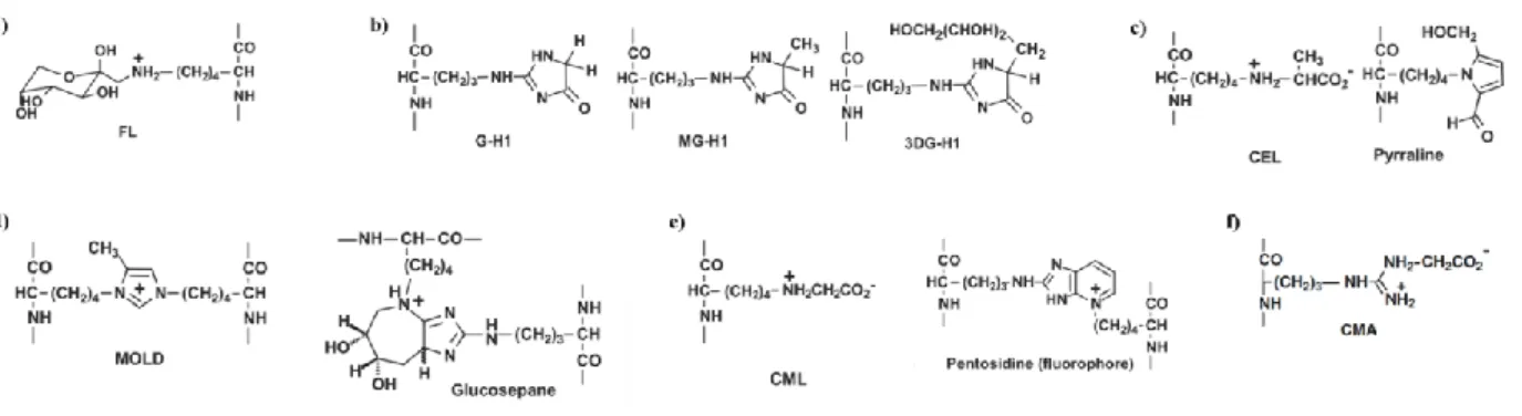
(17) Abbreviations. Abbreviations 3DG-H: 3-deoxyglucosone 3-NT: 3-Nitrotyrosine AAV: adeno-associated virus ACC: acetyl-CoA carboxylase ACOX: acyl-CoA oxidase AdipoR1: adiponectin receptor 1 AdipoR2: adiponectin receptor 2 AGEs: advanced glycation endproducts AgRP: Agouti-related peptide AKT: protein kinase B (PKB) AMPK: AMP-activated protein kinase α-MSH: alpha-melanocyte-stimulating hormone ARC: arcuate nucleous ATGL: adipose tissue triglyceride lipase BMI: body mass index cAMP: cyclic adenosine monophosphate CART: cocaine and amphetamine-regulated transcript CEL: Nε-carboxyethyl-lysine CKM: creatine kinase CMA: Nω-carboxymethylarginine CML: Nε-carboxymethyl-lysine CNS: central nervous system CPT1: carnitine palmitoyltransferase 1 CPT1A: carnitine palmitoyltransferase 1 liver isoform CPT1AM: mutant form of carnitine palmitoyltransferase 1 liver isoform CRH: corticotropic releasing hormone DEX: dexamethasone DMN: dorsomedial nucleous FABP: fatty acid binding protein FAO: fatty acid oxidation FASC: fatty acid synthase complex FASN: fatty acid synthase FATP: fatty acid binding protein FL: Fructosyl-lysine FTO: fat mass and obesity associated GCK: glucokinase G-H1: Nδ-(5-hydro-4-imidazolon-2-yl)ornithine GLUT4: glucose transporter 4 GPAT: glycerol-3-phosphate acyltransferase GSA: glutamic semialdehyde HDL: high-density lipoprotein HF: high fat HSL: hormone-sensitive lipase IDL: intermediate density lipoproteins IL-6: interleukin-6 InsR: insulin receptor VII.
(18) Juana María Torrens García – Doctoral Thesis. IRS: insulin receptor substrate IRX3: iroquois homeobox 3 gene iWAT: inguinal white adipose tissue JAK2: janus-activated tyrosine kinase 2 LCAS: long chain acyl-CoA synthetase LC-MS/MS: liquid chromatography with tandem mass spectrometry detection LDL: low-density lipoprotein LH: lateral hypothalamus LPL: lipoprotein lipase MC3R: melanocortin receptor subtype 3 MC4R: melanocortin receptor subtype 4 MCH: melanin-concentrating hormone MetSO: methionine sulfoxide MG-H1: Nδ-(5-hydro-5-methyl-4-imidazolon-2-yl)ornithine MOLD: bis(lysyl) crosslink derived from methylglyoxal NEFAs: non-esterified fatty acids NFK: N-formylkynurenine NPY: neuropeptide Y ObRb: long form of leptin receptor PBMCs: peripheral blood mononuclear cells PCK1: phosphoenolpyruvate carboxykinase PDK1: 3-phosphoinositide-dependent protein kinase 1 PGC1α: peroxisome-proliferator-activated receptor gamma co-activator 1 alpha PI3K: phosphatidylinositol-3-kinase PIP2: phosphatidylinositol-4,5-biphosphate PIP3: phosphatidylinositol-3,4,5-triphosphate PKA: protein kinase A PKL: liver pyruvate kinase POMC: pro-opiomelanocortin PPARα: peroxisome-proliferator-activated receptor alpha PPARɣ2: peroxisome-proliferator-activated receptor gamma-2 PVN: paraventricular nucleous rWAT: retroperitoneal white adipose tissue SOCS3: suppressor of cytokine signaling SREBP1c: sterol-regulatory-element-binding protein STAT3: signal transducer and activator of transcription 3 TCA: tricarboxylic acid TG: triglyceride TNFα: tumor necrosis factor alpha TRH: thyrotropin releasing hormone VLDL: very low density lipoprotein VMN: ventromedial nucleous WAT: white adipose tissue WHO: World Health Organization. VIII.
(19) Abstract. Effects of moderate maternal energy restriction on the offspring metabolic health, in terms of obesity and related diseases, and identification of determinant factors and early biomarkers Department of Fundamental Biology and Health Sciences. University of the Balearic Islands. Juana María Torrens García. Abstract A growing body of evidence, from epidemiological studies in humans and animal models, indicate that maternal health and nutritional status during gestation and lactation can program the propensity to develop obesity in their offspring. Huge efforts are now being directed toward understanding the molecular mechanisms underlying this developmental programming. Identification of these mechanisms could give some clues about potential strategies to prevent or revert programmed propensity to develop obesity, as well as may help in the identification of early biomarkers. Therefore, the main aim of this PhD Thesis has been: To characterize in rats the programming effects of moderate maternal energy restriction during pregnancy or lactation on the metabolic health of their offspring in terms of obesity and related metabolic alterations, as well as to identify new preventing strategies against programmed obesity and early biomarkers of metabolic health. We have characterized an animal model previously described to exhibit higher propensity to develop obesity and related metabolic alterations ― the offspring of rat dams exposed to moderate food restriction during gestation (CRG) ― to find out some of the potential mechanisms underlying their negative metabolic outcomes. Expression levels of key energy homeostasis-related genes in the hypothalamus and adipose tissue, as well as the measurement of some circulating parameters, showed that these animals were programmed, already from early stages of life, for a lower capacity to respond to insulin and to central leptin action. This could explain the hyperphagia observed in these animals (both genders) and the higher body weight occurring particularly in males. Some of these programmed metabolic disturbances, such as the impaired insulin and leptin sensitivity, and the increased systolic blood pressure, characteristic of CRG animals, were reverted by enhancing hepatic fatty acid oxidation at early ages, through adeno-associated virus (AAV)-mediated gene transference of the cDNA of Cpt1am (encoding for a permanently active form of CPT1A insensitive to its physiological inhibitor malonyl-CoA). AAV-Cpt1am injection in CRG animals was also able to ameliorate inflammatory state and restored the locomotive activity that was diminished in these animals in comparison to their controls. Unlike calorie restriction during gestation, we show here that moderate calorie restriction in rat dams during lactation protects their offspring (CRL) against diet-induced obesity and related metabolic alterations, such as dyslipidemia, insulin resistance and hyperleptinemia. This condition during lactation determines early changes in WAT and liver, affecting lipogenic and oxidative capacity and increasing their sensitivity to the peripheral effects of leptin and insulin, which suggests a better control of energy metabolism. These adaptations occurring in early ages were partially maintained in adulthood and were particularly evident when animals were exposed to an obesogenic environment. Adult CRL animals showed gender-dependent changes at gene expression level in adipose tissue and hypothalamus, suggesting that males were more protected against HF-diet induced peripheral insulin resistance and also showed improved IX.
(20) Juana María Torrens García – Doctoral Thesis. capacity to respond centrally to leptin, while CRL females were programmed for a better sensitivity to the peripheral actions of leptin and to the central action of insulin. We used this animal model to identify early transcriptome-based biomarkers of improved metabolic health by whole-genome microarray analysis in peripheral blood mononuclear cells (PBMCs); these cells may be easily obtained by low invasive techniques and hence represent an attractive source for identifying biomarkers and to extrapolate such biomarkers from animals into humans. Concerning the factors potentially involved in the benefits of maternal calorie restriction during lactation, the analysis by liquid chromatography with tandem mass spectrometry detection of protein damage biomarkers in breast milk from CRL dams, suggests that the diminished content of glycation and oxidation free adducts during the second part of the lactating period, in comparison to control milk, could contribute to the health benefits observed in their adult offspring.. X.
(21) Abstract. Efectos de una restricción energética moderada en las madres sobre la salud metabólica de las crías, en relación a la obesidad y las alteraciones metabólicas asociadas, e identificación de factores determinantes y biomarcadores tempranos. Departamento de Biología Fundamental y Ciencias de la Salud Universidad de las Islas Baleares Juana María Torrens García. Resumen Numerosas evidencias, procedentes tanto de estudios epidemiológicos en humanos como de modelos animales, indican que la salud materna y su estado nutricional durante la gestación y la lactancia pueden programar la propensión a desarrollar obesidad en la descendencia. Se están llevando a cabo grandes esfuerzos para entender los mecanismos moleculares responsables de dicha programación metabólica. La identificación de los mecanismos responsables podría dar ciertas pistas para el desarrollo de estrategias que permitan prevenir o revertir dicha propensión a desarrollar obesidad, así como también podría ayudarnos en la identificación de biomarcadores tempranos de salud metabólica. Por consiguiente, el principal objetivo de esta tesis doctoral ha sido: Caracterizar en ratas los efectos de una restricción energética materna moderada durante la gestación o la lactancia sobre la salud metabólica de la descendencia, en relación a la obesidad y las alteraciones metabólicas asociadas, así como también identificar nuevas estrategias de prevención frente la programación de la obesidad y nuevos biomarcadores tempranos de salud metabólica. Hemos caracterizado un modelo animal, que previamente se había descrito que presentaba una mayor propensión a desarrollar obesidad y alteraciones metabólicas asociadas – las crías de ratas sometidas a una restricción calórica moderada durante la gestación (CRG) – para identificar algunos de los posibles mecanismos responsables de sus efectos negativos. Los niveles de expresión de genes claves relacionados con la homeostasia energética en el hipotálamo y en el tejido adiposo, así como el análisis de ciertos parámetros circulantes, revelaron que estos animales estaban programados, ya desde etapas tempranas de la vida, para una menor respuesta a la insulina y a la acción central de la leptina, en relación con los animales control. Esto podría explicar la hiperfagia observada en estos animales (en ambos sexos), y el mayor peso corporal que presentan, particularmente los machos. Algunas de estas alteraciones metabólicas, tales como la alteración de la sensibilidad a la insulina y a leptina, así como la elevada presión sistólica, característica de los animales CRG, se vieron revertidas al favorecer el incremento de la oxidación hepática de ácidos grasos, en edades tempranas, a través de la transferencia génica mediada por vectores virales adeno-asociados (AAV) del ADNc de la Cpt1am (que codifica para una forma permanentemente activa de la CPT1A, insensible a su inhibidor fisiológico malonil-CoA). La inyección de AAV-Cpt1am en animales CRG también fue capaz de mejorar el estado de inflamación y de restaurar la actividad locomotora que estaba disminuida en estos animales en comparación con los controles. A diferencia de la restricción calórica durante la gestación, observamos que la restricción calórica moderada en ratas madre durante la lactancia, protege a su descendencia (CRL) frente al desarrollo de obesidad inducida por la dieta y el desarrollo de alteraciones metabólicas asociadas, tales como la dislipidemia, la resistencia a la insulina y la hiperleptinemia. Esta condición durante la lactancia determina cambios tempranos en el tejido adiposo y el hígado, XI.
(22) Juana María Torrens García – Doctoral Thesis. afectando la capacidad lipogénica y oxidativa, e incrementando la sensibilidad a la acción periférica de la insulina y la leptina, lo que sugiere un mejor control del metabolismo energético. Dichas adaptaciones, que ocurrieron en edades tempranas, se mantuvieron parcialmente en edad adulta y fueron particularmente más evidentes cuando los animales fueron expuestos a un ambiente obesogénico. Los animales CRL adultos mostraron cambios a nivel de expresión génica en el tejido adiposo y en el hipotálamo que fueron dependientes del sexo, sugiriendo que los machos estaban más protegidos frente a la resistencia periférica a la insulina inducida por una dieta hiperlipídica, así como también mostraron una capacidad mejorada para responder a la leptina a nivel central; en cambio, las hembras CRL estaban programadas para una mejor sensibilidad a la acción periférica de la leptina y a la acción central de la insulina. Utilizamos este modelo animal para identificar marcadores tempranos de transcripción indicadores de salud metabólica mejorada mediante el análisis por microarray de células mononucleares de sangre periférica (PBMCs); estas células se pueden obtener fácilmente mediante técnicas poco invasivas por lo que representan una fuente atractiva para la identificación de biomarcadores y para extrapolar tales biomarcadores de animales a humanos. Con respecto a los factores que podrían estar potencialmente implicados en los efectos beneficiosos de la restricción calórica durante la lactancia, el análisis por cromatografía líquida con sistema de detección por espectrometría de masas en tandem de biomarcadores de daño proteico en la leche de madres CRL, sugiere que el menor contenido de aductos libres de glicación y oxidación durante la segunda mitad de la lactancia, en comparación con la leche de madres control, podría contribuir a explicar los efectos beneficiosos observados en las crías de estos animales en edad adulta.. XII.
(23) Abstract. Efectes d’una restricció energètica moderada a les mares sobre la salut metabòlica de les cries, en relació a l’obesitat i les alteracions metabòliques associades, i identificació de factors determinants i biomarcadors primerencs. Departament de Biologia Fonamental i Ciències de la Salut Universitat de les Illes Balears Juana María Torrens García. Resum Nombroses evidències, procedents tant d’estudis epidemiològics en humans com de models animals, indiquen que la salut materna i el seu estatus nutricional durant la gestació i la lactància, poden programar la propensió de la descendència per desenvolupar obesitat. S’estàn duent a terme grans esforços per entendre els mecanismes moleculars responsables d’aquesta programació metabòlica. La identificació d’aquests mecanismes podria donar-nos certes pistes per al desenvolupament d’estratègies que permetin prevenir o revertir la propensió programada per al desenvolupament d’obesitat, així com també podria ajudar-nos en la identificació de biomarcadors primerencs de salut metabòlica. Per això, el principal objectiu d’aquesta tesis doctoral ha estat: Caracteritzar en rates els efectes d’una restricció energètica moderada a les mares durant la gestació o la lactància sobre la salut metabòlica de la descendència, en relació a l’obesitat i les alteracions metabòliques associades, així com també identificar noves estratègies de prevenció enfront a la programació de l’obesitat i nous biomarcadors primerencs de salut metabòlica. Hem caracteritzat un model animal, que prèviament se va veure que exhibia una major propensió a desenvolupar obesitat i alteracions metabòliques associades – les cries de rates sotmeses a una restricció calòrica moderada durant la gestació (CRG) – per identificar alguns dels possibles mecanismes responsables dels seus efectes negatius. Els nivells d’expressió de gens clau relacionats amb la homeòstasi energètica a l’hipotàlem i al teixit adipós, així com també l’anàlisi de certs paràmetres circulants, varen mostrar que aquests animals estaven programats, ja des d’etapes primerenques de la vida, per una menor resposta a la insulina i a la acció central de la leptina respecte dels animals control. Això podria explicar la hiperfàgia observada en aquests animals (en ambdós sexes), i el major pes corporal, que presenten particularment els mascles. Algunes d’aquestes alteracions metabòliques, com ara l’alteració de la sensibilitat a la insulina i a la leptina, així com l’elevada pressió sistòlica, característica dels animals CRG, es van veure revertides en afavorir l’increment de la oxidació hepàtica dels àcids grassos, a edats primerenques, a través de la transferència gènica mitjançant vectors virals adeno-associats (AAV) de l’ADNc de la Cpt1am (que codifica per una forma permanentment activa de la CPT1A, insensible al seu inhibidor fisiològic, el malonil-CoA). La injecció de AAV-Cpt1am en animals CRG també va ser capaç de millorar l’estat d’inflamació i va restaurar la activitat locomotora que estava disminuïda en aquests animals en comparació amb els controls. A diferència de la restricció calòrica durant la gestació, observarem que la restricció calòrica moderada en rates mare durant la lactància, protegeix a la seva descendència (CRL) enfront del desenvolupament d’obesitat induïda per la dieta i el desenvolupament d’alteracions metabòliques associades a l’adultesa, com ara la dislipèmia, la resistència a la insulina i la hiperleptinèmia. Aquesta condició durant la lactància determina canvis primerencs en el teixit adipós i en el fetge, afectant la capacitat lipogènica i oxidativa, i incrementant la sensibilitat a XIII.
(24) Juana María Torrens García – Doctoral Thesis. la acció perifèrica de la insulina i la leptina, el que suggereix un millor control del metabolisme energètic. Aquestes adaptacions en edats primerenques se varen mantenir parcialment en edat adulta, i varen ser particularment més evidents quan els animals es varen exposar a un ambient obesogènic. Els animals adults CRL varen mostrar canvis en els nivells d’expressió gènica en el teixit adipós i a l’hipotàlem, que varen ser dependents del sexe, suggerint que els mascles estaven més protegits enfront a la resistència perifèrica a la insulina induïda per una dieta hiperlipídica, així com també mostraren una capacitat millorada per respondre a la leptina a nivell central; en canvi, les femelles CRL estaven programades per a una millor sensibilitat a l’acció perifèrica de la leptina i a la acció central de la insulina. Utilitzarem aquest model animal per a la identificació de biomarcadors primerencs de transcripció, indicadors de salut metabòlica millorada, mitjançant l’anàlisi per microarray en cèl·lules mononuclears de sang perifèrica (PBMCs); aquestes cèl·lules se poden obtenir fàcilment mitjançant tècniques poc invasives, pel que representen una font atractiva per a la identificació de biomarcadors i per extrapolar tals biomarcadors d’animals a humans. Pel que fa als factors que podrien estar potencialment implicats en els efectes beneficiosos de la restricció calòrica materna durant la lactància, l’anàlisi per cromatografia líquida amb sistema de detecció per espectrometria de masses en tadem de biomarcadors de dany proteic a la llet de mares CRL, suggereix que el menor contingut d’adductes lliures de glicació i d’oxidació durant la segona mitat de la lactància, en comparació amb la llet de mares control, podria contribuir a explicar els efectes beneficiosos observats en les cries d’aquests animals en edat adulta.. XIV.
(25) List of original articles. List of original articles. The present Doctoral Thesis is based on the following seven original research manuscripts: 1.. 2.. 3.. 4.. 5.. 6.. 7.. Palou M., Konieczna J., Torrens JM., Sánchez J., Priego T., Fernandes ML., Palou A. and Picó C. Impaired insulin and leptin sensitivity in the offspring of moderate caloric-restricted dams during gestation is early programmed. J Nutr Biochem, 23:1627-39, 2012 Torrens JM., Orellana-Gavaldà JM., Palou M., Sánchez J., Herrero L., Picó C., Serra D. and Palou A. Enhancing hepatic fatty acid oxidation as a strategy for reversing metabolic disorders programmed by maternal undernutrition during gestation. Cell Physiol Biochem, 33:1498-515, 2014 Palou M., Priego T., Sánchez J., Torrens JM., Palou A. and Picó C. Moderate caloric restriction in lactating rats protects offspring against obesity and insulin resistance in later life. Endocrinology, 151:1030-41, 2010 Palou M., Torrens JM., Priego T., Sánchez J., Palou A. and Picó C. Moderate caloric restriction in lactating rats programs their offspring for a better response to HF diet feeding in a sex-dependent manner. J Nutr Biochem, 22:574-84, 2011 Torrens JM., Konieczna J., Palou M., Sánchez J., Picó C. and Palou A. Early biomarkers identified in a rat model of a healthier phenotype based on early postnatal dietary intervention may predict the response to an obesogenic environment in adulthood. J Nutr Biochem, 25:208-18, 2014 Konieczna J., Sánchez J., van Schothorst EM., Torrens JM., Bunschoten A., Palou M., Picó C., Keijer J. and Palou A. Identification of early transcriptome-based biomarkers related to lipid metabolism in peripheral blood mononuclear cells of rats nutritionally programmed for improved metabolic health. Genes Nutr, 9:366, 2014 Torrens JM., Palou M., Shaheen F., Pico C., Palou A., Rabbani N. and Thornalley PJ. Moderate maternal calorie restriction in lactating rats affects markers of protein damage by glycation and oxidation in breast milk. To be submitted.. My contribution in each manuscript was: Manuscript 1: I performed the RNA extraction and the gene expression analysis of selected genes in the hypothalamus and rWAT, and the analysis of some circulating parameters. I also collaborated in the statistical analysis and approved the final version of the manuscript. Manuscript 2: I carried out the animal experiment (with the collaboration of other authors for the injection of vectors) and performed all the analyses. I also analyzed all data, participated in the discussion, and wrote a first version of the manuscript. Manuscript 3: I collaborated in the animal experiment. Moreover, I performed the RNA extraction and the analysis of the gene expression levels of leptin in mammary gland and WAT. I also collaborated in the data analysis and approved the final version of the manuscript.. XV.
(26) Juana María Torrens García – Doctoral Thesis. Manuscript 4: I collaborated in the animal experiment. I also performed the RNA extraction and the analysis of the gene expression levels of selected genes in WAT, and I contributed in the analysis of some circulating parameters. I also collaborated in the data analysis. I read and approved the final version of the manuscript. Manuscript 5: I carried out the animal experiment in collaboration with another author. I contributed in the performance of the oral fat tolerance test. I performed the measurement of some circulating parameters, the RNA extraction and the gene expression analysis in WAT and liver. I analyzed the data and wrote a first version of the manuscript. Manuscript 6: I performed the animal experiment in collaboration with another author. I collaborated in the isolation of PBMCs from blood samples. I contributed in the measurement of some circulating parameters, in the RNA extraction from PBMCs and tissues, as well as in the gene expression analysis of selected genes in WAT and liver. I also collaborated in the data analysis. I read and approved the final version of the manuscript. Manuscript 7: I carried out the animal experiment and the collection of maternal plasma and breast milk samples. I collaborated in the analysis of free amino acids and protein damage markers by using the technique of liquid chromatography with tandem mass spectrometry detection. I also collaborated in the analysis of the data, discussion of the results and wrote a first version of the manuscript.. XVI.
(27) Chapter I.. Introduction. -1-.
(28)
(29) Introduction. Introduction 1. Obesity According to World Health Organization (WHO), overweight and obesity are defined as abnormal or excessive fat accumulation that presents a risk to health. Clinicians and epidemiologist usually rely on body mass index (BMI) that is considered a simple index of weight-for-height and commonly used to classify overweight and obesity in adults (WHO·2013). BMI is defined as a person's weight in kilograms divided by the square of his height in meters (kg/m2). The WHO defines: a BMI of 30 or more is obesity; and a BMI equal to or more than 25 is overweight. Nowadays, it is calculated that 400 million adults have a BMI exceeding 30 kg/m2 and this number is forecast to rise to 700 million by 2030, especially in developed countries (Shaw, et al. 2010). Overweight and obesity are the fifth leading risk for global deaths. At least 2.8 million adults die each year as a result of being overweight or obese (WHO·2013). It is due to the fact that obesity is a central factor in development of insulin resistance, Type 2 diabetes and metabolic syndrome, and all of which create an increased risk of cardiovascular disease (Rask-Madsen and Kahn 2012). Metabolic syndrome itself is characterized by central obesity plus a cluster of key risk factors of obesity-related disorders, including impaired glucose tolerance, diabetes mellitus, hypertension and dyslipidemia (Alexander, et al. 2003). The WHO defines that 44% of the diabetes burden and 23% of the ischemic heart disease burden are attributable to overweight and obesity (WHO·2013). Furthermore, a very worrisome fact is the hugely increased level of childhood obesity, that is rising with the same pattern that of the adult population. In the USA, 10% of preschool children are obese and half of these are thought to have impaired glucose tolerance (Rocchini 2002). As childhood obesity is a major risk factor for adult obesity, 20% incidence of childhood obesity portents a further increase in the prevalence of the also early appearance of Type 2 diabetes [reviewed by (Desai, et al. 2013)]. Therefore, obesity and its associated illnesses not only are a rising worldwide health problem, but they are also placing nowadays an enormous financial burden on society for the care and treatment of patients. It has been calculated that over the next 20 years, the healthcare costs attributable to obesity will rise to about 16% of the total of those costs in Western countries (Wang, et al. 2011). In this sense, many attempts have been done by researchers to understand the biology, aetiology and pathogenesis of obesity. The pathogenesis of obesity is very complex. Evidences from twin studies and from adoption and family studies strongly support the concept that genes play a central role in the determination of BMI and, consequently, in the pathogenesis of obesity [reviewed by (Xia and Grant 2013)]. By 2011, nine loci have been recognized to be involved in monogenic forms of obesity (Choquet and Meyre 2011). Among these loci, those that encode for leptin, leptin receptor, proopiomelanocortin (Pomc) and melanocortin 4 receptor (Mc4r) are relevant because a single gene mutation in one of these loci, with a recessive or dominant mode of inheritance, has been associated to extremely severe phenotypes of obesity in childhood (Mutch and Clement 2006). However, genetic predisposition to obesity for most individuals has a polygenic basis, and hence, each single polygene makes a small contribution to the development of obesity. Concretely, until know, 58 loci have been described to contribute to polygenic obesity (Choquet and Meyre 2011). Among them, common genetic variants with replicable effects on obesity susceptibility are: catenin beta like (Ctnnbl1), fat mass and obesity associated (Fto), Mc4r, proprotein convertase subtilisin/kexin type 1 (Pcsk1) and insulin-induced gene 2 -3-.
(30) Juana María Torrens García – Doctoral Thesis. (Insig2). Particularly, the receptor variant with the amino acid isoleucin (wildtype: valine) at position 103 of the MC4R represents the first confirmed polygenic variant with an influence on body mass index. Moreover, variants in the first intron of the gene Fto confer the most pronounced polygenic effect on obesity (Hinney and Hebebrand 2008). Interestingly, recent studies in mice have demonstrated that obesity-associated noncoding sequences within Fto are functionally connected with the iroquois homeobox 3 gene (Irx3). This finding suggests that Irx3 is a functional long-range target of obesity-associated variants within Fto, representing a novel determinant of body mass and composition (Smemo, et al. 2014). However, over the past few years, obesity in developed countries has been increased too rapid to be accounted for only by genetics. Nowadays, it is known that the recent acceleration of obesity and overweight incidence is due to a complex interaction among behavioural, environmental, and genetic factors which ultimately lead to a chronic energy imbalance that favours energy accumulation and excessive weight gain. Obesity is positively associated with dietary patterns of western societies, such as increased fat intake, lower fibre consumption, increased hidden sugars in prepared foods, reduced amounts of unrefined sugars and an inadequate fruit and vegetable intake (Chantel, et al. 2002). Physical activity pattern also plays an important role in obesity. Sedentary behaviours, such as prolonged TV watching, video game playing, computer use, sedentary nature of many forms of work and increasing urbanization have been expanded rapidly in developed countries contributing to obesity development (Ogunbode, et al. 2011). Indeed, technological inventions have created many time- and labor-saving products. As a result, people have reduced the overall energy expenditure in their daily lives, which also contributes to obesity development (Verduin, et al. 2005). In addition to the mentioned interacting factors that lead to obesity development, climbing evidence in human and animal models indicate that certain stimuli experienced during early life may have an important role programming the risk of obesity and its related metabolic disorders (Guilloteau, et al. 2009; Martin-Gronert and Ozanne 2013; Picó, et al. 2012). In particular, it has become apparent that early life environment, especially nutrition during critical periods of development, such as pregnancy and lactation, can have long-lasting effects programming the susceptibility of an individual to develop obesity and other features of the metabolic syndrome in adult life. Such programming causes permanent alterations in the development and organization of energy balance-related tissues and organs that leads to irreversible changes in the structure and function of the body influencing the propensity to obesity and related metabolic alterations in adult life (Martin-Gronert and Ozanne 2013). Therefore, knowing the consequences of obesity for quality of life, morbidity and health costs, it becomes very important to understand the aetiology of obesity as well as to elucidate the mechanisms underlying the threatening increase in obesity rates among the adult population, and even among children. In this regard, identification of these mechanisms could be the basis for the description of early biomarkers which may serve as biological indicators of the propensity of an individual to develop obesity later in life. Moreover, identification of the mechanisms involved in obesity development could be the tool for developing new strategies to prevent or revert the increased risk for obesity. 2. Energy balance regulation As described above, obesity results from a prolonged imbalance between energy intake and energy expenditure, in favour of energy intake. Energy homeostasis is defined as a coordinated balance among food intake, energy expenditure and storage, which maintain stability of the -4-.
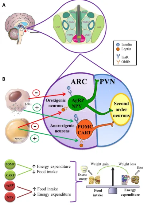
(31) Introduction. metabolic state. It is under the control of an integrated system that has the capacity to rapidly respond to metabolic changes. Energy homeostasis is mainly regulated by the central nervous system (CNS), especially by the hypothalamus that plays an important role in central regulation of appetite and body composition (Brenseke, et al. 2013). In addition, a set of peripheral tissues, such as liver, adipose tissue and skeletal muscle, has also been described as metabolic tissues with an important role in global energy balance regulation under the control of the CNS (Lee, et al. 2003; Pang and Han 2012). 2.1. Main tissues involved in maintenance of energy homoeostasis 2.1.1. Hypothalamus The hypothalamus is the portion of the brain located below the thalamus, just above the brainstem, that contains a number of small nuclei that produce neuropeptides involved in numerous physiological and behavioural functions (Figure 1A). Among them, one of the most important functions of the hypothalamus is to link the nervous system to the endocrine system via the hypophysis. This connection allows the hypothalamus to be informed about the nutritional, energetic and environmental status of the body through peripheral and central orexigenic or anorexigenic messages (Valassi, et al. 2008). Experimental efforts have been made to understand how the brain regulates energy homeostasis and how impaired brain functions contribute to the pathogenesis of obesity, diabetes and related metabolic disorders. The specific hypothalamic structures that are directly involved in energy homeostasis were identified when a set of systematic lesion experiments were performed in rats (Brobeck 1946). In several experiments, various hypothalamic nuclei, including the hypothalamic ventromedial (VMN), paraventricular (PVN) and dorsomedial (DMN) nuclei, were destroyed. The lesion of these nuclei induced hyperphagia and obesity, whereas lesions in the lateral hypothalamus (LH) led to hypophagia. These studies led to the proposal of a “dual center model” that identified the hypothalamic VMN as the “satiety center”, and the LH as the “hunger center” [reviewed by (Gao and Horvath 2008)]. The hypothalamic arcuate nucleus (ARC) is located at the base of the hypothalamus around the third ventricle and is considered a critical region in food intake and energy homeostasis regulation due to various reasons (Elmquist, et al. 1999). Firstly, their neurons, known as “firstorder neurons” have their nerve endings anatomically placed in close proximity to fenestrated capillaries at the base of the hypothalamus, giving them access to humoral signals that are restricted from other regions of the brain. Indeed, they respond rapidly to fluctuations in nutrients and metabolic hormones. Finally, they project broadly to the brain and periphery both directly as well as indirectly [reviewed by (Gao and Horvath 2008)]. ARC neurons are separated in two populations of neurons with opposite effects on feeding behaviour. In this sense, there is a family of anorexigenic neurons that co-express cocaine and amphetamine-regulated transcript (CART) and pro-opiomelanocortin (POMC), which is the precursor for many active neuropeptides including melanocyte-stimulating hormone (α-MSH). These, in turn, reduce food intake and body weight as well as increase energy expenditure in animals and humans by acting on melanocortin receptor subtypes 3 and 4 (MC3R and MC4R, respectively), found to be abundant in the ARC, PVN, LH and DMN. In contrast, there is a family of orexigenic neurons that co-express neuropeptide Y (NPY) and Agouti-related peptide (AgRP). NPY potently stimulates food intake and reduces energy expenditure. AgRP acts as a -5-.
(32) Juana María Torrens García – Doctoral Thesis. natural antagonist of MC3R and MC4R, and hence reduces the anorectic effect of α-MSH [reviewed by (Gao and Horvath 2008)].. Figure 1. A. Anatomical localization of the arcuate nucleus (ARC) and the surrounding hypothalamic nuclei: DMN, dorsomedial nucleus; LHA, lateral hypothalamic area; PVN, paraventricular nucleus and VMN, ventromedial nucleus. B. Schematic diagram of the ARC. Two populations of “first-order” neurons, with opposite effects on food intake regulation and energy expenditure, are located in the ARC. Anorexigenic neurons, which release the neuropeptides Proopiomelanocortin (POMC) and Cocaine and Amphetamine-regulated transcript (CART); and the orexigenic neurons, which release the neuropeptides Agouti-related peptide (AgRP) and the Neuropeptide Y (NPY). The axons of these neurons project to “second-order” neurons, located mainly in PVN and other hypothalamic regions. Both insulin and leptin are secreted into the bloodstream in direct proportion to body fat stores, and both interact with neurons in the ARC. Insulin and leptin activate POMC and CART neurons via their respective receptors, insulin receptors (InsR) and leptin receptor (ObRb). This results in the activation of downstream catabolic neurons which in turn reduce food intake and increase energy expenditure. Conversely, the NPY and AgRP expressing neurons are inhibited by insulin and leptin, reducing the secretion of NPY and AgRP which potently stimulate food intake and reduce energy expenditure. Adapted from (Niswender and Schwartz 2003).. The axons of ARC neurons project to “second-order” neurons located in various parts of the brain, such as PVN and LH, where there are substantial numbers of MCR3 and MCR4. The -6-.
(33) Introduction. projection from the ARC to the PVN is important for the regulation of neurons that produce the anorexigenic substances, such as corticotropic and thyrotropin releasing hormones (CRH and TRH respectively), and for the modulation of sympathetic activity, both of which are significant mechanisms in energy metabolism. The projection from the ARC to the LH is important for the production of orexant molecules, such as melanin-concentrating hormone (MCH) and orexines (Valassi, et al. 2008). Hypothalamus, and particularly the hypothalamic ARC structure, integrates the peripheral information of hormones (insulin, ghrelin and leptin) and nutrients, such as glucose, to maintain energy balance (Breton 2013; Morton, et al. 2006). Concretely, leptin and insulin act at hypothalamic level by binding to its respective receptors, the long form of leptin receptor (ObRb) and insulin receptor (InsR) respectively, which have been localized in the ARC (Baskin, et al. 1998). Both receptors are linked to the phosphatidylinositol 3-kinase (PI3K) pathway and reduce the expression and release of the orexigenic peptides (NPY and AgRP) and activate anorexigenic peptides (CART and POMC) (Figure 1B). In addition, other hypothalamic nuclei, especially the PVN, may in turn also modulate energy expenditure such as lipolysis and/or thermogenesis in adipose tissue, via the sympathetic autonomic nervous system (Fliers, et al. 2003). 2.1.2. White adipose tissue White adipose tissue (WAT) is the major body’s energy storage. The main form of energy storage is represented by high amounts of triglycerides (TGs) in adipocyte lipid droplets (Daval, et al. 2006). The stored TGs are derived from fatty acids which can be either taken up from the diet or synthesized within adipocytes by de novo synthesis from non-lipid substrates (Fliers, et al. 2003). In order to take up fatty acids from plasma, adipocytes synthetize a specific insulin-stimulated enzyme called lipoprotein lipase (LPL), which is exported to the luminal side of vascular endothelium where it can hydrolyse TG-rich lipoproteins such as chylomicrons and very low density lipoproteins (VLDLs) to yield fatty acids and free glycerol. Fatty acids enter into the adipocytes through transporters and are re-esterified with glycerol phosphate to form TGs stored in a single lipid droplet in white adipocytes (Daval, et al. 2006). In addition, glucose also provides the glycerol backbone for TG (Aguilera, et al. 2008). Fatty acids can also be de novo synthesized from glucose, however, the rate of synthesis of fatty acids from glucose in humans is lower than in rodents (Aguilera, et al. 2008). When food is available, via lipogenic pathways, adipocytes store excess of energy as TGs to guarantee the survival in periods of increased energy expenditure or decreased energy availability. Conversely, when energy is needed, TGs are hydrolysed into fatty acids and glycerol, which are exported back into the blood, via activation of lipolytic pathways mainly driven by noradrenergic innervation (Breton, 2013). The key enzymes of norepinephrine-stimulated lipolysis are adipose tissue triglyceride lipase (ATGL), which is the main protein involved in the catalysis of the initial step in TG hydrolysis in adipocyte lipid droplets, and hormone-sensitive lipase (HSL), which hydrolyses stored TGs to free fatty acids as well as several other lipids stored in WAT. Particularly, HSL is regulated by numerous factors and hormones through several mechanisms, including reversible phosphorylation via noradrenergic innervation. This phosphorylation stimulates adenylate cyclase and increases intracellular levels of cyclic adenosine monophosphate (cAMP). Then, cAMP activates protein kinase A (PKA) which in turn phosphorylates and activates HSL. Although ATGL works in conjunction with HSL, its activity is not dependent on PKA phosphorylation (Daval, et al. 2006).. -7-.
(34) Juana María Torrens García – Doctoral Thesis. In addition to its functions related to energy storage and release, WAT plays a key role as an endocrine organ controlling energy homeostasis and substrate partitioning (Daval, et al. 2006). WAT is involved in steroid hormone metabolism and glucocorticoid metabolism. Moreover, WAT produces adipocytokines, such as leptin and adiponectin; other hormones, like resistin; appetite-regulating related peptides, and proteins from the renin-angiotensin system. Among all of them, resistin has been linked with insulin resistance induction while adiponectin has been associated with insulin sensitivity enhancement (Fliers, et al. 2003). Some of these circulating factors act in an autocrine/paracrine manner as adipocyte lipid metabolism regulators, while some of them act as peripheral endocrine signals to regulate energy homeostasis at hypothalamic level, as leptin (Breton 2013). Finally, adipose tissue also secretes other proteins that may be of interest in view of the association between obesity and cardiovascular risk inflammatory. Among them, there are cytokines such as interleukin-6 (IL-6) and tumor necrosis factor alpha (TNFα), and several coagulation and complement factors, such as plasminogen activator inhibitor-1 (the key modulator of the fibrinolytic pathway), tissue factor (the most powerful activator of the coagulation system) and fibrinogen-angiopoietin-related protein (a regulator of angiogenesis and modulator of tumorigenesis) (Fliers, et al. 2003; Trayhurn and Beattie 2001). Concerning, IL-6 and TNFα, these cytokines are involved in systemic inflammation and also appears to participate in the induction and maintenance of the subacute inflammatory state associated with obesity. Moreover, TNFα is overproduced in fatty liver and participates in the development of insulin resistance (Shoelson, et al. 2006) and IL-6 can also favour insulin resistance in insulin sensitive tissues. 2.1.3. Liver Liver is recognized as a metabolic, endocrine and exocrine organ that is responsible for many vital functions, including formation of bile salts, excretion of bilirubin and cholesterol, blood detoxification and purification, immunological processes and production of critical coagulation factors, serum proteins and hormones. In addition, due to the fact that all the blood coming from the digestive system passes through the hepatic portal vein, liver also plays an important role in storage of energy and nutrients obtained from blood, as well as in the metabolism of lipids, proteins and carbohydrates. In this sense, alterations in the ability of the liver to properly regulate lipid metabolism and glucose homeostasis are the heart of the deleterious consequences of obesity and insulin resistance (Brock and Dorman 2007). Regarding lipid metabolism and transport, liver is considered the most dynamic organ of the body and the key site of metabolic integration (Brock and Dorman 2007). Liver is able to acquire fat and cholesterol from the diet. Concretely, dietary TGs and cholesterol are transported from the intestine in the form of chylomicrons. Rapidly, the TGs in the lipid core of the chylomicrons are hydrolysed at the capillary surface by the enzyme LPL, which yields smaller particles known as chylomicrons remnants. The released free fatty acids are used either for storage in adipose tissue or for oxidation in other tissues, while dietary cholesterol is transported in the chylomicrons remnants to liver (Gotto 1990; Sandhofer 1994). Cholesterol and TG are also synthesized in the liver and then secreted into the blood in the form of VLDL. TGs from VLDL particles are also hydrolysed by the enzyme LPL, to yield intermediate density lipoproteins (IDL) which are either taken up by the liver or further catabolized to lowdensity lipoprotein (LDL). These LDL are bound and taken up by specific receptors in the liver and many other tissues; by this pathway, cholesterol is transported from the liver to peripheral tissues (Sandhofer 1994). Moreover, when hepatic glycogen stores are in a state of excess, liver -8-.
(35) Introduction. synthetises de novo fatty acids by deviation of acetyl-CoA to the fatty acid synthase complex (FASC), located within the cytoplasm of hepatocytes. Hepatic fatty acids are stored in the form of TGs, by esterification of acetyl-CoA with glycerol-3-phosphate. Once formed, these TGs are packaged to form VLDL, which are transported out of the liver and back to peripheral adipocytes. In general, there is an equilibrium of fatty acids between the peripheral adipocytes and the liver without any appreciable accumulation of lipids [reviewed by (Brock and Dorman 2007)]. Furthermore, liver influences strategically glucose homeostasis through a delicate balance between hepatic glucose production through glycogenolysis and gluconeogenesis during the fasted state, and hepatic glucose uptake, storage and utilization in the fed state. Transitions between the two states in liver are possible due to the joined physiological action of insulin and glucose promoting the expression of genes normally induced during the fed state and downregulating the expression of genes normally activated during the fasted state (Collier and Scott 2004). On the one hand, representative genes expressed during the fed state are: glucokinase (Gck), liver pyruvate kinase (Pkl), 6-phosphofructo-2-kinase/fructose-2,6-biphosphatase 2 (Pfkfb2), acetyl-coenzyme A carboxylase (Acc), and fatty acid synthase (Fasn). On the other hand, the representative genes that are activated during the fasted state are: phosphoenolpyruvate carboxykinase (Pck1), fructose-1,6-bisphosphatase (Fbp1), and carnitine palmitoyl transferase 1 and 2 (Cpt1 and Cpt2) [reviewed by (Collier and Scott 2004)]. The transcription factor, peroxisome proliferator-activated receptor alpha (PPARα) plays an important role in liver regulating directly the expression of genes involved in fatty acid uptake, such as the gene encoding the fatty acid transport protein (Fatp) and fatty acid binding protein (Fabp). In addition, PPARα also regulates the genes involved in plasma VLDL TG hydrolysis, such as Lpl, and also β-oxidation-related genes, such as Cpt1a and acyl-CoA oxidase 1 (Acox1) (Lee, et al. 2003; Martin, et al. 2009; McIntosh, et al. 2013). While in the fed state the fuel source are carbohydrates and fats, in the fasted state, the main fuel source shift to fats. Fatty acids that were stored during feeding are released from the adipocyte and taken up by the liver. There, they are either reestirified to TGs and assembled into VLDL or broken down through mitochondrial hepatic fatty acid oxidation (FAO) and used to generate ketone bodies (Lee, et al. 2003). Mitochondrial FAO represents a crucial process in energy metabolism. Hepatic FAO is tightly regulated by interaction between the key enzyme CPT1A and the ACC via intermediate malonyl-CoA (McGarry, et al. 1977). Malonyl-CoA, derived from glucose metabolism, is the first intermediate in lipogenesis and regulates FAO by inhibiting CPT1A. CPT1A is localized in the outer mitochondrial membrane and expose its active site at the cytosolic face of the mitochondria (Schreurs, et al. 2010). CPT1A regulates the transfer of long-chain acyl-CoAs from the cytosol into the mitochondria, where they are oxidized. Considering this and taking into account that obesity results from a chronic imbalance between energy intake and energy expenditure, which leads to fat accumulation in lipid deposits, strategies able to tilt the energy balance towards FAO could be considered as potential ways to treat or prevent obesity and related metabolic disorders. In this regard, genetic studies that increased hepatic FAO have been already developed. On the one hand, in vitro studies showed that hepatocytes transduced with adeno-associated viruses (AAV) encoding carnitine palmitoyltransferase 1a (CPT1a) increased the rate of β-oxidation that, in turn, led a decrease in TG content in liver (StefanovicRacic, et al. 2008). In other studies, the gene of Cpt1a was transferred into obese mice by injecting AAV vectors into the tail vein. This led to a nonimmunoreactive long-term increase in lipid oxidation. Concretely, Orellana-Gavaldà et al. used a mutant but permanently active form of CPT1A, the CPT1AM, which is insensitive to malonyl-CoA and therefore leads to a -9-.
(36) Juana María Torrens García – Doctoral Thesis. permanent increase in the rate of FAO, independently of the glucose-derived malonyl-CoA levels. The results of this study showed than an increased hepatic FAO through AAV-mediated gene transference of Cpt1a or Cpt1am reduced obesity-induced hepatic steatosis, weigh gain, inflammation, diabetes, glucose levels and insulin resistance in mice consuming a high-fat (HF) diet (Orellana-Gavaldà, et al. 2011). 2.1.4. Skeletal muscle Skeletal muscle is a major mass peripheral tissue that accounts for approximately 40% of the total body mass. Skeletal muscle serves for overlapping functions, such as movement, posture, stability, communication, heat production, and cold tolerance (Smith and Muscat 2005). As the motor of the body, muscle requires considerable amounts of energy in the form of ATP. Muscle can burn energy faster than can be produced within the cell necessitating a buffer system that uses creatine kinase (CKM) to transfer a high-energy phosphate from phosphocreatine stores to ADP to form ATP. For short bursts of activity, skeletal muscle relies upon glycolysis for ATP production, which is the primary form of energy obtaining (Smith, et al. 2013). In addition to these functions, skeletal muscle is being recognized also as an endocrine organ because expresses and releases into the circulation several cytokines that exert their effect in other parts of the body (Tomas, et al. 2004). These cytokines have been coined “myokines” and are for example, IL-6 and IL-15 (Smith and Muscat 2005). Concretely, IL-6 is able to sense the energy demands in muscle and signal to central and peripheral organs to maintain energy supply. In this sense, sustained and intense exercise induces skeletal muscle to express and release IL-6 into the circulation, especially when glucose and glycogen stores are low. Then, IL-6 stimulates 5' adenosine monophosphate-activated protein kinase (AMPK) activity which activates lipolysis in adipose tissue, inhibits TNFα, and also improves insulin sensitivity [reviewed by (Smith and Muscat 2005)]. Skeletal muscle is also a metabolic flexible tissue and has an essential role in energy balance because is the primary tissue of insulin stimulated glucose uptake, disposal, and storage in form of glycogen, even four-fold the glycogen content of liver. Moreover, skeletal muscle regulates cholesterol efflux and highly influences metabolism via modulation of circulating and stored lipid flux (Smith and Muscat 2006). Fatty acids and glucose are the main energy sources in muscle. Particularly, during initial aerobic exercise, skeletal muscle uses as energy fuel the stored muscle glycogen which is broken down back into glucose via the enzyme glycogen phosphorylase; then, glucose is broken down into pyruvate during glycolysis providing the required energy in the form of ATP. If exercise continues, then, glucose from glycogen and stored muscle TGs become important energy substrates, and muscle must utilize the more efficient oxidative phosphorylation process. In this regard, pyruvate conversion to acetyl-CoA by pyruvate dehydrogenase allows progression through the tricarboxylic acid (TCA) cycle and subsequent oxidative phosphorylation to obtain energy. Concerning stored muscle TGs, the enzyme HLS is responsible for triglyceride breakdown to free fatty acids in muscle; the released free fatty acids are then transported into the mitochondria to undergo β-oxidation and produce acetyl-CoA and NADH (Smith, et al. 2013). If exercise is prolonged over long periods, then fatty acids and lipid mobilization from other tissues are increasingly needed (Smith and Muscat 2005). Therefore, given the fact that skeletal muscle can utilize both carbohydrate and lipid energy substrates and considering the relative mass of the tissue, it is hardly surprising that normal or. - 10 -.
Figure

+7


Documento similar
Correlation analysis between the gene expression of the inflammatory-related NFKB1 and triglyceride levels in the postprandial state at 4 h after 8 weeks of dietary intervention with
Indeed, the variations in all traits related to burrow construction and soil and dung manipulation play a key role in the most important effects of dung beetles on
Effects of Active Video Games on Health-Related Physical Fitness and Motor Competence in Children and Adolescents With Overweight or Obesity: Systematic Review and Meta-Analysis.
For a short explanation of why the committee made these recommendations and how they might affect practice, see the rationale and impact section on identifying children and young
Sarcopenic obesity: prevalence and association with metabolic syndrome in the Korean Longitudinal Study on Health and Aging (KLoSHA). Is central obesity associated with
Herein, we have pur- sued the identification of potential biomarkers in plasma samples and skin biopsies that could define the phenotype of CMT1A patients at mild (Mi), moderate
In this regard, previous studies identified that RBP1 (retinol binding protein 1) was amongst the genes silenced by promoter hypermethylation not only in TCA cycle-mutated
The present review discusses the key findings related to the involvement of TLRs and NLRs in the progression of several vascular and cardiac diseases, with a focus on whether some
