PAPER • OPEN ACCESS
Pulsed x-rays dose measurements from a hundred
joules plasma focus device
To cite this article: J. Jain et al 2018 J. Phys.: Conf. Ser. 1043 012048
View the article online for updates and enhancements.
Related content
In vitro irradiation of colorectal cancer cells by pulsed radiation emitted from a hundred joules plasma focus device and its comparison with continuous irradiation
J. Jain, J. Moreno, R. Andaur et al.
-Potentiality of a small and fast dense plasma focus as hard x-ray source for radiographic applications
Cristian Pavez, José Pedreros, Marcelo Zambra et al.
-the influence of a novel transmission detector on 6 MV x-ray beam characteristics
Sankar Venkataraman, Kyle E Malkoske, Martin Jensen et al.
Pulsed x-rays dose measurements from a hundred joules
plasma focus device.
J. Jain1,2,3,*, J. Moreno1,2,4, R. E. Avila2, G. Avaria1,2,4, C. Pavez1,2,4, B. Bora1,2,4, S.
Davis1,2,4 and L. Soto1,2,4
1Comisión Chilena de Energía Nuclear, Casilla 188-D, Santiago Chile 2
Center for Research and Applications in Plasma Physics and Pulsed Power, P4 3Universidad de Talca, Chile
4
Universidad Andres Bello, Departamento de Ciencias Físicas, República 220, Santiago, Chile
*E-mail: [email protected], [email protected]
Abstract. Present work is aimed to perform dosimetric measurements to characterize dosis obtained from pulsed x-rays emitted from a hundred joules plasma focus device PF-400J using thermoluminescent dosimeters (TLD-100). Two dosimeter arrays (containing 21 dosimeters in each) were used. One of the arrays was kept inside the PF-400J vacuum chamber and other outside the vacuum chamber, simultaneously. It was found that dosis obtained from the inside array (~200.7 mGy) were hundred times larger than the outside array (~1.1 mGy) for hundred pulses of x-rays. Later, the vacuum window of PF-400J, which was made of 1 mm aluminum, was replaced by a plastic window and a similar dosimeter array was kept outside the chamber over the plastic window. With this arrangement, the obtained doses (100 pulses of x-rays) were of the same order of magnitude (~106 mGy) as it was inside the vacuum chamber. Later, a lead piece was inserted inside the hollow anode of PF-400J, which increased dose (~250 mGy) per hundred pulses of x-ray outside the vacuum chamber using plastic vacuum window. Our results suggest that PF-400J could be a useful device to study low dose pulsed radiation effects on cancer cell lines in in vitro experiments.
1. Introduction
2
voltage, at first discharge takes place over the insulator and forms a plasma current sheet (PCS), which develops a current density between anode and cathode. Later, due to self-generated magnetic field the PCS expands and runs over the effective length of the anode under the action of Lorentz force. At the open end of the anode, the PCS compresses neutral gas and forms a plasma column, typically known as pinch. During compression and pinch phases, electromagnetic forces are induced that accelerate charged particles. Electrons impinge the anode and produce x-rays via bremsstrahlung. Plasma dynamics and radiation emission under the framework of PF devices can be found in [20].
PF devices emit radiation in pulsed form, which can be used to study the effects of pulsed radiation on cancer cells in in vitro experiments, which has importance to study post-irradiation effects on cells at cellular level. Indeed, pulsed low-dose-rate radiation therapy is used to treat the recurrent cancer [21]. Cancer research has advanced a lot from technology point of view but at cellular level, it requires more studies. For instance, studies about the distinct effects of pulsed and continuous radiation on cells. In order to use pulsed radiation (x-rays) emitted from PF devices for cancer cells irradiation, it is mandatory to characterize doses. S. Zapryanov et al [15]used a 3 kJ PF device in order to irradiate live microorganisms by soft x-rays. Dose measurement was performed using thermoluminescent detectors (TLD). Inside the PF chamber at 15 cm from the plasma column, dosis were ~11 mSv for 4 shots using 20 m Al foil and ~ 65 mSv for 14 shots using 100 m Al foil. In addition, it was mentioned that the total energy released in hard x-rays region is lower than the total energy released in soft x-rays region.
Pavez et al [22]used a hundred joules PF device, PF-400J, in order to study x-rays emission and equivalent dosis. Dose measurements were performed using TLDs (TLD-100). Based on the observations, dose per shot of the order of 17 Sv around the symmetry axis was reported. In the present work, TLD-100 dosimeters are used in order to measure the doses inside and outside the PF-400J [3, 22-25] simultaneously. These measurements were performed keeping in mind the use of pulsed x-rays to irradiate cancer cells in in vitro experiments.
In section 2 experimental setup for dose measurements is presented. The results are presented and discussed in section 3. Work is concluded in section 4.
2. Experimental setup
In all the experiments, annealed dosimeters were used prior to use them for pulsed x-rays dose measurement emitted from PF-400J. Annealing is useful to re-configure the internal structure of the dosimeters. Sensitivity of TLD-100 dosimeters used in these experiments showed a 5% standard deviation. To reduce this effect, 100 dosimeters were uniformly irradiated by a calibrated x-ray source, and 60 of them were selected, with deviation less than 3% from the common average. Further, the response from that irradiation was used for the individual calibration of the selected dosimeters, so, later, their signals from exposure to PF-400J radiation was scaled with that calibration factor.
3. Results and discussion
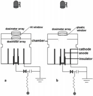
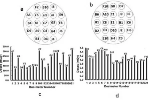
Figures 2(a), 2(b), 2(c), and 2(d) show schematic of the dosimeter arrays and accumulated dosis obtained from 100 pulses of x-rays inside (figures (2a), (2c)) and outside (figures (2b), 2(d)) of PF-400J vacuum chamber, respectively. The average dose in 100 x-ray pulses was found ~ 200.7 ± 72.69 mGy and 1.1 ± 0.23 mGy, inside and outside the vacuum chamber respectively. In addition, relatively higher dose zones inside the chamber were identified. In figures (2a) and (2c) it can be seen that the central dosimeters (F5, H5, J8, G5, C7, I4, B9, C4, and G4), shown by italic font style, acquire relatively higher doses.
4
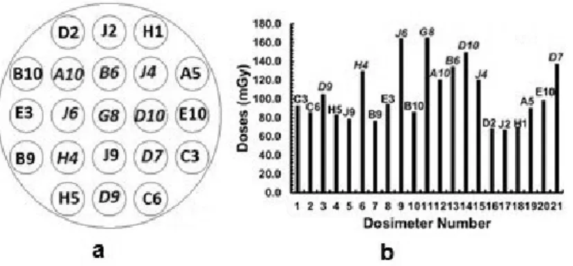
These results suggest that most of the x-rays would have been attenuated during their passage through aluminum window. Keeping in mind this observation, a vacuum window made of petri dish material, that is plastic of width 1.2 mm was used, please see figure 1(b). A dosimeter array (as shown in figure 2) was kept over this window. Figures 3(a) and 3(b) show the schematic arrangement of dosimeter arrays and obtained results, respectively with this arrangement. In this case, average dose (100 pulses of x-rays) outside the chamber was 106 ± 31.18 mGy, which has same order of magnitude as was inside the chamber. From figures 2(a) and 2(b) it can be seen that the central dosimeters (A10, B6, J4, J6, G8, D10, H4, and D7), shown by italic font, acquire relatively higher dosis. This pattern is similar as found inside the chamber, as mentioned earlier.
A lead piece was inserted inside the hollow anode of PF-400J to measure the dosis outside the vacuum chamber. In this case, 100 pulses of the x-rays provide dosis ~ 250 mGy. Please note the plastic window was used for this experiment. Insertion of lead increases dose per hundred x-ray pulses that are almost two times than without insertion of lead.
In the first experiment, dosis outside the vacuum chamber were found hundred times smaller than inside the chamber. Please note, the dosimeter array that was kept outside had various obstacles in the path of x-rays to reach it. X-rays first interact with 15 m aluminum foil then plastic of petri dish (~1.2 mm) which contains the dosimeter array inside the chamber. Later, x-rays will interact with the upper cap of petri dish, made of plastic that was kept inside the chamber and after with 1 mm Al window, over which the dosimeter array was kept outside the chamber. It has been reported that the emission of low energy x-rays is higher than the higher energy x-ray in PF devices [13]. Since transmission percentage of rays below 15 keV through 1 mm Al window is less than 10%, therefore low energy x-rays (say below 15 keV) are strongly attenuated by those various materials. Hence, much higher dosis inside the chamber, two order of magnitude higher than outside the chamber. These observations are consistent with the concept that low energy x-ray emission dominates in PF discharges [13].
It was observed that the use of plastic vacuum window provides similar dosis outside the vacuum chamber of PF-400J as was inside, see figure 3. Nonetheless, the dosis in this configuration were almost half the dosis inside the chamber. The decrement in dosis with the use of plastic window might be due the fact that x-rays have to pass through 1.2 mm window material and later through the box in which the dosimeter array was arranged. X-rays will be attenuated while passing through these obstacles.
Increment in dose per hundred x-ray pulses was observed with the insertion of a lead piece inside the hollow anode. Prior to discuss the effects of lead insertion inside the hollow anode on x-rays emission, let us first discuss how x-rays produce in PF devices. During compression and pinch phases of PF devices, there is generation of various kinds of instabilities and electromagnetic fields. Charged particles accelerate in the presence of the induced electromagnetic fields in various directions. Electrons accelerate toward the central anode and produce x-rays via bremsstrahlung upon impinging the anode. Insertion of lead may have following cause that could affect the doses. Interaction of electron beams with lead target can sputter lead material and introduce impurities in pinch phase. These impurities increase the resistivity of the pinch that further allow the induced electromagnetic fields to penetrate inside the pinch. This penetration of electric fields will allow relatively large number of electrons within the pinch to accelerate and produce x-rays via bremsstrahlung. In addition, these impurities themselves may work as target. Nonetheless, in the present work only an experimental observation that insertion of lead inside the hollow anode increases dose per hundred pulses of x-ray is presented. More computational and experimental efforts are needed in order to explain x-rays emission from PF-400J.
6 4. Conclusion
Two experiments were carried out in order to measure the dosis inside and outside the vacuum chamber of plasma focus device PF-400J, simultaneously (first experiment). In this case, average dosis were ~ 200.7 mGy and 1.1 mGy inside and outside the vacuum chamber respectively. Note that, in the first experiment the vacuum window over which the dosimeter array was kept outside the chamber was made of aluminum of thickness 1mm. Based on these observations, it was realized that most of the x-rays would have been attenuated while passing through aluminum window. In the second experiment, a vacuum window that was made of plastic was used in order to have similar dosis outside the vacuum chamber as was inside. In this case, the dosis outside the chamber were ~106 mGy. Later, insertion of a lead piece inside the hollow anode increased dose per hundred x-ray pulses that were ~ 250 mGy, outside the PF-400J vacuum chamber while using plastic vacuum window. These findings suggests that PF-400J could be a useful device for in vitro experiments to study the effects of low dose pulsed radiation on cancer cells.
Acknowledgement
The work is supported by grant ACT-1115, CONICYT, Chile.
References
[1] Castillo F, Milanese M, Moroso R and Pouzo J 2000 Journal of Physics D: Applied Physics30 1499-1997 [2] Michel L, Schonbach K H and Fischer H 1974 Applied Physics Letter 24 57
[3] Silva P, Moreno J, Soto L, Birtein L, Mayer R, Mayer E and Kies W 2003 Applied Physics Letter83 3269 [4] Wang X, Han M, Wang Z and Kun L 1999 China Technological Sciences42 83-87
[5] Verma R, Rawat S R, Paul L; Augustine T L T, Shariff H, Ying G J, Springham S V, Talebitaher A, Ilyas U and Shyam A 2012 IEEE Transactions on Plasma Science40 3280 – 3289
[6] Castillo F, Herrera J J E, Rangel J, Milanese M, Moroso R, Pouzo J, Golzarri J I and Espinosa G 2003 Plasma Physics and Control Fusion45 289
[7] Zambra M, Moreno J, Silva P, Soto L, Sylvester G and Pavez C 2008 Journal of Physics: Conference Series134 012047
[8] Kato Y and Be S H 1986 Applied Physics Letter48 686
[9] Beg F N, Ross I, Lorenz A, Worley J F, Dangor A E and Haines M G 2000 Journal of Applied Physics88 3225
[10] Moreno C, Vénere M, Barbuzza R, Del Fresno M, Ramos R, Bruzzone H, González Florido P J and Clausse A 2002 Brazilian Journal of Physics32 20-25
[11] Hussain S, Ahmad S, Khan M Z, Zakullah M and Waheed A, 2003 Journal of Fusion Energy22 195-200 [12] Gemishev O, Zapryanov S, Blagoev A, Markov M and Savov V 2014 Biotechnology & Biotechnological
Equipment28 850-854
[13] Dubrovsky V, Gazaryan I, Gribkov G V, Ivanove A, Yu P, Kost O A, Orlova M A and Troshina N N 2003 Journal of Russian Laser Research24 289-300
[14] Virelli A, Zironi I, Pasi F, Ceccolini E, Nano R, Facoetti A, Gavoc E, Fiore M R, Rocchie, F, Mostacci D, Cucchi G, Castellani G, Sumini M and Orecchia R 2015 Radiation Protection Dosimetry166 1–5 [15] Zapryanov S, Goltsev V, Galutsov B, Gelev M and Blagoev A 2012 European Physical Journal of Applied
Physics58 11201
[16] Lee S, Lee P, Zhang G, Feng X, Gribkov V A, Liu M, Serban A and Wong T K S 1998 IEEE Transactions on Plasma Science26 1119-1126
[17] Gribkov V A, Srivastava A, Keat P L C, Kudrryashov V and Lee S 2002 IEEE Transactions on Plasma Science30 1331-1338
[18] Inestrosa-Izurieta M J, Ramos-Moore E and Soto L 2015 Nuclear Fusion55 093011
[19] Bernard A, Bruzzone H, Choi P, Chuaqui H, Gribkov V, Herrera J, Hirano K, Krejci A, Lee S, Luo C, Mezzetti F, Sadowski M, Schmidt H, Ware K, Wong C S and Zoita V 1998 J. Moscow Phys. Soc.8 93 – 170
[20] Kang S, Lang J, Wang P, Li J, Lin M, Chen X, Guo M, Chen F, Chen L and Ming Ma C 2014 Journal of Applied Clinical Medical Physics15 102 – 113
Physics and Control Fusion54 105018 (9pp)
[22] Silva P, Moreno J, Pavez C, Soto L and Arancibia J 2006 AIP Conf. Proc875 442
[23] Jain J, Moreno J, Pavez C, Bora B, Inestrosa-Izurieta M J, Avaria G and Soto L 2016 Journal of Physics: Conference Series720 012042
[24] Jain J, Moreno J, Avaria G, Pavez C, Bora B, Inestrosa-Izurieta M J, Diez D, Alvarez O, Tapia J, Marcelain K, Armisen R and Soto L 2016 Journal of Physics: Conference Series720 012043
[25] Soto L, Pavez C, Moreno, J, Inestrosa-Izurieta, M J, Veloso F, Gutierrez G, Vergara J, Clausse A, Bruzzone H, Castillo F and F Delgado-Aparicio L 2014 Physics of Plasmas21 122703
[26] Tarifeño A, Pavez C, Moreno J and Soto L 2011 IEEE Trans. Plasma Science39 756 [27] P. Silva, J. Moreno, L. Soto, L. Birstein, R. Mayer, W. Kies 2003 Applied Physics Letters83
3269
[28] Soto L, Silva P, Moreno J, Zambra M, Kies W, Mayer R E, Clausse A, Altamirano L, Pavez C, and Huerta L 2008 J. Phys. D: App. Phys. 41 205215