Wnt 5a Modulates Recycling of Functional GABA(A) Receptors on Hippocampal Neurons
10
0
0
Texto completo
(2) 8412 • J. Neurosci., June 23, 2010 • 30(25):8411– 8420. choline receptor (␣7-nAChR) clusters (Farías et al., 2007). Interestingly, ␣7-nAChR is a member of the ligand-gated ion-channel superfamily that also comprises GABAA-Rs. Owing to the fact that the cellular mechanisms that neurons use to regulate GABAA-Rs cell surface stability and activity has been of considerable interest, we focused on studying the role of the Wnt pathway on the modulation of inhibitory synapses, particularly in at the clustering and surface expression of GABAA-Rs.. Materials and Methods Materials Culture media, 2-amino-5-phosphonovaleric acid (APV), 6-cyano-7nitroquinoxaline-2,3-dione (CNQX), tetrodotoxin (TTX), insulin, antiGABAA (␥2 subunit receptor) and fumagillin from Sigma; anti ␣1 subunits of GABAA-Rs, anti-synapsin-1 and anti-Wnt-5a (Santa Cruz Biotechnology Inc), mouse anti-PSD-95 (UC Davis/NINDS/NIMH, CA); Foxy-5 was obtained from Genemed Synthesis; Lithium, BDNF, 3,3⬘,5,5⬘-tetramethylbenzidine (TMB), Immuno pure ABC Peroxidase Staining Kit (Pierce). The dynamin blocking P4 peptide was purchased from Tocris Bioscience. KN-93, TAT-TI-JIP153-163 and Gö6976 (12-(2cyanoethyl)-6,7,12,13-tetrahydro-13-methyl-5-oxo-5H-indolo(2,3-a) pyrrolo(3,4-c)-carbazole) were obtained from Calbiochem.. Hippocampal neuronal cultures Hippocampal neurons were obtained from 18-d-old Sprague Dawley rat embryos. Hippocampi were aseptically dissected and trypsinized for 20 min. After centrifugation for 1 min, cells were seeded in phenol-red-free DMEM plus 10% horse serum into 1% poly-L-lysine-coated plates. After 120 min, the medium was removed and Neurobasal medium was added containing 1% B27 supplement from Invitrogen. On day 3 of culture, hippocampal neurons were treated with 2 M 1--D-arabinofuranosylcytosine (AraC) for 24 h. Fifteen- to 18-d-old neuron cultures were used for various experiments; the average number of neurons in each experiment was ⬃95% of the cells present in the cultures (Alvarez et al., 2004; Farías et al., 2007, 2009).. Conditioned medium containing Wnt ligand To generate secreted Wnt ligand, HEK-293 cells were stably transfected by Lipofectamine 2000 (Invitrogen) according to manufacturer’s instructions with constant and equal amounts of empty vector pcDNA or pcDNA containing sequences encoding Wnt-ligand or soluble Frizzled receptor protein (sFRP-1) coupled to the sequence encoding a hemagglutinin (HA) tag. Wnt-7a was a gift from Dr. Patricia Salinas (University College London, UK), Wnt-5a was a gift from Randall T. Moon (University of Washington, Seattle, WA), Wnt-3a a gift from Dr. Roel Nusse (Stanford University, Palo Alto, CA) and sFRP was a gift from Dr. Jeremy Nathans (John Hopkins University School of Medicine, Baltimore, MD). For Wnt-conditioned, control media or media containing sFRP transfected HEK-293 cells were grown to 85% confluence and maintained in Neurobasal medium without supplements by 60 h. Wnt secretion was verified by Western blot using a HA-specific antibody (Millipore). For the electrophysiological studies the media containing Wnt ligands was dialyzed against ACSF for 16 h at 4°C.. Quantification of cell surface GABAA-Rs by a colorimetric ELISA assay (Wang et al., 2003) Briefly, hippocampal neurons were washed with PBS and starved with Neurobasal without supplement. At the end of each experiment, the cells were fixed with 4% paraformaldehyde in PBS for 3 min at room temperature. The fixation was then neutralized by incubation with 1% glycine at 4°C for 10 min. To determine the total amount of GABAA-Rs proteins, the cells were permeabilized by incubation with 0.2% Triton X-100. The wells were blocked with 3% BSA at 4°C for at least 3 h. A primary antibody for GABAA-Rs ␣1 (Santa Cruz Biotechnology Inc.) or ␥2 (Sigma) subunits was then added to the cultures at a dilution of 1:250 and maintained at 4°C for 12 h. The cells were extensively washed and the antibody was detected using the Ultra-Sensitive ABC Peroxidase Staining Kits (Pierce). The colorimetric reaction using TMB substrate was measured at. Cuitino et al. • Wnt-5a Modulates GABAA-R Recycling. 450 nm or stopped by addition of 0.25 ml of 3 N HCl for 10 min at room temperature.. Quantification of cell surface GABAA-Rs by biotinylation assay (Cuitino et al., 2005) Briefly, hippocampal neurons were biotinylated with sulfo-NHS-LCbiotin (Pierce) to a final concentration of 0.5 mg/ml for 45 min at 4°C. After biotinylation steps, the free biotin was quenched by incubation with 50 mM NH4Cl for 10 min. Cells were lysed in ice-cold SA buffer (150 mM NaCl, 20 mM Tris pH 8.0, 5 mM EDTA, 1% Triton X-100, 0.2% BSA and protease inhibitors). Nuclear and cellular debris was removed by centrifugation at 14,000 ⫻ g for 5 min at 4°C and the biotinylated cellsurface proteins were then adsorbed to streptavidin agarose beads for 16 h at 4°C. Beads were washed and the bound proteins were analyzed by SDS-PAGE followed by immunoblotting. The values for biotinylated cargo proteins were normalized to total cargo proteins expressed in the cells.. Image analysis and quantification Hippocampal neurons were cultivated at a density 30,000 cells/coverslip. For cell surface GABAA-Rs staining, cells were fixed with 4% paraformaldehyde/4% sucrose for 20 min at room temperature. Fixed cells were washed and incubated with an antibody to ␣1 or ␥2 GABAA-Rs subunit (Santa Cruz Biotechnology Inc. and Sigma, respectively). For the staining of other proteins, cells were fixed as described above and permeabilized by incubation in PBS-0.2% Triton X-100, and stained with the following antibodies: synapsin I (Santa Cruz Biotechnology Inc.) and PSD-95 (UC Davis/NINDS/NIMH). Finally, cells were incubated with Alexa 543, Alexa 488, and/or Alexa 633 (Pierce) for 30 min at 37°C. To determine which Wnt signaling was involved in the increase of GABAA-Rs on the cell surface, the hippocampal neurons were preincubated with fumagillin (50 nM) for 16 h. Then, KN-93 (10 M), Gö6976 (200 nM) and TAT-TIJIP (1 M) were coincubated with Foxy-5 for different times. To analyze receptor clustering, we quantified the number of clusters per neurite length with ImageJ program [National Institutes of Health (NIH), Bethesda, MD]. Neurons on coverslips were imaged using a confocal microscope LSM 5 Pascal with a 63⫻/1.4 numerical aperture oil-immersion objective. Images used for quantification were taken with identical microscope settings and analyzed using ImageJ software (NIH). GABAA-Rs images from 10 microscope fields for each condition, of three independent experiments, were registered. Each field containing processes for 1 neuron were studied, in which 3 neurites per neuron were selected using the phalloidin staining to label neuronal processes. To quantify GABAA-R clusters, images of individual neurites were isolated, background for neurite free fields were subtracted and adjusted to the threshold. GABAA-R cluster number and size were obtained with the Particle Analysis tool using a size particle limit of 0.05–1 m 2. Cluster number was normalized against neurite length to obtain cluster density.. Slice preparation and electrophysiological analysis Procedures for animal care, surgery, and slice preparation were in accordance with the guidelines for the care and use of laboratory animals adopted by the Society for Neuroscience. The procedures will be described briefly because they have been extensively detailed previously (Fuenzalida et al., 2007). Slice preparation. Young Wistar rats (15–20 d of age) were decapitated, and the brain was removed and submerged in cold (⬃4°C) artificial CSF (ACSF; in mM: 124.00 NaCl, 2.69 KCl, 1.25 KH2PO4, 2 Mg2SO4, 26 NaHCO3, 2.50 CaCl2 and 10.00 glucose). The pH was stabilized at 7.4 by bubbling the ACSF with carbogen (95% O2, 5% CO2). Transverse hippocampal slices (300 –350 m thick) were cut with a Vibroslice microtome (VSL, WPI, Sarasota, FL) and incubated in ACSF for ⬎1 h at room temperature and incubated in the ACSF (⬃1 h, at room temperature, 20 –22°C). Slices were transferred to a 2 ml chamber fixed to binocular stereo microscope (MSZ-10, Nikon). Slices were superfused with carbogen-bubbled ACSF (2 ml/min) and maintained at room temperature. All recordings were made under CNQX (20 M) and APV (50 M) (Sigma) were added to ACSF perfusion media to suppress excitatory ␣-amino-3-hydroxy-5-methyl-4-isoxazolepropionic acid receptor (AMPA) and N-methyl D-aspartate receptor (NMDAR)-transmission..
(3) Cuitino et al. • Wnt-5a Modulates GABAA-R Recycling. Stimulation, recording, and analysis. Single cell recordings were made in the whole-cell configuration with fire-polished pipettes (3–5 M⍀) filled with intracellular solution (see below), connected to an EPC-7 patch-clamp amplifier (Heka Instruments), filtered at 3.0 kHz, sampled at 4.0 kHz using an A/D converter (ITC-16, InstruTech), and stored with Pulse FIT software (Heka Instruments). Single-electrode voltage-clamp recordings were obtained from pyramidal neurons of CA1. In the voltage-clamp configuration, the series resistance was compensated to ⬃70%, and neurons were accepted only when the seal resistance was ⬃1G⍀ and the series resistance (7–14 M⍀) did not change ⬃10% during the experiment. The intracellular solution contained (in mM): 97.5 K-gluconate, 32.5 KCl, 10.0 HEPES, 1 MgCl2, 5 EGTA, and 4 sodium salt (Na-ATP), pH 7.2. Experiments started after a 5–10 min stabilization period following entry into the intracellular compartment with patch electrodes. The voltage-clamp recordings were rejected when the access resistance (7–15 M⍀) increased 20% during the experiment. The spontaneous or mini IPSPs, sIPSC and mIPSC respectively, were analyzed offline, using an analysis software (Minianalysis, Synaptosoft), which allowed visual detection of events. Considering the intra- and extracellular chloride concentration, the reversal potential of the IPSC was ⬃60 mV. Then, to dissecting IPSC from EPSC, all the cells were voltageclamped at 0 mV (holding potential). The dynamin blocking Peptide P4 (Tocris Bioscience) was dissolved at 50 M in the internal solution described above, as described (Kittler et al., 2000). To determine whether Wnt-5a induces the surface expression of receptor by JNK, PKC, or CaMKII, we dissolved the inhibitors TAT-TI-JIP (1 M), Gö6976 (200 nM) and KN-93 (10 M) in the internal solution. The evoked IPSC was elicited using concentric electrodes (platinum/iridium, FHC Inc.), placed at stratum radiatum close to the pyramidal layer (⬃10 –20 m). The GABAergic neurons were activated by bipolar cathodic stimulation through an isolation unit (Isoflex, A.M.P.I.). Voltage-clamp data were high-pass filtered at 3.0 kHz and sampled at rates between 6.0 and 10.0 kHz, through a Digidata 1322A (Molecular Devices).. Recycling assay of GABAA-Rs on hippocampal neurons Hippocampal neurons DIV 18 were biotinylated with 0.5 mg/ml SulfoNHS-SS-Biotin (Pierce) in Neurobasal medium at 37°C for 60 min. Dishes were placed on ice and the remaining biotin on the cell surface was stripped with 2–5 ml of ice-cold cleaving buffer (in mM: 50 glutathione, 75 NaCl, 10% bovine serum albumin, 0.1 CaCl2, 1 MgCl2, and 0.075 N NaOH) at 4°C for 30 min and quenched with 5 mg/ml iodoacetamide at 4°C for 15 min. Afterward, neurons were washed and returned to the 37°C incubator for 15 min, with and without the Wnt-5a ligand, to allow recycling of endocytosed cargo proteins back to cell surface. Then newly appeared cell surface biotin was again stripped with cleaving buffer at 4°C for 30 min and quenched again with iodoacetamide. Finally neurons were washed and lysed in ice-cold SA buffer at 4°C. Nuclear and cellular debris was removed by centrifugation at 14,000 ⫻ g for 5 min at 4°C and the supernatants were precipitated with streptavidin agarose beads (Pierce) for 16 h at 4°C. The beads were washed and the samples were prepared for immunoblot analysis. The values for biotinylated cargo proteins protected from glutathione treatment were normalized to total cargo proteins expressed in the cells (Morimoto et al., 2005). In an alternative approach, the recycling of the receptor was evaluated by immunofluorescence. For these studies, neurons were incubated for 30 min at 37°C with an antibody against ␥2 subunits (Sigma). Then, the cells were acid stripped for 5 min with 0.1 M glycine-0.1 M NaCl, pH 3.0 at 4°C, before the treatment with Wnt-5a. The neurons were treated with Wn-5a by the times indicated at 37°C, allowing that the prelabeled subunit recycled to the cell surface. The neurons were fixed with 4% paraformaldehyde/4% sucrose for 10 min at 4°C and 10 min at room temperature. The surface receptors were detected in the absence of detergent using Alexa-conjugated secondary antibody, to detect receptors that reappear in the plasma membrane from the intracellular pool (Vargas et al., 2008). Then, the neurons were permeabilized by incubation in PBS-0.2% Triton X-100 for staining gephyrin (BD Transduction Laboratories). The image analysis for determinate the number and size of the GABAA-R clusters was accomplished as described above.. J. Neurosci., June 23, 2010 • 30(25):8411– 8420 • 8413. Figure 1. A–D, The insertion of the ␣1- and ␥2-GABAA receptor in the neuronal surface is stimulated by Wnt-5a in a time-dependent manner. Hippocampal neurons 15 DIV were incubated at 37°C with Wnt-5a ligand (A, B) or Wnt-5a/sFRP (C, D) Foxy-5 (50 M). Neurons were labeled with an antibody against GABAA-Rs (␣1 or ␥2 subunits, extracellular epitopes). The ABC system was used with TMB as substrate, the final reaction was measured at 450 nm. Error bars indicate SEM (n ⫽ 4). *p ⬍ 0.05.. Statistical analysis Statistical analysis was performed using statistical software Prism 5 (GraphPad Software Inc.). Values are expressed as mean ⫾ SEM. Statistical significance of differences was assessed with the nonpaired Student’s t test or ANOVA, and non-normally distributed data were analyzed using the Mann–Whitney or Kruskal–Wallis test ( p ⬍ 0.05 was considered significant).. Results Wnt-5a ligand induces neuronal surface expression of GABAA-Rs Previous studies suggest that an important factor in regulating the efficacy of GABAergic inhibition is the number and stability of postsynaptic GABAA-Rs. Thus, a direct relationship between the number of synaptic GABAA-Rs and the strength of the inhibitory synapses has been demonstrated (Otis et al., 1994; Collingridge et al., 2004). On account of that in the brain are expressed ⬎19 subunits of the GABAA-Rs, combinatorial subunit composition leads to different subtypes of receptors with different pharmacological properties and subcellular localization (Rudolph and Möhler, 2006; Jacob et al., 2008). We analyzed the effect of Wnt-5a in receptors containing the ␣1 and ␥2 subunits. Importantly, ␥2 is critical for the expression, trafficking, clustering and synaptic localization of the major heteropentameric receptor expressed in brain (Essrich et al., 1998). We used a quantitative colorimetric assay to calculate the surface expression of GABAA-Rs (Wang et al., 2003). We evaluated the effect of Wnt5a, a ligand that leads to the activation of the Wnt/PCP and Wnt/ Ca 2⫹ pathways in hippocampal neurons (Farías et al., 2009). We observed that Wnt-5a induced a rapid increase of ⬃25% of surface expression of GABAA-Rs, reaching ⬃40% after 15 min of treatment. This effect was maintained for up to 60 min, measured by the detection of GABAA-Rs ␣1 and ␥2 subunits (␣1GABAA-Rs and ␥2-GABAA-Rs) (Fig. 1 A, B). To test the specificity of the Wnt-5a effect, the Wnt ligand was incubated with a.
(4) 8414 • J. Neurosci., June 23, 2010 • 30(25):8411– 8420. soluble frizzled receptor protein antagonist (sFRP). sFPR binds to the Wnt ligand, thereby preventing the interaction with cellular membrane-bound Frizzled receptor (Rattner et al., 1997). The coincubation of the Wnt-5a with sFRP prevented the increase of the surface expression of the GABAA-Rs triggered by the ligand (Fig. 1 A, B). To further establish that the observed effects were due to Wnt-5a and not to other proteins present in the conditioned medium, we used a formylated hexapeptide (Foxy-5) derived from the sequence of the Wnt-5a ligand (Genemed Synthesis). This peptide mimics the full molecule in cultures of hippocampal neuron (Farías et al., 2009; our unpublished data). Treatment with the Foxy-5 (50 M) increases the surface expression of ␣1-GABAA-Rs by 40% at 15 min of treatment and continues for 60 min (Fig. 1C). In addition, we observed a similar effect in ␥2 containing GABAA-Rs (Fig. 1 D). We confirmed that treatment with Insulin (0.5 M) produces a significant increase in cell surface GABAA-Rs after 5 min (supplemental Fig. S1 A, available at www.jneurosci.org as supplemental material), which then decrease staying above control levels for the remaining 60 min (Wang et al., 2003). Previously, our laboratory had demonstrated that Wnt-7a, a canonical Wnt ligand, was able to regulate the presynaptic region (Farías et al., 2007; Cerpa et al., 2008). Therefore, we evaluated the potential contribution of this pathway in the surface expression of GABAA-Rs. We used Wnt-7a ligand and lithium, an activator of canonical Wnt pathway that inhibits glycogen synthase kinase-3 (GSK-3) a key enzyme of this signaling pathway (Inestrosa and Arenas, 2010). However, neither of both substances changed the surface expression of GABAA-Rs (supplemental Fig. S1 B, C, available at www.jneurosci.org as supplemental material). This result suggests that Wnt-5a, but not Wnt-7a, specifically regulates the surface expression of the GABAA-Rs in neurons. To corroborate these observations we performed experiments of surface biotinylation in cultured hippocampal neurons. As it was expected, treatment with Wnt-5a for 15 min increased the surface expression of ␥2 containing GABAA-Rs without affecting the total amount of this GABAA-Rs subunit (Fig. 2 A, B). We did not observe any effect of Wnt-5a on transferrin receptor, TfR, another constitutively expressed receptor in hippocampal neurons (Parton et al., 1992). Treatment with sFRP abolished the effect of Wnt-5a, however, Wnt-3a, a canonical Wnt ligand (Alvarez et al., 2004), did not trigger any effects (Fig. 2 A, B). Taken into account these results, they indicate that Wnt-5a, but not canonical ligands, such as Wnt-3a and Wnt-7a, increase the surface expression of GABAA-Rs composed of ␣1 and ␥2 subunits in neurons. To study the clustering of GABAA-Rs at the cell surface, hippocampal pyramidal neurons were analyzed by immunofluorescence using specific antibodies to GABAA-Rs subunits, in particular directed toward the external epitopes of the ␥2 or ␣1 subunits (Fig. 2C; data not shown). The neurons were incubated with Wnt-5a or Foxy-5 at the time indicated. The number of GABAA-R clusters in dendritic networks increase during the first 5 min after Wnt-5a or Foxy-5 treatment and remain elevated for the rest of the experiment (Fig. 2 Da). Interestingly the size of the clusters did not increase (Fig. 2 Db). Together, these results indicate that Wnt-5a and Foxy-5 increases the number but not the size of the GABAA-R clusters. Wnt-5a increases the efficacy of GABA synapses at the postsynaptic level To determine whether the increase in GABAA-Rs surface expression induced by Wnt-5a leads to a concomitant increase in the. Cuitino et al. • Wnt-5a Modulates GABAA-R Recycling. number of functional receptors, we analyzed the effect of Wnt-5a over evoked GABAA receptor-mediated IPSC (eIPSC) in hippocampal CA1 pyramidal neurons. In whole-cell configuration at holding potential of 0 mV (close to the reversal potential of the glutamatergic current), the eIPSC evoked by the paired-pulse protocol were isolated in the presence of the AMPA and NMDA ionotropic glutamatergic receptor antagonists CNQX (20 M) and APV (50 M), respectively (Fig. 3A, top recordings). After stable baseline responses obtained for at least 10 min, the bath applications of Wnt-5a ligand induced a strong, fast and longlasting increase of eIPSC peak amplitude (179.8 ⫾ 24.9%; p ⬍ 0.05; n ⫽ 10) (Figs. 3A, filled circle, 3B, black bar). This effect is illustrated by a representative neuron in Figure 3A, top recordings. The superimposed recordings showed the changes in the amplitude of eIPSC before (gray trace) and during perfusion of Wnt-5a (black line). In contrast, the bath application of control media failed to induce any significant changes in the inhibitory synaptic efficacy (93.5 ⫾ 10.6%; p ⬎ 0.05; n ⫽ 4; Fig. 3A, open circles). To establish the specificity of the Wnt-5a effect, we used an antibody against Wnt-5a. Treatment with this antibody abolished the effect of Wnt-5a ligand (data no shown). It has been established that the induction and expression of long-lasting changes in the synaptic efficacy can be determined by pre and/or postsynaptic mechanism (Citri and Malenka, 2008). Thus, to establish whether the Wnt-5a-dependent eIPSC potentiation is due to presynaptic or postsynaptic mechanisms, we analyzed the paired-pulse index in presence of Wnt-5a. According to previous studies (Murthy et al., 1997) all of the tested eIPSCs showed paired-pulse depression (38.6 ⫾ 4.0%; n ⫽ 10; Fig. 3C, white column) that did not change with the application of Wnt-5a (35.5 ⫾ 3.0%; p ⬍ 0.05; n ⫽ 10; Fig. 3C, black column) or control media (40.1 ⫾ 1.7%; p ⬎ 0.05; n ⫽ 4). The scaled superimposed recordings in Figure 3C shows that the Wnt-5a failed to induce changes in the time course and paired-pulse relationship of eIPSC. The above results indicate that the potentiation of eIPSC induced by Wnt-5a is exclusively mediated by postsynaptic mechanisms. In addition, short- or long-term changes in the presynaptic excitability of inhibitory neurons that increase the spontaneous firing of inhibitory interneurons, may result in the increase of efficacy of GABA-mediated inhibitory current. To analyze whether Wnt-5a increases the amplitude and frequency of GABAA-mediated postsynaptic currents simultaneously, we analyzed the spontaneous and miniature postsynaptic inhibitory current (sIPSC and mIPSC, respectively). Similarly to experimental conditions to the evoked IPSC, sIPSC and mIPSC were recorded in whole-cell configuration at a holding potential of 0 mV, in the presence of CNQX (20 M) and APV (50 M). The GABAergic mIPSCs were recorded in the presence of TTX (0.5 M). Wnt-5a application induces a strong increase of the amplitude without affecting the frequency of sIPSC and mIPSCs (Fig. 4). Figure 4 illustrates single recordings of representative neurons of sIPSC and mIPSCs before and during application of Wnt-5a (15 min). Wnt-5a increase the amplitude of both spontaneous and miniature synaptic current. In average, the sIPSC amplitude increased from 33.1 ⫾ 2.1– 47.1 ⫾ 2.3 pA (n ⫽ 10) while mIPSC amplitude increased from 20.0 ⫾ 1.5–28.0 ⫾ 1.7 pA (n ⫽ 10). However, the frequency of sIPSC and mIPSC was similar before and during bath application of Wnt-5a. The frequency sIPSC in control conditions was 8.7 ⫾ 0.8 Hz and 9.1 ⫾ 0.4 Hz in the presence Wnt-5a (15 min of treatment). Similarly, in controls the mIPSC frequency was of 1.5 ⫾ 0.1 Hz and 1.45 ⫾ 0.1 Hz under Wnt-5a. In addition, rise time and decay time constant of mIPSC.
(5) Cuitino et al. • Wnt-5a Modulates GABAA-R Recycling. J. Neurosci., June 23, 2010 • 30(25):8411– 8420 • 8415. 2004; Jacob et al., 2008). Internalized GABAA-Rs are then subjected to either rapid recycling or targeted for lysosomal degradation. To understand the mechanism by which Wnt-5a regulates the surface expression of GABAA-Rs, first, we investigated whether the endocytic process of receptor was modified. To test this, we performed whole-cell patch-clamp electrophysiological experiments using P4, a peptide that interferes with the function of the GTPase dynamin, thus blocking clathrin-dependent endocytosis of GABAA-Rs (Kittler et al., 2000). P4 produced an increase in the amplitude and frequency of mIPSCs and increased synaptic GABAA-Rs in cultured cortical neurons (Kittler et al., 2000). We reasoned that if the P4 peptide and Wnt-5a mediate their effects by targeting different components of the same endocytic pathway, their effects should not be additive on GABAA-Rs-mediated current. Interestingly, we observed an additive effect during cotreatment with P4 peptide and Wnt-5a (15 min of treatment). A single recording of a representative neuron of mIPSCs before and during application of Wnt-5a is illustrated (Fig. 5). Treatment with the P4 peptide induces a clear increase in the amplitude of GABAA-Rs current without affecting the frequency of mIPSCs (Fig. 5). We observed that the cotreatment with P4 peptide/Wnt-5a inFigure 2. The clustering of the ␥2-GABAA receptor on the neuronal surface is specifically induced by Wnt-5a. A, Neurons (18 duces a further increase in the amplitude DIV) were treated with control media, Wnt-3a or with Wnt-5a ligand for 15 min, or coincubating with Wnt-5a/sFRP. Then neurons of mIPSCs (P4: 36.0 ⫾ 3.2 pA, n ⫽ 6; P4 ⫹ were labeled with Sulfo-NHS-LC-Biotin and processed at 4°C. The GABAA-Rs present in neuronal surface and total protein was Wnt-5a: 48.4 ⫾ 2.8, n ⫽ 6) without afanalyzed using SDS-PAGE and Western blot with the antibody against the ␥2 subunit. Transferrin (TfR), another receptor, was also fecting the frequency of mIPSCs (P4: detected as control. B, Quantification of the blots (n ⫽ 3). C, GABAA-Rs were immunostained with antibody to ␥2 subunits in 1.1 ⫾ 0.3 Hz, n ⫽ 6; P4 ⫹ Wnt-5a: 1.0 ⫾ nonpermeabilized cells showing clustering of the receptor. Representative images of control hippocampal neurons or those treated 0.2 Hz, n ⫽ 6). These results suggest that with Wnt-5a or Foxy-5, at the time indicated, are shown. D, The number of clusters in 20 m of neurite (Da) and the size (m 2, the P4 peptide and Wnt-5a act in different normalized to the control) (Db) were analyzed (n ⫽ 5). Error bars indicate SEM. *p ⬍ 0.05. pathways to increase the neural surface expression of GABAA-Rs. were unaffected by Wnt-5a (data not shown). Because an increase Afterward, we investigated whether the recycling process was in the frequency but not in the amplitude of mIPSCs is generally modified by Wnt-5a treatment. To identify the recycling of this thought to reflect a presynaptic increase in probability of transreceptor, we used labeling with Sulfo-NHS-SS-Biotin (cleavable mitter release (Malenka and Nicoll, 1999), both the paired-pulse biotin), which allows the receptors to follow reinsertion from protocol and the frequency measurements of spontaneous events internal compartments to the cell surface (Morimoto et al., indicate that the presynaptic excitability and probability of GABA 2005). Neurons were labeled with cleavable biotin and incubated release was unaffected by exposure to Wnt-5a. Additionally, the using different periods of times with Wnt-5a, allowing the recyevoked, spontaneous and mini IPSCs were GABAA-mediated becling of endocytic cargo proteins back to the cell surface. The cause they were blocked by picrotoxin (data no shown). Toneurons were lysed, a part of the extract was prepared for Western gether, these results suggest that the noncanonical Wnt ligand, blot analysis, and the other part was precipitated with streptaviWnt-5a, induces a rapid increase in the response mediated by din. We observed that a fraction of receptor is precipitated with GABAA-Rs, due to an increase in the number of functional recepstreptavidin before and after the treatment with glutathione (Fig. tors present on the surface plasma-membrane of hippocampal 6 Aa), indicating that there is an intracellular pool of the labeled neurons. receptor which is protected from the reduction with glutathione (0 min). Treatment with Wnt-5a induces a rapid recycling of the Wnt-5a modulates the recycling of GABAA-Rs receptor toward the cell surface, as it is demonstrated by a minor Previous studies have revealed that neuronal GABAA-Rs undergo protection from the reduction of the biotinylated receptor (Fig. significant rates of constitutive endocytosis (Kittler et al., 2000; 6 Ab). The quantification of the receptor that returns to the surCollingridge et al., 2004; Jacob et al., 2008), a process that has face (⫹glutathione) is expressed as a percentage of the total rebeen established to regulate synaptic inhibition (Kittler et al., ceptor labeled with cleavable biotin (⫺glutathione). In basal.
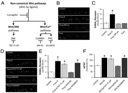
(6) 8416 • J. Neurosci., June 23, 2010 • 30(25):8411– 8420. Cuitino et al. • Wnt-5a Modulates GABAA-R Recycling. conditions, the basal recycling is ⬃40%. That is, 60% of the receptor does not recycle to the surface and thus remains intracellular linked to biotin (Fig. 6 Ab, line ⫹glutathione in Biotinylated proteins). Wnt-5a treatment induces a strong increase of recycling reaching 90%. In other words, only 10% of the labeled GABAA-Rs was protected from the reduction, indicating that a greater number of biotinylated receptors are in the cell surface when the neurons were incubated with glutathione (Fig. 6 Ab, line ⫹glutathione in Biotinylated proteins). These results clearly indicate that the treatment with Wnt-5a induces an increase in the recycling of the GABAA-Rs. One alternative approach to evaluate the recycling is prelabeled neurons with ␥2-specific antibody and then incubated neurons with Wnt-5a. Neurons were returned at 37°C for a different period of time in the presence of Wnt-5a, allowing recycling of marked receptor to the surface of the cell. The appearance of the receptor previously marked on the cell surface indicates the rescue of neuronal receptors from degradation. Image of representative neuron before and after the treatment with Wnt-5a is illustrated (Fig. Figure 3. Wnt-5a ligand increases the amplitude of evoked IPSC, without affecting the paired-pulse index. Intracellular recording of a CA1 pyramidal cells after Wnt-5a ligand treatment at a holding membrane potential of 0 mV and in the presence of 50 M 6 B). Initially, GABAA-Rs clusters are few APV and 20 M CNQX. A, Averaged evoked IPSCs by paired-pulse protocols (eIPSCs), before (Aa) and after 15 min (Ab) of continued and small (cluster/20 m: 2.65 ⫾ 0.29; perfusion with Wnt-5a. Time course of the effect of Wnt-5a (black circles) on eIPSC amplitudes is shown. B, Quantification of the cluster area was considerate as 1). However, average amplitude of eIPSCs before (media) and after 15 min of treatment (control or Wnt-5a). C, Determination of the PPR index after 5 min of treatment with Wnt-5a, before (media) and after 15 min of treatment (control or Wnt-5a). “Media” corresponds to the values of eIPSCs when the cells have GABAA-Rs clusters increase significantly not been treated. “Control” corresponds to the values of eIPSCs when the cells were treated with vehicle. Error bars indicate SEM in number (control: 6.14 ⫾ 0.64; Wnt-5a: (n ⫽ 10). *p ⬍ 0.01. 11.04 ⫾ 0.49), but after 15 min of treatment the clusters numbers increase even more (control: 15.80 ⫾ 0.49; Wnt-5a: 21.55 ⫾ 0.69). In addition, the size of GABAA-Rs clusters increased significantly at 5 min (control: 1.22 ⫾ 0.03; Wnt-5a: 1.40 ⫾ 0.03) and at 15 min after treatment with Wnt-5a (control: 1.68 ⫾ 0.1; Wnt-5a: 1.91 ⫾ 0.02). The number of GABAA-R clusters increase nearly twice in control conditions and nearly four times in neurons treated with Wnt-5a (Fig. 6 B). The size of the clusters (the clusters area) increase ⬃68% in control conditions and 90% in cells treated with Wnt-5a. Both the size and the number remained high during 60 min of Wnt-5a treatment (data not show). The localization, the Figure 4. Wnt-5a ligand increases the amplitude of spontaneous and miniature IPSCs. number and size of the clusters, and the total levels of synaptic Events were recorded from voltage-clamped (0 mV) CA1 pyramidal cells in the presence of 50 protein gephyrin did not change in the presence of Wnt-5a (supM APV and 20 M CNQX. Recording of spontaneous and miniature IPSCs, sIPSC and mIPSC plemental Fig. S2, available at www.jneurosci.org as supplemenrespectively, in cells treated with control or Wnt-5a. tal material). These results indicate that the treatment with Wnt-5a increases the recycling of GABAA-Rs. It happens because, al., 2009; our unpublished observations). To determine whether a significant proportion of internalized GABAA-Rs are rapidly the noncanonical Wnt pathways are involved in the regulation of recycled back to the plasma membrane and virtually no the surface expression of the GABAA-Rs induced by Wnt-5a or GABAA-R degradation could be detected within the first hour Foxy-5, we used fumagillin (Fum), a synthetic inhibitor of the (Kittler et al., 2004). noncanonical Wnt pathways. This inhibitor acts downstream of the Wnt receptors, but upstream of CaMKII and JNK without The noncanonical Wnt/Ca 2ⴙ pathway regulates the increase affecting the canonical Wnt/-catenin pathway (Farías et al., of the surface expression of GABAA-Rs induced by Wnt-5a 2009) (Fig. 7A). Previously, we described that Fum blocked the Previously, we demonstrated that Wnt-5a and Foxy-5 act as nonactivation of JNK and CaMKII induced by Wnt-5a or Foxy-5 in canonical ligands in mature hippocampal neurons, activating mature hippocampal neurons (Farías et al., 2009; our unpubboth Wnt signaling pathway, Wnt/JNK and Wnt/Ca 2⫹ (Farías et.
(7) Cuitino et al. • Wnt-5a Modulates GABAA-R Recycling. Figure 5. Wnt-5a ligand does not affect the endocytosis of GABAA-Rs. Events were recorded from voltage-clamped (0 mV) CA1 pyramidal cells in the presence of 50 M APV, 20 M CNQX and 0.5 M TTX. The P4 peptide was injected by the patch pipette. Illustrates the single recordings of representative neurons of mIPSCs in the absence or in the presence of Wnt-5a (15 min).. J. Neurosci., June 23, 2010 • 30(25):8411– 8420 • 8417. sion of GABAA-Rs (supplemental Fig. S3 A, B, available at www. jneurosci.org as supplemental material). To confirm that the Wnt/Ca 2⫹ pathway is implicated in the increase of functional GABAA-Rs, we analyzed the effect of TAT-TI-JIP, KN-93 and Gö6976 over evoked GABAA-R-mediated eIPSCs, in hippocampal CA1 pyramidal neurons. We observed that the effect of Wnt-5a (Fig. 7F ) or Foxy-5 (supplemental Fig. S3C, available at www.jneurosci.org as supplemental material) is not blocked by TAT-TI-JIP or by Gö6976. Consistently with the results obtained by the immunofluorescence assay, the effect of Wnt-5a and Foxy-5 is completely inhibited by KN-93 (Fig. 7F; supplemental Fig. S3C, available at www.jneurosci.org as supplemental material). These results suggest that CaMKII is the main mediator of the effect of Wnt-5a over GABAA-Rs. The inhibitors alone do not affect the amplitude of the eIPSCs mediated by GABAA-Rs (supplemental Fig. S3D, available at www.jneurosci.org as supplemental material). Together, these results indicate that the noncanonical Wnt/Ca 2⫹ pathway is required to modulate the recycling of functional GABAA-Rs on hippocampal neurons.. Discussion GABAA-Rs are critical mediators of synaptic inhibition in the brain (Macdonald and Olsen, 1994; Jacob et al., 2008). At synapses, GABAA-Rs constitutively undergo significant rates of constitutive endocytosis, via clathrin-coated pits in a dynamin-dependent process; the internalized GABAA-Rs are then subjected to either rapid recycling or targeted for lysosomal degradation (Kittler et al., 2004; Jacob et al., 2008). Therefore, changes in the rates of GABAA-R endocytosis and/or endocytic sorting represent potentially powerful mechanisms to regulate GABAA-R cell surface number and inhibitory synFigure 6. GABAA Receptors are recycled on the neuronal surface under the effect of Wnt-5a ligand. A, Neurons were incubated aptic transmission (Collingridge et al., for 1 h at 37°C with Sulfo-NHS-SS-Biotin. Aa, Control cells at 0 min were reduced or not with glutathione. Ab, Neurons were 2004; Kittler et al., 2004). A direct relastimulated for 15 min with Wnt-5a at 37°C. Surface Sulfo-NHS-SS-Biotin was reduced or not with glutathione, biotinylated and tionship between the number of postsyntotal ␥2-GABAA-Rs were detected. B, Neurons were labeled with an antibody against ␥2 subunits at 37°C and then incubated with aptic GABAA-Rs and the strength of the Wnt-5a. The ␥2 subunits were detected in nonpermeabilized neurons and correspond to the receptor recycling to the surface cell. synapse has been demonstrated (Nusser et al., 1997, 1998). Therefore, to maintain a stable cell-surface receptor number, continlished observations). Consistent with our previous studies, the ual membrane insertion of newly synthesized or recycled receptors is effect induced by Foxy-5 (50 M) at 15 min on the surface clusrequired (Kennedy and Ehlers, 2006). However, how neurons faciltering of GABAA-Rs is inhibited by Fum (Fig. 7 B, C). To dissect itate the insertion of GABAA-Rs into synaptic membranes remains whether Wnt-5a induces the surface expression of receptor by the to be determined. This issue is not only of importance for inhibitory Wnt/JNK or the Wnt/Ca 2⫹ pathways, we used inhibitors for the synaptic transmission, in fact, the major sites of excitatory synaptic three well known effectors of these pathways: TAT-TI-JIP to intransmission in the brain, the AMPA-type glutamate receptors also hibit JNK activity of the Wnt/JNK pathway and KN-93 and cycle between the plasma membrane and intracellular compartGö6976 to inhibit CaMKII and PKC activities of the Wnt/Ca 2⫹ ments playing a role in synaptic plasticity (Citri and Malenka, 2008). pathway (Fig. 7A). Hippocampal neurons were incubated with To address the mechanisms underlying GABAA-R membrane trafFoxy-5 for 15 min in the presence of the inhibitors mentioned ficking we have studied the role of the Wnt signaling pathway. Our and the clusters number per m of neurite was measured. TATresults demonstrate that treatment with the noncanonical Wnt-5a TI-JIP did not affect significantly the cluster number of GABAA-Rs ligand significantly increases the amount of the functional induced by Foxy-5 (Fig. 7 D, E). However, when we analyzed GABAA-Rs on the neuronal cell surface, increasing the number of the contribution of the CaMKII and PKC, we found that only in clusters and the amplitude of the inhibitory currents. the presence of the CaMKII inhibitor, the effect of Foxy-5 on the Since the presynaptic and postsynaptic regions strongly intersurface expression of the GABAA-Rs was completely blocked (Fig. 7 D, E). The inhibitors alone do not affect the surface expresact, alterations in structuring the presynaptic terminal or the.
(8) 8418 • J. Neurosci., June 23, 2010 • 30(25):8411– 8420. Cuitino et al. • Wnt-5a Modulates GABAA-R Recycling. postsynaptic region are accompanied by a parallel change in the opposite synaptic site (Ahmad-Annuar et al., 2006; Citri and Malenka, 2008; Salinas and Zou, 2008). However, when we analyzed the scaffold protein gephyrin we did not observed any change in the presence of Wnt-5a treatment, during the same time frame where Wnt-5a increased the surface expression of GABAA-Rs. These results suggest that Wnt-5a does not affect the organization of the whole inhibitory postsynaptic region, at least on the same time scale. In fact, our studies were performed after short-term exposure to Wnt ligands, therefore we do not know whether a long-term exposure to Wnts will affect the presynaptic counterpart as a consequence of the postsynaptic differentiation. Previous studies in our laboratory demonstrated that Wnt-5a does not affect the clustering of different presynaptic proteins until 60 min (Cerpa et al., 2008, Farías et al., 2010; Inestrosa Figure 7. Noncanonical Wnt/Ca 2⫹ pathway signaling is involved in the surface expression of GABAA-Rs induced by Wnt-5a. and Arenas, 2010). Therefore, although A, The scheme indicates the two noncanonical Wnt pathways described for Wnt-5a, Wnt/Ca 2⫹ and Wnt/JNK. The effectors for each an increase in cluster number could indi- pathway: CaMKII, PKC and JNK are shown as well as the drugs used to inhibit these kinases: TAT-TI-JIP, KN-93 and Gö6976. cate new, unsilenced synapses, it is more Fumagillin (Fum) is a general inhibitor of the noncanonical Wnt pathways. B, Representative neurite images of hippocampal likely that this result from an increase in neurons exposed to Foxy-5 for 15 min in the presence or absence of Fum. C, Quantification of ␥2-GABAA-R clusters number/20 m receptor levels above the immunocyto- neurite shown in B). D, Representative neurite images of hippocampal neurons incubated with Foxy-5 or coincubated with Foxy-5 chemical detection threshold at previously and inhibitors indicated, for 15 min. E, Quantification of ␥2-GABAA-R clusters number/20 m neurite of the treatments indicated existing synapses. Furthermore, because we in D). F, Quantification of average amplitude of eIPSCs in presence of Wnt-5a or coincubated with the inhibitors indicated for 15 min do not observed changes in paired-pulse re- of treatment. The inhibitors were injected by the patch pipette. Error bars indicate SEM (n ⫽ 10). *p ⬍ 0.05. lationship or mIPSC frequency in CA1 neureceptor stability in the cell surface. Thus, the PKC activation rons in hippocampal slices after Wnt-5a or Foxy-5 application, it promotes GABAA receptor endocytosis and decreases cell surface appears that the increase in surface receptors happens at preexisting expression of the receptor. This phenomenon is accompanied by synapses. strong decreases in GABA-gated chloride currents (Herring et al., In addition, we demonstrate that the regulation of the expres2005). In addition, it has been demonstrated that CaMKII actision of GABAA-Rs induced by Wnt-5a on the cell surface is due to vation promotes the recruitment of postsynaptic GABAA-Rs, enthe fact that this ligand increases the recycling of the receptor hancing the amplitude of GABA whole-cell currents and IPSCs without affecting the endocytic process or the total protein level. (Churn and DeLorenzo, 1998; Wei et al., 2004). In accordance These effects induced by the Wnt-5a are specific since they are with the studies mentioned above, we demonstrated that the blocked by sFRP, a soluble antagonist of Wnt signaling. Morenoncanonical Wnt/Ca 2⫹ pathway, particularly CaMKII, is reover, the increase induced by Wnt-5a in the surface expression of quired to modulate the effect of Wnt-5a on GABAA-Rs. the GABAA-Rs was reproduced by a formylated hexapeptide that GABAA-Rs not only function as chloride channels that also mimics Wnt-5a effect (Foxy-5). These results suggest that Wnt-5a regulate membrane voltage and conductance, but also play a crufacilitates the membrane insertion of GABAA-Rs. However, the cial role in the establishment of functional synapses, as well as its precise mechanisms that mediate the stabilization of GABAA-Rs maturation and stabilization (Ben-Ari, 2002; Ben-Ari et al., on the neuronal surface induced by Wnt-5a remains to be estab2004). In addition, they are involved in the control of the excitlished. But, at least an increase in the recycling of GABAA-Rs is ability of the brain, circadian rhythms, cognition, sleepingtriggered by the Wnt-5a ligand. wakening cycle, learning and memory (Rudolph and Möhler, During the last years, it has been observed that the phosphor2006). Functional adaptation of GABAergic synapses can generylation of GABAA-Rs subunits is an important mechanism that ally be achieved by changes in either the neurotransmitter release dynamically modulates GABAA-Rs trafficking at synapses (Brandon properties of GABAergic neurons or changes in gene expression, et al., 2002) The GABAA-Rs is phosphorylated by diverse kinases cellular distribution, or function of postsynaptic GABAA-Rs. including the cAMP-dependent protein kinase (PKA), PKC, However, experimental evidences suggest that the synaptic effiCaMKII, Protein kinase B (Akt) and tyrosine kinases of the Src cacy of GABAergic synapses is tightly correlated with the number family. On this context, Brandon and coworkers described that of postsynaptic GABAA-Rs (Kittler et al., 2000, Jacob et al., 2008). GABAA receptor function, dependent upon the subtype anaTherefore, changes in the trafficking of these receptors could lyzed, and it can be differentially modulated by phosphorylation regulate neuronal plasticity and contribute to the manifestation of key residues within the intracellular loop of receptor 1–3 and of a wide range of neurological and psychiatric disorders includ␥2 subunits. Interestingly, PKC and CaMKII are involved in the ing epilepsy (Naylor et al., 2005), mood disorders such as anxiety noncanonical Wnt signaling pathway initiated by Wnt-5a (Farías and depression (Brambilla et al., 2003; Tunnicliff and Malatynska, et al., 2009, 2010; Inestrosa and Arenas, 2010). Previous studies 2003), and alcoholism (Kumar et al., 2003). have demonstrated that these kinases modulate differentially the.
(9) Cuitino et al. • Wnt-5a Modulates GABAA-R Recycling. There are a wide variety of receptors that can be distinguished based on their pharmacologic profile, in their subcellular localization or simply by the combination of their subunits. The main GABAA-Rs expressed in the brain are composed by 2 ␣1 subunits, 2  subunits and a ␥2 subunit (Jacob et al., 2008). The ␥2 subunit has been described as the responsible for the sensitivity to benzodiazepines, the synaptic localization, and the trafficking modulation of the receptors (Essrich et al., 1998; Connolly et al., 1999). But the current view is that gephyrin stabilizes receptor clusters in the postsynaptic membrane by preventing their lateral diffusion and/or internalization. In the present study we have described that Wnt-5a modulates the surface expression of the ␣1 as ␥2 subunit in mature hippocampal neurons, increasing the number of the clusters and the colocalization with gephyrin, conferring to the Wnt signaling pathway a key role in the maintenance of the GABAergic synapses in the brain. The Wnt signaling pathway has been involved in various cellular processes, including functions in the neuronal development and maintenance of the nervous system (Lie et al., 2005; AhmadAnnuar et al., 2006; Chen et al., 2006; Salinas and Zou, 2008; Inestrosa and Arenas, 2010). Wnt proteins signal through at least three different pathways. In the canonical pathway, Wnt ligand increases cytoplasmic -catenin levels, allowing -catenin to enter the nucleus where it co-activates the transcription of Wnt target genes (Logan and Nusse, 2004; Toledo et al., 2008). Several “noncanonical” Wnt signaling pathways do not affect gene transcription through -catenin, they mediate other cellular processes through different molecular intermediates instead, including the regulation of monomeric GTPases of the Rho/Rac family and changes in intracellular calcium levels (Montcouquiol et al., 2006; Salinas and Zou, 2008). Our laboratory has demonstrated that the Wnt signaling regulates the presynaptic localization of ␣7-nAChRs (Farías et al., 2007), it induces recycling and exocytosis of synaptic vesicles (Cerpa et al., 2008) and the clustering of PSD-95 at the postsynaptic region (Farías et al., 2009). However, little is known about its role in mature neurons, even though Wnt ligands and proteins that mediate their signaling are expressed in the mature nervous system (Inestrosa and Arenas, 2010). In the present work, we are proposing that the Wnt signaling pathway has a key role in the homeostasis of inhibitory neuronal synapses, suggesting a possible effect on the plasticity of the inhibitory synapses. Receptor translocation has important implications for synaptic function and given that GABAA-Rs cycle between synaptic sites and intracellular endocytic structures (Kittler et al., 2000, 2004; Jacob et al., 2008). The capacity of neurons to modulate the removal and/or insertion of GABAA-Rs in synaptic membranes may have profound effects on the efficacy of synaptic transmission (Otis et al., 1994). Thus, the rapid increase of GABAA-Rs induced by Wnt-5a in the postsynaptic domain of inhibitory synapses provides an additional mechanism for the induction of synaptic plasticity in these synapses.. References Ahmad-Annuar A, Ciani L, Simeonidis I, Herreros J, Fredj NB, Rosso SB, Hall A, Brickley S, Salinas PC (2006) Signaling across the synapse: a role for Wnt and Dishevelled in presynaptic assembly and neurotransmitter release. J Cell Biol 174:127–139. Alvarez AR, Godoy JA, Mullendorff K, Olivares GH, Bronfman M, Inestrosa NC (2004) Wnt-3a overcomes -amyloid toxicity in rat hippocampal neurons. Exp Cell Res 297:186 –196. Ben-Ari Y (2002) Excitatory actions of GABA during development: the nature of the nurture. Nat Rev Neurosci 3:728 –739.. J. Neurosci., June 23, 2010 • 30(25):8411– 8420 • 8419 Ben-Ari Y, Khalilov I, Represa A, Gozlan H (2004) Interneurons set the tune of developing networks. Trends Neurosci 27:422– 427. Brambilla P, Perez J, Barale F, Schettini G, Soares JC (2003) GABAergic dysfunction in mood disorders. Mol Psychiatry 8:721–737, 715. Brandon N, Jovanovic J, Moss S (2002) Multiple roles of protein kinases in the modulation of ␥-aminobutyric acid (A) receptor function and cell surface expression. Pharmacol Ther 94:113–122. Brünig I, Penschuck S, Berninger B, Benson J, Fritschy JM (2001) BDNF reduces miniature inhibitory postsynaptic currents by rapid down regulation of GABAA receptor surface expression. Eur J Neurosci 13:1320 – 1328. Cerpa W, Godoy JA, Alfaro I, Farías GG, Metcalfe MJ, Fuentealba R, Bonansco C, Inestrosa NC (2008) Wnt-7a modulates the synaptic vesicle cycle and synaptic transmission in hippocampal neurons. J Biol Chem 283:5918 –5927. Chen J, Park CS, Tang SJ (2006) Activity-dependent synaptic Wnt release regulates hippocampal long term potentiation. J Biol Chem 281:11910 – 11916. Churn SB, DeLorenzo RJ (1998) Modulation of GABAergic receptor binding by activation of calcium and calmodulin-dependent kinase II membrane phosphorylation. Brain Res 809:68 –76. Ciani L, Salinas PC (2005) WNTs in the vertebrate nervous system: from patterning to neuronal connectivity. Nat Rev Neurosci 6:351–362. Citri A, Malenka RC (2008) Synaptic plasticity: multiple forms, functions, and mechanisms. Neuropsychopharmacology 33:18 – 41. Collingridge GL, Isaac JT, Wang YT (2004) Receptor trafficking and synaptic plasticity. Nat Rev Neurosci 5:952–962. Connolly CN, Uren JM, Thomas P, Gorrie GH, Gibson A, Smart TG, Moss SJ (1999) Endocytosis of homomeric ␥2 subunit splice variants of ␥-aminobutyric acid type A subcellular localization and receptors. Mol Cell Neurosci 13:259 –271. Cuitino L, Matute R, Retamal C, Bu G, Inestrosa NC, Marzolo MP (2005) ApoER2 is endocytosed by a clathrin-mediated process involving the adaptor protein Dab2 independent of its Rafts’ association. Traffic 6:820 – 838. Essrich C, Lorez M, Benson JA, Fritschy JM, Lüscher B (1998) Postsynaptic clustering of major GABAA receptor subtypes requires the ␥2 subunit and gephyrin. Nat Neurosci 1:563–571. Farías GG, Vallés AS, Colombres M, Godoy JA, Toledo EM, Lukas RJ, Barrantes FJ, Inestrosa NC (2007) Wnt-7a induces presynaptic colocalization of ␣7-nicotinic acetylcholine receptors and adenomatous polyposis coli in hippocampal neurons. J Neurosci 27:5313–5325. Farías GG, Alfaro IE, Cerpa W, Grabowski CP, Godoy JA, Bonansco C, Inestrosa NC (2009) Wnt-5a/JNK signaling promotes the clustering of PSD-95 in hippocampal neurons. J Biol Chem 284:15857–15866. Farías GG, Godoy JA, Cerpa W, Varela-Nallar L, Inestrosa NC (2010) Wnt signaling modulates pre- and postsynaptic maturation: therapeutic considerations. Dev Dyn 239:94 –101. Fuenzalida M, Fernandez de Sevilla D, Buño W (2007) Changes of the EPSP waveform regulate the temporal window for spike-timing-dependent plasticity. J Neurosci 27:11940 –11948. He P, Shen Y (2009) Interruption of -catenin signaling reduces neurogenesis in Alzheimer’s disease. J Neurosci 29:6545– 6557. Herring D, Huang R, Singh M, Dillon GH, Leidenheimer NJ (2005) PKC modulation of GABAA receptor endocytosis and function is inhibited by mutation of a di-leucine motif within the receptor 2 subunit. Neuropharmacology 48:181–194. Inestrosa NC, Arenas E (2010) Emerging roles of Wnts in the adult nervous system. Nat Rev Neurosci 11:77– 86. Jacob TC, Moss SJ, Jurd R (2008) GABAA receptor trafficking and its role in the dynamic modulation of neuronal inhibition. Nat Rev Neurosci 9:331–343. Kennedy MJ, Ehlers MD (2006) Organelles and trafficking machinery for postsynaptic plasticity. Annu Rev Neurosci 29:325–362. Kittler JT, Delmas P, Jovanovic JN, Brown DA, Smart TG, Moss SJ (2000) Constitutive endocytosis of GABAA receptors by an association with the adaptin AP2 complex modulates inhibitory synaptic currents in hippocampal neurons. J Neurosci 20:7972–7977. Kittler JT, Thomas P, Tretter V, Bogdanov YD, Haucke V, Smart TG, Moss SJ (2004) Huntingtin-associated protein 1 regulates inhibitory synaptic transmission by modulating ␥-aminobutyric acid type A receptor membrane trafficking. Proc Natl Acad Sci U S A 101:12736 –12741..
(10) 8420 • J. Neurosci., June 23, 2010 • 30(25):8411– 8420 Kumar S, Kralic JE, O’Buckley TK, Grobin AC, Morrow AL (2003) Chronic ethanol consumption enhances internalization of ␣1 subunit-containing GABAA receptors in cerebral cortex. J Neurochem 86:700 –708. Lie DC, Colamarino SA, Song HJ, Désiré L, Mira H, Consiglio A, Lein ES, Jessberger S, Lansford H, Dearie AR, Gage FH (2005) Wnt signalling regulates adult hippocampal neurogenesis. Nature 437:1370 –1375. Logan CY, Nusse R (2004) The Wnt signaling pathway in development and disease. Annu Rev Cell Dev Biol 20:781– 810. Macdonald RL, Olsen RW (1994) GABAA receptor channels. Annu Rev Neurosci 17:569 – 602. Malenka RC, Nicoll RA (1999) Long-term potentiation—a decade of progress? Science 285:1870 –1874. Montcouquiol M, Crenshaw EB 3rd, Kelley MW (2006) Non-canonical Wnt signaling and neural polarity. Annu Rev Neurosci 29:363–386. Morimoto S, Nishimura N, Terai T, Manabe S, Yamamoto Y, Shinahara W, Miyake H, Tashiro S, Shimada M, Sasaki T (2005) Rab13 mediates the continuous endocytic recycling of occludin to the cell surface. J Biol Chem 280:2220 –2228. Murthy VN, Sejnowski TJ, Stevens CF (1997) Heterogeneous release properties of visualized individual hippocampal synapses. Neuron 18:599 – 612. Naylor DE, Liu H, Wasterlain CG (2005) Trafficking of GABAA receptors, loss of inhibition, and a mechanism for pharmacoresistance in status epilepticus. J Neurosci 25:7724 –7733. Nusser Z, Cull-Candy S, Farrant M (1997) Differences in synaptic GABAA receptor number underlies variation in GABA mini amplitude. Neuron 19:697–709. Nusser Z, Hájos N, Somogyi P, Mody I (1998) Increased number of synaptic GABAA receptors underlies potentiation at hippocampal inhibitory synapses. Nature 395:172–177. Otis TS, De Koninck Y, Mody I (1994) Lasting potentiation of inhibition is. Cuitino et al. • Wnt-5a Modulates GABAA-R Recycling associated with an increased number of gamma-aminobutyric acid type A receptors activated during miniature inhibitory postsynaptic currents. Proc Natl Acad Sci U S A 91:7698 –7702. Parton RG, Simons K, Dotti CG (1992) Axonal and dendritic endocytic pathways in cultured neurons. J Cell Biol 119:123–137. Rattner A, Hsieh JC, Smallwood PM, Gilbert DJ, Copeland NG, Jenkins NA, Nathans J (1997) A family of secreted proteins contains homology to the cysteine-rich ligand-binding domain of frizzled receptors. Proc Natl Acad Sci U S A 94:2859 –2863. Rudolph U, Möhler H (2006) GABA-based therapeutic approaches: GABAA receptor subtype functions. Curr Opin Pharmacol 6:18 –23. Salinas PC, Zou Y (2008) Wnt signaling in neural circuit assembly. Annu Rev Neurosci 31:339 –358. Toledo EM, Colombres M, Inestrosa NC (2008) Wnt signaling in neuroprotection and stem cell differentiation. Prog Neurobiol 86:281–296. Tunnicliff G, Malatynska E (2003) Central GABAergic systems and depressive illness. Neurochem Res 28:965–976. Vargas KJ, Terunuma M, Tello JA, Pangalos MN, Moss SJ, Couve A (2008) The availability of surface GABAB receptors is independent of ␥-aminobutyric acid but controlled by glutamate in central neurons. J Biol Chem 283: 24641–24648. Wan Q, Xiong ZG, Man HY, Ackerley CA, Braunton J, Lu WY, Becker LE, MacDonald JF, Wang YT (1997) Recruitment of functional GABAA receptors to postsynaptic domains by insulin. Nature 388:686 – 690. Wang Q, Liu L, Pei L, Ju W, Ahmadian G, Lu J, Wang Y, Liu F, Wang YT (2003) Control of synaptic strength, a novel function of Akt. Neuron 38:915–928. Wei J, Zhang M, Zhu Y, Wang JH (2004) Ca 2ⴙ-calmodulin signalling pathway up-regulates GABA synaptic transmission through cytoskeletonmediated mechanisms. Neuroscience 127:637– 647..
(11)
Figure


Documento similar
Hippocampal neurons treated with Aβ oligomers in the presence of the Wnt-5a ligand, showed a decrease in the SynGAP clusters Figure 3B, very similar to the observed by the
In the present work, in hippocampal neurons, Wnt3a ligand modulates AMPK activation through a mechanism dependent on GSK-3β inhibition, and this effect is specific to the Wnt
