IL 21 signaling pathway modulation in B lymphocytes by vitamin D in systemic lupus erythematosus
74
0
0
Texto completo
(2) ©2019, Daniela María Hirigoyen Pérez. Se autoriza la reproducción total o parcial, con fines académicos, por cualquier medio o procedimiento, incluyendo la cita bibliográfica que acredita al trabajo y a su autor. ii.
(3) PONTIFICIA UNIVERSIDAD CATÓLICA DE CHILE Programa de Doctorado en Ciencias Médicas Dirección de Investigación y Doctorado Escuela de Medicina. La Comisión Examinadora, constituida por los Profesores abajo firmantes, aprueba la Defensa Pública de la Tesis Doctoral titulada:. “IL-21 SIGNALING PATHWAY MODULATION IN B LYMPHOCYTES BY VITAMIN D IN SYSTEMIC LUPUS ERYTHEMATOSUS” Aprobación Defensa Pública:. SRA. DANIELA HIRIGOYEN PEREZ Calificándose el trabajo realizado, el manuscrito sometido y la defensa oral, con nota (…..…) ……………... _____________________ Dr. Mauricio Cuello Director de Investigación y Doctorado Escuela de Medicina, PUC ______________________________ Dra. Claudia Sáez Sub-Directora de Investigación y Doctorado Escuela de Medicina, PUC _______________________________ Dra. Andrea Leiva Sub-Jefe Programa Doctorado en Ciencias Médicas Escuela de Medicina, PUC _______________________________ Dra. Paula Burgos Co-directora de Tesis Escuela de Medicina, PUC. _____________________________ Dr. Felipe Heusser Decano Facultad de Medicina, PUC ______________________________ Dr. Jorge Carvajal Jefe Programa Doctorado en Ciencias Médicas Escuela de Medicina, PUC _______________________________ Dr. Alfonso González Director de Tesis Facultad de Medicina, USS _______________________________ Dr. Bruno Nervi Profesor Evaluador Interno Escuela de Medicina, PUC. _______________________________ Dr. Rodrigo Pacheco Profesor Evaluador Externo Fundación Ciencia y Vida. iii Santiago, 30 de enero de 2019.
(4) 2. DEDICATORY. To my husband Javier and to my parents, Carlos y Beatriz, for your unconditional support. To my son Clemente.
(5) 3. ACKNOWLEDGMENT First, to my supervisor, Dr. Paula Burgos, thank you for giving me the opportunity to work with you, opening your laboratory doors to me and allowing me to do my research. Thank you for everything I have learnt from you. To Dr. Alfonso González, for being my supervisor and helping me with my experiment design and results discussion. To Mirentxu Iruretagoyena, for being with me during all my thesis, helping me with experiment design and results discussion. To Rebeca Montalva, for being an excellent work partner and, helping me with the experiments. To Dr. Alejandra Alvarez for examined patients that participate in the study. To Dr. Rodrigo Naves, for helping me with experiment design and welcoming me to your laboratory. To Eric Acuña and Payton Ottum, for helping me with the assays with T lymphocytes. To Aracely Alcoholado, Jocelyn Muñoz, Paulina Tapia and Nancy Vásquez for their friendship and be always ready to help me with blood samples extraction. To Marta Quiñones and Mauricio Castro for your friendship. To my thesis committee, for the advice given during my thesis. To Dr. Jaime Pereira, Dr. Claudia Sáez, Dr. Silvana Zanlungo, for being there during my thesis process and the support given all these years. To patients and healthy controls who participated in this study. To my family: my parents Carlos and Beatriz for being there every time I needed and all that you have taught me. To my sister Carolina and my brother Rodrigo. To Alejandra Vielma and Jorge Torres, for their friendship and help during this thesis. To my friends, Paula, Isabel, Jocelyn, Javier, Francisca, Javiera, Claudia, Stephanie, Matías and Leonardo. To my husband Javier, for being a substantial support during my PhD studies, for the patience and love that you give me every day. To my son Clemente.. v.
(6) Finally, to my funding: CONICYT fellowship 21150777 and Medicine School and Vicerrectoría de Investigación UC fellowship. To Fondecyt 1141211. Thank you. vi.
(7) 4. INDEX. 1. THESIS COMMITTEE................................................................................................. iii 2. DEDICATORY .............................................................................................................. iv 3. ACKNOWLEDGMENTS.............................................................................................. v 4. INDEX............................................................................................................................. vii 5. TABLE INDEX............................................................................................................... ix 6. FIGURE INDEX............................................................................................................. x 7. ABBREVIATIONS LIST……....................................................................................... xi 8. RESUMEN....................................................................................................................... 1 9. ABSTRACT..................................................................................................................... 3 10. INTRODUCTION.......................................................................................................... 5 10.1 Systemic Lupus Erythematosus.…..................................................................... 5 10.2 T-B lymphocyte interaction.…............................................................................ 6 10.3 Molecular mechanisms in SLE ........................................................................... 7 10.4 Vitamin D ............................................................................................................. 7 10.5 Vitamin D supplementation in SLE patients .................................................... 9 10.6 Interleukin 21 (IL-21) ....................................................................................... 10 10.7 IL-21 signaling ................................................................................................... 11 vii.
(8) 10.8 Vitamin D and Interleukin 21 .......................................................................... 13 11. HYPOTHESIS............................................................................................................. 15 11.1 Main objective ................................................................................................... 16 11.2 Specific objectives .............................................................................................. 17 12. METHODS................................................................................................................... 19 12.1 Participants ........................................................................................................ 19 12.2 IL-21 and IL-21R detection ……...................................................................... 20 12.3 IL-21 detection in activated T cells .................................................................. 21 12.4 IL-2 and IL-21 detection in activated PBMC ................................................. 22 12.5 B cell activation .................................................................................................. 22 12.6 STAT3 phosphorylation ................................................................................... 23 12.8 IL-21 polymorphisms …………….................................................................... 23 12.8 Statistical analysis.……………......................................................................... 24 13. RESULTS………………............................................................................................. 25 CHAPTER 1: VITAMIN D SUPPLEMENTATION IN SLE PATIENTS .............. 25 CHAPTER 2: VITAMIN D EFFECT ON IL-21/IL-21 SIGNALING IN B CELLS IN VITRO…....................................................................................................................... 34 CHAPTER 3: IL-21 POLYMORPHISMS AND SLE .............................................. 45 14. DISCUSSION .............................................................................................................. 50 15. CONCLUSION ............................................................................................................ 56 16. BIBLIOGRAPHY …................................................................................................... 57 viii.
(9) 5. TABLE INDEX. Table 1.. Baseline characteristics of the studied population: placebo. 26. and vitamin D groups Table 2.. Characteristics of placebo and vitamin D groups before. 27. (visit 1) and after supplementation (visit 2 – 3 months) Table 3.. Allele and genotype frequencies of rs907715 (C/T) in controls and SLE patients. 45. Table 4.. Allele and genotype frequencies of rs6822844 (G/T) in controls and SLE patients. 46. Table 5.. Characteristics of SLE patients according to rs907715 polymorphism genotype. 48. Table 6.. Characteristics of SLE patients according to rs6822844. 49. polymorphism genotype. ix.
(10) 6. FIGURE INDEX. Figure 1.. Vitamin D supplementation protocol in patients with Systemic Lupus Erythematosus with hypovitaminosis D.. 19. Figure 2.. Serum IL-21 levels in healthy controls and in SLE patients.. 28. Figure 3.. Serum IL-21 levels in SLE patients before and after supplementation.. 29. Figure 4.. IL21 mRNA expression in PBMCs from healthy controls and SLE patients.. 30. Figure 5.. Vitamin D supplementation does not modify IL-21 mRNA expression on PBMC in SLE patients.. 31. Figure 6.. IL-21R expression in B cells from healthy controls and SLE. 32. patients. Figure 7.. Vitamin D supplementation does not modify IL-21R expression on CD19+ lymphocytes in SLE patients.. 33. Figure 8.. CD8- T cells positive for IL-21 are higher in SLE patients.. 35. Figure 9.. 1α,25-(OH)2D3 reduces IL2 expression in activated PBMC from healthy controls and SLE patients.. 38. Figure 10. 1α,25-(OH)2D3 has no effect on IL-21R expression in activated B cells from healthy controls.. 40. Figure 11. 1α,25-(OH)2D3 exerts no effect on IL-21 induced STAT3 phosphorylation of B cells from healthy controls.. 43. Figure 12. Changes in vitamin D levels after supplementation according to IL-21 polymorphism genotype.. 47. x.
(11) 7.. ABBREVIATION LIST. 1α,25-(OH)2D3: 1α,25-dihydroxyvitamin D3 APC: Allophycocyanin BCR: Lymphocyte B Receptor BFA: Brefeldin A EDTA: Ethylenediaminetetraacetic Acid FBS: Fetal Bovine Serum FITC: Fluorescein isothiocyanate HEPES: 4-(2-hydroxyethyl)-1-piperazineethanesulfonic acid IgG: Immunoglobulin G IgM: Immunoglobulin M IMDM: Iscove's Modified Dulbecco's Medium IL-21: Interleukin 21 IL-21R: Interleukin 21 Receptor PE: Phycoerythrin PBMC: Peripheral Blood Mononuclear Cells PGA: Physician Global Assessment PMA: Phorbol-12-myristate 13-acetate pSTAT3: phosphorylated STAT3 RPMI: Roswell Park Memorial Institute medium FBS: Fetal Bovine Serum SELENA-SLEDAI: Safety of Estrogens in Systemic Lupus Erythematosus National Assessment - Systemic Lupus Erythematosus Disease Activity Index SLE: Systemic Lupus Erythematosus xi.
(12) STAT3: Signal Transducers and Activators of Transcription-3 TH: T helper lymphocytes UVB: Ultraviolet light B VDR: Vitamin D Receptor. xii.
(13) 8. RESUMEN. El Lupus Eritematoso Sistémico (LES) es una enfermedad autoinmune, caracterizada por la activación de linfocitos B y la generación de autoanticuerpos, con una variedad de manifestaciones clínicas. Se ha reportado que los pacientes con LES tienen niveles más bajos de vitamina D que la población sana, lo que podría estar dado, en parte, a que estos pacientes deben evitar la exposición solar. La deficiencia de vitamina D puede jugar un rol en SLE, ya que recientemente, se ha descrito que esta vitamina puede modificar la respuesta inmune, reduciendo la proliferación y activación de linfocitos B, disminuyendo la producción de anticuerpos. Los mecanismos implicados en la acción de la vitamina D no están completamente dilucidados, pero si se demuestra que es efectiva como tratamiento, sería una droga segura y de bajo costo. La vitamina D podría disminuir la expresión de la interleuquina-21 (IL-21), citoquina tipo I que participa en la activación y diferenciación de linfocitos B, y alteraciones en su vía de señalización podrían inducir la generación de linfocitos B auto reactivos y producción de autoanticuerpos. Nuestra hipótesis fue que la vitamina D reduce la función de IL-21, definida como la expresión, la señalización y los niveles de IL-21, en una cohorte de mestiza de LES. El objetivo principal fue determinar si la suplementación con vitamina D tiene un efecto en los niveles de IL-21 y su vía de señalización en Linfocitos B de pacientes con LES. Para el primer objetivo, reclutamos pacientes con LES con hipovitaminosis que participaron en un protocolo de suplementación aleatorizado, doble ciego, controlado por placebo. No encontramos diferencias en las variables clínicas ni en la expresión del receptor de IL-21 (IL-21R) en linfocitos B en pacientes que recibieron vitamina D o placebo. Detectamos la IL-21 en linfocitos T activados, encontrando que la población de linfocitos T IL-21+ es mayor en pacientes con Lupus que en controles sanos. Para los otros objetivos, evaluamos el efecto de la vitamina D en la función de IL-21 en linfocitos T y B de controles sanos in vitro. La exposición a vitamina D no modificó la población de linfocitos T IL-21+ in vitro, también analizamos la expresión de IL-21R y la fosforilación de STAT3 (pSTAT3) en 1.
(14) linfocitos B activados in vitro, expuestos a vitamina D, y observamos que la vitamina D no reduce la expresión de IL-21R o la fosforilación de STAT3 inducida por IL-21. Finalmente, caracterizamos dos polimorfismos de IL-21, rs907715 y rs6822844 en pacientes con Lupus, y observamos que están asociados con la fotosensibilidad y edad, y uso de metotrexato en pacientes con Lupus, respectivamente. Pese a que la vitamina D no influencia la señalización de IL-21 en linfocitos B, esta es la primera vez que la expresión de IL-21R y pSTAT3 se estudian en linfocitos B expuestos a vitamina D. Estos hallazgos son relevantes para ayudar a dilucidar los mecanismos de inmunomodulación de la vitamina D.. 2.
(15) 9. ABSTRACT. Systemic Lupus Erythematosus (SLE) is an autoimmune disease, characterized by autoantibodies generation and B lymphocyte activation, with a variety of clinical manifestations. It has been reported that SLE patients have lower vitamin D levels that to some extent is due to avoidance to sun exposure in these patients. Vitamin D deficiency could play a role in SLE, as recently it has been described that this vitamin could modify the immune response, by reducing B lymphocyte proliferation and activation and decreasing antibodies production. Vitamin D mechanisms are not fully elucidated but if this vitamin is shown to be successful as a treatment, this is a safe and cheaper drug. One possible target is interleukin-21 (IL-21), type I cytokine participates in B cell activation and differentiation, and whereby signaling pathway disturbances could induce auto reactive B cells generation and autoantibodies production. We hypothesized that vitamin D could reduce IL-21 function; define as expression, signaling and levels of IL-21; in a SLE Mestizo cohort. The main objective of this study was to determine if vitamin D supplementation has an effect on IL-21 levels and its signaling pathway in B-lymphocytes from SLE patients. For the first objective we used supplemented SLE patients with hypovitaminosis D from a randomized, doubleblinded, placebo-controlled trial. No differences in clinical variables or in interleukin-21 receptor (IL-21R) expression in B cells in patients who received vitamin D or placebo were found. We detected IL-21 in activated T lymphocytes, finding that IL-21+ T lymphocytes are higher in SLE patients than in healthy controls. For the other objectives, we evaluated vitamin D effect on IL-21 function in T and B cells from healthy controls in vitro. Vitamin D exposure does not modify IL-21+ T lymphocytes in vitro, we also analyzed IL-21R expression and STAT3 phosphorylation (pSTAT3) in. 3.
(16) activated B cells in vitro exposed to vitamin D and it was observed that vitamin D does not reduce IL-21R expression or IL-21-induced pSTAT3. Finally, two IL-21 polymorphisms, rs907715 and rs6822844 in lupus patients were characterized and it was observed that rs6822844 and rs907715 were associated with photosensitivity and age and methotrexate use respectively in SLE patients. Moreover, vitamin D did not influence IL-21 signaling in B cells, this is the first time IL-21R expression and pSTAT3 have been studied in B cells exposed to vitamin D, these findings are relevant to help elucidate the vitamin D mechanism.. 4.
(17) 10. INTRODUCTION. The immune system protects our organism from agents that could be harmful to our body, and has developed refined regulation mechanisms, which let it distinguish between harmful or safe molecules for our organism (1). Autoimmune diseases are the result of regulation mechanisms failure and lead to the destruction of healthy tissues. Autoimmune trigger mechanisms are not fully understood and there are no effective therapies to cure these diseases, so important efforts are required to understand cellular and molecular bases that participate in these pathologies. 10.1. Systemic Lupus Erythematosus. Systemic Lupus Erythematosus (SLE) is a chronic autoimmune systemic disease with diverse and heterogeneous clinical manifestations that mainly affects young women. Depending to the cohort studied, its prevalence varies from 3.2 to 517.5 per 100,000 inhabitants. It mainly affects women at a 9:1 proportion compared to men (2). SLE etiology is not clear and genetic and epigenetic factors have been identified that could predispose disease and hormonal and environmental factors that trigger or exacerbate it (3). There are only a few studies that have been carried out in Latin America. GLADEL (Grupo Latinoamericano de Estudio del Lupus) created a SLE registry in the sub-continent, analyzing a cohort of 1,500 patients from 9 countries, finding that Latin-American ethnicities impact disease development, where Mestizo patients (born in America with European ancestry) present a more severe disease compared to white ones (4). Moreover, there are no epidemiologic registries in Chile on this disease. For this reason, it is important to develop epidemiologic and basic studies in order to understand the mechanisms implied in SLE pathology in our population. Our laboratory is recording/registering SLE patients attending Pontificia Universidad 5.
(18) Catolica de Chile’s Rheumatology service to characterize socioeconomic, genetic, clinical and laboratory traits of this population. On the other hand, as SLE is a heterogeneous disease, many biological therapies have been developed to treat this pathology, but they are expensive, not 100% effective and have diverse adverse side effects. In the last 50 years only belimumab has been approved for SLE treatment, so it is relevant to find new, cheaper and safer therapies to decrease SLE clinical activity, thus vitamin D is a good alternative that has been suggested by several studies because it can inhibit or module the adaptive immune response. 10.2 T-B lymphocyte interaction The immune system has two branches: innate and adaptive immunity and both participate in the pathology of molecular mechanisms in SLE. In innate immunity, neutrophils, macrophages and activated dendritic cells produce type I interferon with the subsequent activation of the adaptive immunity (5). Lymphocytes T and B are two cellular types which integrate adaptive immunity. B lymphocyte activation requires antigenic recognition by lymphocyte B receptor (BCR) and to differentiate to plasma cells and produce high affinity antibodies it requires help from T helper lymphocytes (TH) (CD4+ lymphocytes). Recently, a new type of TH lymphocytes, follicular T helper lymphocytes (TFH) (6), has been described that expresses co-stimulator molecules, as CD28, CD40L and ICOS that interact with their counterpart in B lymphocytes to provide the necessary help. Cytokines produced by T cells, IL-6 and IL-21 also participate in this process. A deregulation in the interaction between B lymphocytes and follicular T cells can lead to a loss of tolerance and development of autoimmune diseases.. 6.
(19) 10.3 Molecular mechanisms in SLE SLE is characterized by autoantibody production, which generates immune complexes that accumulate in different organs, leading to tissue damage. These autoantibodies are produced by B lymphocytes, which are deregulated in SLE patients, playing a fundamental role in disease development (7). As follicular T cells help in B cell activation, its deregulation could lead to exacerbated production of antibodies. Certain adhesion and co-stimulatory molecules, like CD44, SLAM and CD40L are up-regulated in T lymphocytes from SLE patients. CD40L participates in B cell activation, which in turn, increases the expression of this molecule in T cells (8). Signaling pathways in T lymphocytes are also deregulated in SLE and transcription factor STAT3 is overexpressed in this pathology (8). Finally, circulating follicular T helper cells are increased in SLE patients (9). In SLE, B cells are altered, with a loss of tolerance in central and peripheral levels, allowing the presence of auto reactive B cells capable of recognizing auto antigens, whose frequency in augmented in this pathology. There is also an increased expression of cytokines that stimulate B lymphocyte function (7, 10). Due to this evidence it is important to study B cell mechanisms in SLE. 10.4 Vitamin D One of the environmental factors implied in SLE is UVA and UVB exposure, as it triggers photosensitive rashes and disease flares, as it increases keratinocytes apoptosis, displaying antigens as DNA (11, 12). Sun avoidance is highly recommended in patients suffering from SLE, which contributes to vitamin D deficiency (hypovitaminosis D) in these patients (13, 14). Therapies used in SLE, as corticosteroids and antimalarials, and renal insufficiency could also influence vitamin D levels (15). Several studies have demonstrated that vitamin D serum levels are lower in SLE patients compared to healthy subjects (16, 17). 7.
(20) The 1,25-dihydroxyvitamin D3 (vitamin D) is a secosteroid produced in the skin by the action of UV-radiation on 7-dehydrocolesterol and it experiences two hydroxylations that will produce the active form in the liver (enzyme CYP2R1) and in the kidney (enzyme CYP27B1) (13). Vitamin D has diverse functions mediated by vitamin D receptor (VDR), a member of the steroid receptor superfamily. This receptor heterodimerizes for retinoid X receptor (RXR) and acts like a transcription factor, binding to the promoter of vitamin D target genes in vitamin D response elements (VDRE). Vitamin D can induce or suppress gene expression (18). One of the main functions of vitamin D is calcium homeostasis, sustaining the plasma concentration of calcium, regulating bone mineralization and intestinal absorption of this mineral (19, 20). Recently, it has been described that vitamin D can modify immune response, in an endocrine and autocrine manner, as its receptor and the hydroxylating enzyme CYP27B1 are expressed in immune cells (21, 22). Vitamin D deficiency could play a role in this pathology, as this hormone acts on innate and adaptive immunity, which are deregulated in SLE pathogenesis (22). Vitamin D has effects on T lymphocytes and B lymphocytes, inducing VDR expression in TH lymphocytes from healthy controls and SLE patients. It inhibits CD4+ T lymphocytes, activation, by reducing signaling molecules expression (PKCδ, ERK1/2), activation (CD11a, CD70) and co-stimulatory molecules (CD40L), which are upregulated in SLE (23). Several in vitro studies have been performed analyzing the vitamin D effect on B lymphocytes where it was found that vitamin D inhibits B cell proliferation, differentiation to plasma cells in response to anti-IgM, anti-CD40 and IL-21, and reduces class-switch and antibody production (24). B lymphocyte activation induces 8.
(21) VDR expression, which is further increased when cells were exposed to vitamin D. This hormone can regulate its own signaling in B cells, as it induces 1,25-dihydroxyvitamin D3 24-hydroxylase (CYP24A1) expression, enzyme that degrade vitamin D to an inactive form (25). This evidence shows that vitamin D acts on B cells activation processes, but the exact mechanisms are not dilucidated, so it is interesting to study the molecules involved in vitamin D action. 10.5 Vitamin D supplementation of SLE patients Due to fact that vitamin D deficiency could contribute to the generation and maintenance of auto reactive lymphocytes present in SLE, several groups have sought to restore normal values of vitamin D and analyzed this to see how the disease course was modified, intending to use vitamin D as a safe therapy in these patients. Normal range of vitamin D, 25(OH)D serum levels is between 30 and 100 ng/ml, insufficiency is defined as levels between 20 and 30 ng/ml and deficiency is described as levels below 20 ng/ml (13, 26). Petri et al monitored 1006 SLE patients and those with hypovitaminosis D were supplemented orally with vitamin D, where a 20ng/ml increase in vitamin D serum levels was associated to a decrease in SELENA-SLEDAI and Physician Global Assessment (PGA) (27). There are a few randomized studies where vitamin D supplementation is compared to placebo, which differ in the ethnicity of the cohort, time and dose of vitamin D exposure. In 2007, Abou-Raya performed the large randomized, controlled by placebo trial in 267 patients, where they supplemented SLE patients with vitamin D, finding that those patients who received vitamin D had augmented serum levels. This increase correlated with a reduction in the SLE clinical score SLEDAI and in the antibody production, this did not happen in the placebo group (28). Despite these results, there are two studies where no differences were seen in the SLEDAI score in patients receiving vitamin D (29, 30), so there are not enough studies. 9.
(22) and conclusive evidence on the effect of vitamin D on the disease course in SLE and the recommendation on vitamin D use is still controversial. Due to this evidence it is relevant to study the response and mechanism of vitamin D supplementation in a Mestizo SLE population. 10.6 Interleukin 21 (IL-21) As previously described, circulating follicular TH lymphocytes are increased in SLE patients, which correlate with disease activity, together with augmented secretion of interleukin 21 (IL-21), the main cytokine produced by this cell type (31). IL-21 belongs to cytokine type I family and its functions are mediated through its receptor, IL-21R, which is composed of common γ chain (γc) and specific receptor (32, 33). IL-21 is mainly produced by CD4+ T lymphocytes, while its receptor is expressed in a variety of cells, such as NK cells, T and B lymphocytes, myeloid and endothelial cells (34). IL-21 has plenty of functions, modulating proliferation and survival processes. This cytokine is essential in follicular TH lymphocytes differentiation from TH naïve lymphocytes, and it participates in germinal centers formation, where T lymphocytes promote B lymphocytes activation (35). IL-21 is involved in T-B cells cooperation, helping in B lymphocyte development, expansion and activation, with plasma cell generation and inducing immunoglobulin secretion (36, 37). As B lymphocytes are deregulated in SLE and IL-21 is important in B cell activation and differentiation and it could play a role in SLE pathogenesis. Even though IL-21 serum levels in SLE patients have been studied by several groups, there is no conclusive evidence to date. In a Chinese cohort and in a Korean cohort, IL-21 serum levels were higher in SLE patients than in healthy controls (38, 39). In accordance to this evidence, T lymphocytes positive for IL-21 are increased in SLE patients compared to healthy controls (40, 41). However, no significant results have been described in IL-21 plasma 10.
(23) levels in SLE patients in a cohort from the United States (42) and in addition, in a new diagnosed Chinese cohort, IL-21 was lower in SLE patients than controls (43). The controversial differences shown in these studies could be due to the diversity of characteristics and ethnic groups of SLE patients enrolled, thus as SLE disease varies according to the genetics it is relevant to analyze IL-21 in our cohort of patients. The role of IL-21 in SLE has also been studied in murine models, where mice that spontaneously developed a lupus-like pathology, MRL/MpJ-Fas(lpr/lpr)/J (MRLlpr), and knock-out for IL-21 receptor, showed an improvement in clinical variables, as proteinuria, lymphadenopathy and skin lesions, compared to wild-type mice (44). There was a reduction in IgG deposition in kidneys, CD4-CD8-, CD4+ and B lymphocytes in the spleen and autoantibodies characteristic of SLE. In vitro studies showed that B cells from knock-out mice have impaired antibody production when stimulated with CD40 and IL-21 (44). Several IL-21 polymorphisms have been described and some studies have associated them to SLE, which could also explain the IL-21 role in this pathology. Two of them are polymorphism rs907715 and rs6822844, which have been associated to SLE in European-American, European and Afro-American cohorts (45, 46) and also in a Colombian cohort (47), respectively. In a Mestizo population like ours, no study has been done on the association of IL-21 polymorphism with SLE. 10.7 IL-21 signaling IL-21 binds to its type I cytokine receptor, a heterodimeric complex between the specific receptor and the common chain (48). IL-21 binding triggers an intracellular signaling pathway, mainly activating Jak-STAT, inducing signal transducers and activators of transcription-3 (STAT3), to a lesser extend STAT1 y STAT5; also stimulating MAPK y PI3K/Akt pathways (49). 11.
(24) IL-21 signaling has been studied in SLE patients, but its role in this pathology has not been clarified. IL-21R expression in immune cells was studied in SLE patients, but studies show divergences: no difference was found in the frequency of T and B lymphocytes positive for IL-21R (40), receptor expression is down-regulated in B cells (38) or is augmented in peripheral blood mononuclear cells (PBMC) in SLE patients compared to healthy controls (39). Recently, De la Varga et al. reported that the frequency of antibody secreting cells positive for IL-21R and receptor expression in this cell type were increased in SLE patients compared to healthy controls (50). Due to this background, no conclusion can be reached about role of IL-21R in SLE pathogenesis. STAT3 transcription factor participates in follicular TH lymphocytes generation, as a STAT3 mutation leads to a 50% decrease in this cell type. STAT3 contributes in IL-21 expression, as IL-12 inducted IL-21 expression in T lymphocytes is reduced in STAT3deficient patients (51). STAT3 plays a role in IL-21 function in B cells, as it has been described that a lack of STAT3 reduces proliferation and differentiation of B cells induced by IL-21. Finally, STAT3 induces Blimp-1 expression, which is involved in antibody secreting cells generation (52). STAT3 could play a role in SLE pathogenesis, Harada et al. studied SLE patientsderived T lymphocytes, finding that STAT3 expression and phosphorylation at serine and tyrosine are increased in SLE patients compared to healthy controls (53). IL-21 activates STAT3 in T lymphocytes from SLE patients and healthy controls, but no difference was found between subjects (54). Nakou et al. reported, using microarrays, that STAT3 is associated with SLE, and its expression and phosphorylation are upregulated in B lymphocytes isolated from murine model of SLE, NBZ/NZWF1, compared to healthy mice (55).. 12.
(25) 10.8 Vitamin D and Interleukin 21 Vitamin D deficiency could be associated with B cell alterations in SLE, as in an increase in plasma cells and antibodies production, since this vitamin can modulate the immune system, and the lack of vitamin D could influence clinical activity in this disease. IL-21 participates in activation and differentiation processes in B cells and could play a role in SLE pathogenesis, thus there could be a relationship between these molecules. Breuer et al. performed a UV-B therapy in Multiple Sclerosis patients with hypovitaminosis, which increased vitamin D serum levels and reduced IL-21+ CD4+ T lymphocytes (56). In vitro studies demonstrated a relationship between vitamin D and IL-21, as T lymphocytes exposed to vitamin D reduced IL-21 mRNA expression (57) and IL-21+ T lymphocytes frequency (58). No studies are reported where the effect of vitamin D on IL-21 signaling is analyzed in SLE patients or B cells. Vitamin D could reduce STAT3 phosphorylation, but it has only been examined in lymphocytes isolated from mice or using cell lines (59). As described above, the evidence shows a discrepancy in the results from different trials and vitamin D has a controversial role in SLE disease as an immunomodulator with great potential benefits such as low cost, safe and easy access, qualities that make this vitamin a possible therapy. Thus, it is essential to understand the mechanisms to explain the role of different parts of immune system that could influence the response, especially in our population (Mestizo). On the other hand, the role of IL-21 signaling on SLE patients has not been completely elucidated and the relationship with immunomodulators, specifically vitamin D is about to be discovered. This thesis postulates that vitamin D has an immunomodulatory role decreasing the IL-21 function, defined as IL-21 levels, IL-21R expression and IL-21 induced STAT3 phosphorylation. Patients with vitamin supplementation could reduce IL-21 serum 13.
(26) levels and the expression of IL-21R in B cells. Vitamin D could also decrease STAT3 phosphorylation in B cells in vitro.. 14.
(27) 11. HYPOTHESIS. Vitamin D reduces IL-21 function in B lymphocytes in a Systemic Lupus Erythematosus patient cohort. 15.
(28) 11.1 MAIN OBJECTIVE. Determine the effect of vitamin D supplementation on IL-21 levels and its signaling pathway in B lymphocytes from SLE patients.. 16.
(29) 11.2 SPECIFIC OBJECTIVES. 1. Evaluate vitamin D supplementation on IL-21/IL-21 signaling in B cells derived from SLE patients. 1.1. To determine IL-21 serum levels and mRNA expression in healthy controls and SLE patients at baseline (Visit 1) and after receiving vitamin D (Visit 2). 1.2. To analyze IL-21R expression in B lymphocytes from SLE patients before (Visit 1) and after vitamin D or placebo supplementation (Visit 2). 2. Evaluate vitamin D effect on T lymphocytes IL-21+ in vitro. 2.1. To compare IL-21 expression in activated T lymphocytes from SLE patients and healthy controls. 2.2. To determine IL-21 expression in activated T lymphocytes from healthy controls exposed to distinct concentrations of vitamin D. 3. Determine vitamin D effect on IL-21 signaling pathway in B lymphocytes from healthy controls in vitro. 3.1. To evaluate IL-21R expression in activated B lymphocytes from healthy controls exposed to vitamin D at different concentrations and time exposure. 3.2. To analyze STAT3 phosphorylation in activated B lymphocytes from healthy controls exposed to vitamin D at different concentrations and time exposure. 4. Evaluate an association between IL-21 polymorphisms and vitamin D supplementation in SLE patients. 4.1. To analyze IL-21 polymorphisms, rs907715 y rs6822844, association to SLE.. 17.
(30) 4.2. To determine the relationship between IL-21 polymorphisms and response to vitamin D supplementation.. 18.
(31) 12. METHODS 12.1 Participants SLE patients for objective 1 and 4: SLE patients attending Red Salud UC-Christus who fulfilled at least four of the American College of Rheumatology's revised criteria for SLE were recruited (60, 61). Patients were women, with no renal disease, using stable medication dose of no more than 10 mg/day of prednisone. Pregnant patients, patients who had previously received B cell-targeted therapy or with overlaying syndrome were excluded. A total number of 39 SLE patients were recruited with hypovitaminosis D (vitamin D serum levels below 30 ng/ml) , all of them were receiving either placebo or vitamin D dose 50,000 UI weekly for 12 weeks, according to Endocrinology Society guidelines (62), as Figure 1 shows. Measurements of vitamin D levels were made on visit 1 (baseline) and on visit 2 (after 12 weeks).. Figure 1: Vitamin D supplementation protocol in. patients. with. Systemic. Lupus. Erythematosus with hypovitaminosis D. SLE patients for objective 2: SLE patients attending Red Salud UC-Christus who fulfilled at least four of the American College of Rheumatology's revised criteria for SLE were recruited (60, 61). Pregnant patients, patients who had previously received B cell-targeted therapy or with overlaying syndrome were excluded. 19.
(32) Healthy controls: women, age ≥ 18 years, without autoimmune diseases or symptoms and without relatives with any autoimmune diseases. All samples were obtained after the patients and control subjects signed the informed consent in accordance with the Declaration of Helsinki. The ethics committees of the Pontificia Universidad Catolica de Chile and Comision Nacional de Investigacion Cientifica y Tecnologica (CONICYT) approved the entire study protocol. Clinical variables: Disease activity was assessed using the Safety of Estrogens in Lupus Erythematosus National Assessment — Systemic Lupus Erythematosus Disease Activity Index (SELENA-SLEDAI). Damage was evaluated by SLICC (Systemic Lupus International Collaborating Clinics) Damage Index for Systemic Lupus Erythematosus. Laboratory variables: dsDNA antibodies, complements, leukocytes, lymphocytes, platelets count, and vitamin D levels were analyzed at Red Salud UC-Christus laboratory. 12.2 IL-21 and IL-21R detection Serum was separated from peripheral blood and stored at -20°C for later cytokine assay. Serum IL-21 was analyzed using commercially available ELISA from Affimetrix (Santa Clara, CA, USA), according to the manufacturer´s instructions. For IL-21 and IL-21R mRNA expression, RNA was extracted using TRIzol reagent (Invitrogen, Carlsbad, California, USA) from peripheral blood mononuclear cells (PBMC) isolated from heparinized blood from patients and healthy controls using Ficoll Histopaque (GE Healthcare, Buckinghamshire, England) or B cells purified by negative selection using MACS columns following the manufacturer’s instructions 20.
(33) (Miltenyi Biotec, Bergisch-Gladbach, Germany). Reverse transcription was performed using 1 ug RNA using High-Capacity cDNA Reverse Transcription Kit (Applied Biosystems, Foster City, California, USA). IL-21 and IL-21R expression was analyzed by quantitative PCR (qPCR) using specific TaqManTM probes for IL-21 (Hs00222327) and IL-21R (HS00222310) and TaqMan TaqMan Universal Master Mix II. TM. , in the StepOneTM PCR System (Applied Biosystems). IL-21R expression. was examined in PBMC stained with anti-CD19 FITC-conjugated and anti-IL-21R PE-conjugated antibodies and fixed with 0.5% paraformaldehyde. Cells were analyzed from FACSCalibur cytometer and FlowJo software. 12.3 IL-21 detection in activated T cells PBMC were isolated from heparinized blood from patients and healthy controls using Ficoll Histopaque (GE Healthcare, Buckinghamshire, England), cultured in IMDM medium with 10% fetal bovine serum (FBS), 2 mM L-glutamin, 0.1 mM non-essential amino acids, 1 mM sodium pyruvate, 0.05 mM β-mercaptoethanol and 100 U Penicillin/Streptomycin Gibco® (Life Technologies, Carlsbad, CA, USA). PBMC were activated for 4 hours using 50 ng/ml phorbol-12-myristate 13-acetate (PMA), 1 µM Ionomycin and 5 µg/ml brefeldin A (Sigma Aldrich, St Louis, MO, USA) at 37°C and 5% CO2, with or without distinct 1α,25-dihydroxyvitamin D3 (Sigma Aldrich) concentrations. Activated PBMC were fixed using BD Fixation buffer (BD Biosciences, San José, CA, USA), and stored in BD Pharmingen Stain Buffer (FBS) (BD Biosciences) at 4°C overnight. Cells were permeabilized BD Perm/Wash (BD Biosciences) and stained with an anti-CD8 FITC-conjugated (eBioscience, San Diego, CA, USA), anti-IL-21 PE-conjugated (BD Pharmingen, San José, CA, USA) and anti-CD3 APC-conjugated (BioLegend, San Diego, CA, USA) antibodies following the manufacturer’s instructions. Cells were analyzed with a BD Accuri Cytometer using FlowJo software (FlowJo, LLC, Ashland, OR, USA). 21.
(34) 12.4 IL-2 and IL-21 detection in activated PBMC PBMCs were isolated from heparinized blood from patients and healthy controls using Ficoll Histopaque (GE Healthcare), cultured in RPMI medium with 10% fetal bovine serum (FBS),. and 100 U Penicillin/Streptomycin. Gibco® (Life. Technologies). PBMC were activated for 24 hours using 10 ng/ml PMA, and 1 µM Ionomycin (Sigma Aldrich) at 37°C and 5% CO2, with or without distinct 1α,25dihydroxyvitamin D3 (Sigma Aldrich). Cell-free culture supernatants were harvested, aliquoted and frozen at –80°C. IL-21 was detected using a commercially available ELISA from Biolegend according to the manufacturer´s instructions. For IL-2 detection, RNA was extracted from activated PBMC using TRIzol reagent and cDNA was prepared as previously described. IL-2 expression was analyzed by quantitative PCR using specific primers for IL-2 and Fast SYBRTM Green Fast Master Mix (Applied Biosystems) in the StepOneTM PCR System (Applied Biosystems).. 12.5 B cell activation B cells were isolated from PBMC derived from healthy controls by negative selection using MACS columns following the manufacturer’s instructions (Miltenyi Biotec, Bergisch-Gladbach, Germany). B cells were cultured in RPMI medium with GlutaMAX supplemented with 10% FBS, with 10% fetal bovine serum (FBS), 10 mM HEPES, 1 mM sodium pyruvate, 0.05 mM β-mercaptoethanol (Life Technologies), 100 U Penicillin and 0.1 mg/ml Streptomycin (Biological Industries, Cromwell, CT, USA). Lymphocytes were activated using 3 μg/ml F(ab’)2 anti-IgM (Jackson InmunoResearch Laboratories), 1 μg/ml anti-CD40 (R&D Systems) for 3 days, at 37°C and 5% CO2, according to the protocol described before by Chen (24), with or without 1α,25-dihydroxyvitamin D3 at different concentrations. Cells were 22.
(35) stained with anti-CD19-FITC conjugated (BD Biosciences) and anti-IL-21R PEconjugated (BD Pharmingen) antibodies and fixed with 0.5% paraformaldehyde. Cells were analyzed with a FACSCalibur Cytometer using FlowJo software (Tree Star Inc., Ashland, OR, USA). 12.6 STAT3 phosphorylation Isolated B cells were cultured as described above and stimulated with 3 μg/ml F(ab’)2 anti-IgM, 1 μg/ml anti-CD40 for 18 hours, at 37°C and 5% CO2. Then, 50 ng/ml IL-21 (BioVision, Milpitas, CA, USA) and 1α,25-dihydroxyvitamin D3 at different concentrations were added for 30 minutes. Cells were fixed with Fixation Buffer, permeabilized with Perm Buffer III (BD Biosciences) and stained with antiCD20-Alexa Fluor® 488-conjugated, anti-pSTAT3 PE-conjugated and anti-STAT3 APC-conjugated (BD Phosphoflow, San José, CA, USA), according to the manufacturer’s instructions. Cells were analyzed with a BD Accuri Cytometer using FlowJo software. 12.7 IL-21 polymorphisms Total DNA was extracted from epithelial cells from healthy controls and SLE patients using BuccalAmp™ DNA Extraction Kits (Epicentre, Madison, WI, USA) according to the manufacturer’s instructions. For genotyping, TaqmanTM assays for rs907715 (C_8949748_10) and rs6822844 (C_28983601_10) were performed using TaqManTM Universal Master Mix IITM, StepOne™ PCR system (Applied Biosystems). Allelic discrimination was analyzed in StepOne™ Software (Applied Biosystems).. 23.
(36) 12.8 Statistical analysis Statistical analyses were performed using GraphPad Prism 6.01 software (GraphPad Software, San Diego, CA, USA). Data are shown as mean ± standard deviation or median and range. A two-sided non-parametric Mann-Whitney U test was used for independent samples, Wilcoxon matched-pairs signed rank test was used for paired samples and Friedman test was used to detect differences in treatments. Grubbs' test for outliers was used to detect outliers. A value of p<0.05 was considered statistically significant.. 24.
(37) 13. RESULTS. CHAPTER 1: VITAMIN D SUPPLEMENTATION IN SLE PATIENTS. OBJECTIVE 1: Evaluate vitamin D supplementation on IL-21/IL-21 signaling in B cells derived from SLE patients. As the aim of this study was to analyze the effect of vitamin D on IL-21 signaling, for this objective SLE patients were supplemented with vitamin D. Thirty-nine SLE patients with hypovitaminosis D were recruited, who entered a supplementation protocol, double-blinded controlled with placebo. Baseline characteristics from patients included in this study are shown in Table 1. No significant differences between groups were seen at the beginning of the protocol. Patients received either placebo or 50,000 IU vitamin D3 for 3 months, according to guidelines (62). This dosage schedule was enough to increase vitamin D levels to normal levels (higher than 30 ng/ml), because 25(OH)D serum levels increased from 18.1 ± 6.3 ng/ml to 43.8 ± 9.8 ng/ml in patients who received the vitamin D orally. This was not seen in the placebo group where levels decreased from 19.1 ± 5.2 ng/ml to 17.1 ± 6.5 ng/ml (Table 2). Even when vitamin D levels reached the normal range, no changes were seen on SLEDAI score in patients receiving vitamin D (Table 2).. 25.
(38) 26.
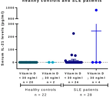
(39) Clinical parameters did not modify after supplementation however we wanted to examine if IL-21 serum levels or IL-21R expression changed with vitamin D supplementation in patients. We examined IL-21 serum levels in 28 patients and 22. 27.
(40) healthy controls, but we could only detect IL-21 in 5 patients (Figure 2), 2 of which entered the supplementation protocol (Figure 3). As we could not detect IL-21 in the serum from most of the individuals, we activated PBMC derived both from healthy controls and SLE patients for 24 hours with PMA/Ionomycin in the presence of different vitamin D concentrations and evaluated the supernatants for IL-21 by ELISA. This assay was performed from 4 healthy controls and 3 SLE patients. IL-21 was not detected in any of the supernatants, even recombinant IL-21 was detected in the same ELISA assay. IL-21 mRNA expression in 14 SLE patients and 6 healthy donors was studied however no differences were found between groups (Figure 4). IL-21 mRNA did not change after supplemented patients received either vitamin D or placebo (Figure 5). S e r u m IL - 2 1 le v e ls in h e a lt h y c o n t r o ls a n d S L E p a t ie n t s S e r u m IL - 2 1 le v e ls (p g /m l). 1000. 900. N o s e d e t e c tó IL - 2 1 e n. 300. 2 2 c o n tr o le s s a n o s 2 3 p a c ie n te s. 200. 100. 0. V it a m in D. V it a m in D. V it a m in D. V it a m in D. < 3 0 n g /m l. 3 0 n g /m l n = 2. < 3 0 n g /m l. 3 0 n g /m l n = 4. n = 20. n = 24. H e a lth y c o n tro ls. S L E p a tie n ts. n = 22. n = 28. Figure 2: Serum IL-21 levels in healthy controls and in SLE patients. Serum IL-21 was analyzed by ELISA in 22 healthy controls and 28 patients, IL-21 was only detected in 5 patients and no 28.
(41) differences were found between patients with normal or low serum vitamin D levels.. Figure 3: Serum IL-21 levels in SLE patients before and after supplementation. Serum IL-21 was analyzed by ELISA in SLE patients supplemented with placebo (n =11) or vitamin D (n=11). We detected IL-21 in one patient who received placebo, 225.24/256.17 pg/ml (visit 1/visit 2), and in one patient that received vitamin D, 52.84/56.85 pg/ml (visit 1/visit 2). Mean ± SD is shown.. 29.
(42) IL 2 1 m R N A e x p r e s s i o n i n. R e la t iv e e x p r e s s io n IL 2 1 /R P L 3 2. c o n t r o ls a n d S L E p a t ie n t s p = 0 .7 7 6. 1200 1000 800 600 400 200 0. H e a l th y c o n tr o l s. S L E p a ti e n ts. n = 6. n = 14. Figure 4: IL21 mRNA expression on PBMCs from healthy controls and SLE patients. We analyzed IL21 mRNA expression by qPCR on peripheral mononuclear cells from 6 controls and 14 SLE patients, and we found no differences between groups. Mann Whitney U test, p = 0.529. Mean ± SD is shown. p < 0.05 for significance.. 30.
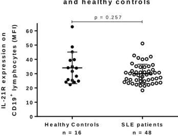
(43) IL 2 1 m R N A e x p r e s s io n o n P B M C s fr o m S L E p a t ie n t s. R e la t iv e e x p r e s s io n IL 2 1 /R P L 3 2. s u p p le m e n te d w ith v ita m in D o r p la c e b o p = 0 .1 2 5. 2500 p = 0 .8 7 5 2000 1500 1000 500 0. P la c e b o V ita m in D P la c e b o V ita m in D n=4 n=5 n=4 n=5. T0. T3m. S U P P L E M E N T A T IO N P R O T O C O L. Figure 5: Vitamin D supplementation does not modify IL-21 mRNA expression on PBMC in SLE patients. IL21 mRNA expression was examined in PBMC isolated from SLE patients supplemented with placebo (n = 4) or vitamin D (n = 5). There is no difference in IL21 expression before and after receiving vitamin D or placebo. Significance was tested by Wilcoxon matched-pairs signed rank Test. Mean ± SD is shown. p < 0.05 for significance. IL-21R expression in B lymphocytes in SLE patients compared to healthy subjects was also analyzed, as the evidence does not provide any conclusion to whether this receptor is up-regulated in B cells from SLE patients. We found no differences in IL-21R 31.
(44) expression in B cells between SLE patients and healthy controls (29.8 ± 6.7 and 34.0 ± 11.1) (Figure 6a). When we compared if IL-21R expression changed with vitamin D supplementation, no differences were found in the vitamin D group (13.1 ± 5.5 vs. 13.9 ± 5.9) or placebo group (14.8 ± 6.6 vs. 15.4 ± 6.6) (Figure 6b). In this objective, we found that IL-21 is not up-regulated in SLE patients. Vitamin D supplementation does not modify IL-21 or its signaling in SLE patients.. IL - 2 1 R e x p r e s s io n in S L E p a t ie n t s a n d h e a lth y c o n tr o ls. ly m p h o c y t e s (M F I). 60 50 40 30 20. +. C D 19. IL - 2 1 R e x p r e s s io n o n. p = 0 .2 5 7. 10 0 H e a lt h y C o n t r o ls. S L E p a t ie n t s. n = 16. n = 48. Figure 6: IL-21R expression on B cells from healthy controls and SLE patients. PBMC isolated from healthy patients and SLE patients were stained with FITC-anti-CD19 and PE-anti-IL-21R, fixed and acquired with BD FACSCalibur cytometer. There were no significant differences in IL-21R expression in B cells between patients and controls (29.8±6.7 and 34.0±11.1) Mean intensity fluorescence (MFI) for IL-21R on B cells is shown. Significance was tested by MannWhitney U Test. 32.
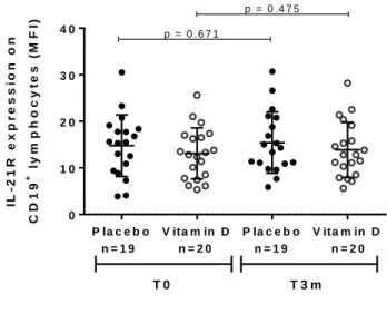
(45) IL - 2 1 R e x p r e s s io n o n C D 1 9. +. ly m p h o c y t e s f r o m. S L E p a t i e n t s s u p p le m e n t e d w it h v it a m in D o r p l a c e b o ly m p h o c y te s (M F I). p = 0 .6 7 1. 30. 20. 10. +. CD19. IL - 2 1 R e x p r e s s i o n o n. p = 0 .4 7 5. 40. 0 P la c e b o n=19. V ita m in D n=20. P la c e b o n=19. T0. V ita m in D n=20. T3m. S U P P L E M E N T A T IO N P R O T O C O L. Figure 7: Vitamin D supplementation does not modify IL-21R expression on CD19+ lymphocytes in SLE patients. PBMC were isolated from SLE patients supplemented with placebo or vitamin D, stained with FITC-anti-CD19 and PE-anti-IL-21R, fixed and acquired with BD FACSCalibur cytometer. There was no difference in IL-21R expression before and after receiving vitamin D (13.1 ± 5.5 vs. 13.9 ± 5.9) or placebo (14.8 ± 6,6 vs 15.4 ± 6.6). Mean intensity fluorescence (MFI) for IL-21R on B cells is shown. Significance was tested by Wilcoxon matched-pairs signed rank Test.. 33.
(46) CHAPTER 2: VITAMIN D EFFECT ON IL-21/IL-21 SIGNALING IN B CELLS IN VITRO. OBJECTIVE 2: Evaluate vitamin D effect on T lymphocytes IL-21+ in vitro. In this objective, the aim was to analyze IL-21 in T lymphocytes, as we could not previously detect IL-21 in serum from healthy controls and SLE patients, and it has been reported that CD4+ T cells positive for IL-21 are increased in SLE patients compared to healthy individuals (40, 41). We activated PBMC from healthy controls and SLE patients with PMA, Ionomycin and Brefeldin A for 4 hours and we found that CD8- T cells positive for IL-21 were higher in SLE patients compared to healthy individuals (4.5±1.7% vs. 1.8±0.4%) (Figure 7b). Vitamin D has immunomodulatory effects (22), so it could modulate CD8- IL-21+T cells. As previous results were evaluated in SLE patients and local effects may not be seen in peripheral blood, we analyzed the vitamin D effect on PBMC from healthy donors. To evaluate this, PBMC were exposed to 1α,25-dihydroxyvitamin D3 (1α,25(OH)2D3) simultaneously with PMA/Ionomycin and brefeldin A for 4 hours. 1α,25(OH)2D3 at 10 and 100 nM concentrations did not change the frequency of T cells positive for IL-21 (Figure 7c).. 34.
(47) Figure 8. CD8- T cells positive for IL-21 are higher in SLE patients. PBMC were isolated from healthy and SLE subjects and stimulated with PMA/ionomycin and brefeldin A in vitro. The cells were fixed, and permeabilized, followed by staining with FITC-anti-CD8a, PE-anti-IL-21 and APC-anti-CD3. Due to the down-regulation of CD4 after stimulation with PMA/Ionomycin, cells were gated for CD3+CD8- T-cell subset. The frequency of peripheral blood CD3+CD8-IL-21+ cells was determined by flow cytometry. A. Representative data from a patient sample is shown. B. Percentage of CD3+CD8-IL-21+ cells in healthy controls and SLE patients is represented. Significance was tested by Mann-Whitney U test. C. PBMC isolated from healthy donors were stimulated with PMA/ionomycin and brefeldin A in vitro in the presence of different concentrations of 1α,25-(OH)2D3 or vehicle control for 4 hours. CD3+CD8-IL21+population was analyzed for each condition and normalized for CD3+CD8-IL-21+ population in PMA/ionomycin condition (control). Data from 5 independent. 35.
(48) experiments is represented. Mean ± SD is shown. Significance was tested by Friedman test, * p<0.05 for significance.. 36.
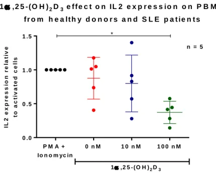
(49) Vitamin D effect IL-2 expression in activated PBMC To test vitamin D, we analyzed its suppressive effect on IL-2 mRNA expression, as it can reduce IL-2 expression in activated lymphocytes has been described (63). PBMC from 3 healthy controls and 3 SLE patients were activated for 24 hours with PMA and Ionomycin, and IL-2 mRNA expression was assessed by qPCR. We found that in all, but one individual, vitamin D reduced IL-2 expression, so Grubbs' test for outliers was performed, detecting this subject as an outlier (Grubbs test, α = 0.01). When this data point was excluded, 100 nM vitamin D significantly reduced IL-2 expression in activated PBMC (Figure 9).. 37.
(50) 1 ,2 5 -(O H ) 2 D 3 e f f e c t o n IL 2 e x p r e s s io n o n P B M C f r o m h e a lt h y d o n o r s a n d S L E p a t ie n t s * n = 5 t o a c t iv a t e d c e lls. IL 2 e x p r e s s io n r e la t iv e. 1 .5. 1 .0. 0 .5. 0 .0 PM A +. 0 nM. 10 nM. 100 nM. Io n o m yc in 1 ,2 5 - (O H ) 2 D 3 F r ie d m a n te s t, p = 0 .0 1 7. Figure 9: 1α,25-(OH)2D3 reduces IL2 expression on activated PBMC from healthy controls and SLE patients. PBMC from 3 controls and 2 SLE patients were activated with PMA/Ionomycin in vitro in the presence of different concentrations of 1α,25-(OH)2D3 or vehicle control for 24 hours. We analyzed IL2 mRNA expression by qPCR finding that 100 nM 1α,25-(OH)2D3 significantly reduced IL2 expression, p = 0.017. 5 independent experiments are shown. Mean ± SD is shown. Significance was tested by Friedman test, p<0,05 for significance. *post test: Dunn's multiple comparisons test significant: PMA + Ionomycin vs PMA + Ionomycin + 1α,25-(OH)2D3 100 nM, p<0.05. 38.
(51) OBJECTIVE 3: Determine vitamin D effect on IL-21 signaling pathway in B lymphocytes from healthy controls in vitro. 1α,25-(OH)2D3 effect on IL-21R expression To explore the effect of vitamin D on IL-21 signaling, we analyzed IL-21R expression on activated B cells purified from PBMC of healthy controls, this was stimulated for 3 days with αIgM and αCD40, as described by Chen (24). Compared with day 0 expression, the mean fold increase of IL-21R by B cells after 3 days of activation was 276%, but vitamin D had no effect on IL-21R expression at concentrations of 10 and 100 nM (Figure 10b). This is consistent with the findings in patients, where vitamin D supplementation did not modify IL-21R expression in B cells after 3 months.. 39.
(52) 40.
(53) Figure 10. 1α,25-(OH)2D3 had no effect on IL-21R expression in activated B cells from healthy controls. B cells were purified from PBMC isolated from healthy subjects. Purified B were stimulated with αIgM and αCD40 for 3 days in vitro. The cells were stained with FITC-anti-CD19 and PE-anti-IL-21R, fixed and acquired using BD Accuri C6 flow cytometer at day 0, 1 and 3. A. Cells were gated for CD19 positive cells (B cells) and MFI for IL-21R in B cells was analyzed. Negative control is unstimulated B cells with isotype PE conjugated antibody. Representative data from one subject is shown. B. IL-21R expression changed at day 3 compared to day 0 after stimulation, IL-21R expression for each time points and condition was normalized with IL-21R expression (MFI) at day 0. C. Stimulated B cells were exposed to 1α,25-(OH)2D3 at different concentrations for three days. Data from 7 independent experiments is represented. D. IL-21R expression changed at day 3 compared to day 0 after stimulation, but vitamin D did not change it. Significance was tested by Friedman test at day 3 (p = 0.801), p<0.05 for significance. Mean ± SD is shown.. 41.
(54) 1α,25-(OH)2D3 effect on IL-21 induced STAT3 phosphorylation. To evaluate vitamin D effect on STAT3 phosphorylation (pSTAT3) in B cells, we had to achieve the best conditions to stimulate it, because previous reports used different protocols to assess IL-21 induced STAT3 phosphorylation. Ding et al. described that overnight incubation of B cells with fibroblasts expressing CD40L improved IL-21 induced STAT3 phosphorylation on B cells (64). According to this information, we stimulated B cells from healthy controls overnight with αIgM/αCD40 and then exposed them to IL-21 during different time points. As previously reported, we found that IL-21 induces STAT3 phosphorylation at 30 minutes, compared to B cells stimulated only with αIgM/αCD40 (Figure 11a and b) (54). So, we used this system to evaluate 1α,25(OH)2D3 effect on pSTAT3, incubating B cells with 1α,25-(OH)2D3 for the same period as IL-21 stimulus. We found that vitamin D did not change IL-21 induced STAT3 phosphorylation at 10 and 50 nM (Figure 11c).. 42.
(55) 43.
(56) Figure. 11.. 1α,25-(OH)2D3 exerts. no. effect. on. IL-21. induced. STAT3. phosphorylation of B cells from healthy controls. B cells were purified from PBMC isolated from healthy subjects. Purified B were stimulated overnight with αIgM and αCD40, following 30 minutes with IL-21 in vitro. The cells were fixed, and permeabilized, followed by staining with FITC-anti-CD20, PE-anti-pSTAT3 and APCanti-STAT3 and acquired using BD Accuri C6 flow cytometer. A. CD20 positive cells (B cells) and MFI for pSTAT3 were analyzed. Representative data from one subject is shown. MFI pSTAT3/ MFI STAT3 was evaluated. B. Purified B were stimulated with αIgM and αCD40 with IL-21 for different times. C. Purified B were stimulated with αIgM, αCD40 and IL-21 in the presence of different concentrations of 1α,25-(OH)2D3 or vehicle control for 30 minutes. MFI pSTAT3/ MFI STAT3 was analyzed for each condition. Four independent experiments are represented. Significance was tested by Friedman Test, p= 0.006, *post test: Dunn's multiple comparisons test significant: Basal vs. αIgM + αCD40 + IL-21 and αIgM + αCD40 vs. αIgM + αCD40 + IL-21, p<0.05 for significance. Mean ± SD is shown.. 44.
(57) CHAPTER 3: IL-21 POLYMORPHISMS AND SLE OBJECTIVE 4: Evaluate an association between IL-21 polymorphisms and vitamin D supplementation in SLE patients. Association between IL-21 polymorphisms and SLE. We aimed to analyze if there is an association between IL-21 polymorphisms and SLE, as this has been described before in European descendants, also in African Americans for rs907715 polymorphism (45, 46) and in a Colombian cohort for rs6822844 (47). We analyzed rs907715 polymorphism in 30 control subjects and 62 SLE patients and we could not find an association of genotype or allele (χ2=0.901, p=0.636; Fischer’s test, p=1) (Table 3). For rs6822844 polymorphism, we could not find any association (χ2=0.396, p=0.529; Fischer’s test, p=0.635) (Table 4).. 45.
(58) Then, we wanted to address if the genotype of each patient could influence the change of vitamin D serum levels in patients who received vitamin D. For rs907715 polymorphism, we found no differences in vitamin D supplementation between genotypes, but when we compared heterozygote genotype vs. homozygote genotypes, serum vitamin D levels increased more in patients with CT genotype than in patients with CC genotype (Δ 33.6 ± 3.2 vs. 23.4 ± 9.6) (Figure 12a).. 46.
(59) Figure 12: Changes in vitamin D levels after supplementation according to IL-21 polymorphism genotype. A: rs907715. Vitamin D levels increased in patients with CC genotype 23.4 ± 9.6 ng/ml, those with CT genotype improved in 33.6 ± 13.2 ng/ml and patients with TT genotype augmented in 20.7 ± 9.9 ng/ml. No differences were found between groups. Kruskal-Wallis test, p=0.102. B: rs6822844.. Patients with GG. genotype improved vitamin D levels in 23.2 ± 10.9 ng/ml, those with heterozygote GT genotype augmented in 31.1 ± 19.9 ng/ml and patients with TT genotype improved in 23.9 ± 11.7 ng/ml. No differences were found between heterozygote and homozygote GG genotype. Mann-Whitney test, p=0.594. p < 0.05 for significance. Finally, disease features were analyzed to establish any association with IL-21 polymorphisms, which is reported in Tables 5 and 6. We found that rs907715 TT genotype was associated with older patients when the sample was taken (p = 0.005) and methotrexate exposure (p = 0.025). rs6822844 was associated with photosensitivity (p = 0.036) and a tendency to older aged patients when the sample was taken was seen (0.065). 47.
(60) 48.
(61) 49.
(62) 14. DISCUSSION There is growing evidence that vitamin D has a controversial role in the regulation of the immune system, which can influence SLE pathology. Also, vitamin D mechanisms are not fully understood, but one candidate molecule could be IL-21, which has been implied in SLE pathology. We found that vitamin D did not modulate IL-21R in supplemented SLE patients and IL-21R expression and pSTAT3 in B cells from healthy controls in vitro. First, to address the role of IL-21 in SLE, we analyzed its serum levels in SLE patients and healthy controls by ELISA, but we could not detect the cytokine in the serum from most of our subjects. This finding differs from what has been described previously by Wong and Lee, where IL-21 was detected and increased in SLE patients compared to healthy controls (38, 39). Those studies used both in-house and a discontinued ELISA assay, making it difficult for us to replicate their results. It is worth mentioning that previous studies that analyzed the role of IL-21 in SLE patients, evaluating IL-21 serum levels, used ELISA assays from different manufacturers, which varies in the detection limit and range for detection (65, 66). As we could not reproduce data from previous reports, we evaluated two different ELISA for IL-21, but we could not detect IL-21. As IL-21 participates in germinal center reaction in follicles, it could be undetectable in serum, making it necessary to study the source of this cytokine, T helper lymphocytes. Due to these results, we evaluated T cell population positive for IL-21, as the studies performed by Terrier and Dolff reported that this subset also increased in SLE patients compared to healthy individuals (40, 41). We found that in a Mestizo population, the fraction of CD8- T cells positive for IL-21 was higher in SLE patients compared to healthy individuals, consistent with Terrier’s findings (Figure 3b). Another reason why we could not detect IL-21 in serum from SLE patients could be disease activity, as it has 50.
(63) been reported that IL-21 mRNA is higher in patients with active disease, and we analyzed IL-21 in serum from patients with mild to moderate disease. For our second objective, we recruited patients with a mild to severe disease, but we did not find any correlation between SLE Disease Activity Index (SLEDAI) and CD8-IL-21+, as Dolff had also described. Palandra and colleagues detected IL-21 from activated T cells from healthy donors, but they did not detect IL-21 in several hundred serum samples from healthy and autoimmune disease patients using LC-MS/MS (67), comparable to what we describe in this study. Finally, the lack of detection could be due to the population studied, as genetics play a role in SLE disease and most of the groups that reported increased IL-21 serum levels in SLE were performed in Oriental (or Asian) cohorts and in this ethnic group a higher prevalence of hematogical manifestations has been described which correlated with IL21(68, 69). Many studies where T cell populations positive for IL21+ are compared between healthy controls and SLE patients are European, and they do not report IL-21 serum levels. It has been shown that vitamin D exerts an immunomodulatory effect on lymphocytes, so we wanted to address the effect of vitamin D on IL-21 expression in T cells. No differences in IL-21+ T cells were found. Previously, Jeffery found that 1α,25-(OH)2D3 reduces T CD4+IL-21+ at 5 days of culture, which contrasts with our results, which could be due to the time lymphocytes were exposed to the vitamin (58). Also, the expression of vitamin D receptor (VDR) may explain the lack of effect of 1α,25-(OH)2D3 in this study, as it has been reported that VDR must be induced in T cells, through the activation of this cell type. So, at 4 hours, vitamin D could only exert its function in a non-genomic way (70). There is only one study by Ikeda and colleagues, where human IL-21 mRNA levels were analyzed in the presence of this vitamin, where 1α,25-(OH)2D3 reduced IL-21 and IL-21R mRNA in CD4+ T cells polarized to TH17 conditions for 6 51.
(64) days (57).There are two main differences between our study and the one carried out by Ikeda: we analyzed protein expression, which may not correlate with mRNA expression. The second one is that Ikeda only analyzed TH17 subset and IL-21 is not only expressed in this TH subgroup, as it is also expressed in T follicular helper cells (TFH), the subset that specifically provides help for B cell differentiation (5). We also analyzed vitamin D effect in IL-21 mRNA expression in PBMC from supplemented SLE patients, finding that vitamin D did not reduce the cytokine expression neither in patients who received vitamin D or placebo. Seven individuals were recruited where IL-21 could be detected in T lymphocytes by flow cytometry and PBMC was activated for 24 hours in the presence of vitamin D. Supernatants were analyzed for IL-21 by ELISA and the cytokine was not detected, which corroborated our previous results where IL-21 was undetectable in serum from patients and controls. Next, we analyzed if the lack of effect of vitamin D on IL-21 expression was specific by evaluating its effect on IL-2 mRNA expression, which has been described as downregulated in lymphocytes exposed to vitamin D (63). We activated PBMC derived from 5 subjects in the presence of vitamin D for 24 hours and we found that 100 nM vitamin D reduces IL-2 expression, which indicates that vitamin D has an inhibitory effect on IL2, but not on IL-21. Vitamin D has been implicated in plasma cell differentiation (24), and reduces B cell proliferation but the mechanism is not fully understood. IL-21 plays a role in these two processes, so vitamin D can exert its function by modifying the cytokine receptor, IL21R, expression. To analyze what happens with IL-21 signaling in patients after receiving vitamin D, we examined IL-21R expression in B cells. This parameter was not modified after patients received either vitamin D or placebo. This may be due to the population that expresses IL-21R, as Le Coz demonstrated that this receptor is mostly 52.
(65) expressed by mature naïve CD19+ B cells, the population does not change after vitamin D supplementation (71). On the other hand, antibody secreting cells population is decreased after vitamin D supplementation (72), which may explain the differences found by Abou-Raya after supplementation in ds-DNA antibodies title and SLEDAI (28). No differences in IL-21R expression in B cells from SLE patients and healthy controls were found, which is not consistent with two previous studies, where it has been reported that IL-21R expression is down-regulated in SLE patients. This difference could be due to the same reason that we could not detect IL-21 serum levels as these studies were also performed in Asian cohorts (38, 73). Then, vitamin D immunomodulatory effects directly on B cells from healthy donors in vitro were evaluated, analyzing IL-21R expression and IL-21 induced STAT3 phosphorylation in B cells exposed to vitamin D. IL-21R expression is up-regulated when B cells were activated for 3 days, but remained unchanged when B cells were exposed to vitamin D. This is consistent with the findings in SLE patients, where vitamin D supplementation did not modify IL-21R expression in B cells after 3 months. We analyzed IL-21R expression at day 3, as vitamin D receptor is expressed in unstimulated B cells, and up-regulated when they are activated or exposed to 1α,25(OH)2D3 for 3 days in vitro (24). Previously, there is only one report that described that vitamin D could decrease IL-21R mRNA expression in T cells in TH17 polarizing conditions (57). This is the first report where IL-21R expression is analyzed in B cells exposed to vitamin D. Chen et al. examined the expression of several genes involved in plasma differentiation in the presence of 1α,25-(OH)2D3, but. did not find any. difference, which is in concordance with our findings (24). IL-21 predominantly signals through the STAT3 pathway. This transcription factor has been implied in SLE pathogenesis, as its phosphorylation is augmented in T and B cells derived from SLE patients compared to healthy controls (53) and because STAT3 is 53.
(66) necessary for plasma cell differentiation (74). We show that Vitamin D did not change STAT3 phosphorylation induced by IL-21 stimulation. This is the first study that examines the effect of vitamin D on STAT3 signaling in B cells, moreover, there are no studies on the role of vitamin D on STAT3 signaling in human immune cells. Previously, Muthian reported that IL-12 induced STAT3 phosphorylation is inhibited by 1α,25-(OH)2D3 in murine T cells in a short time period, even though the hormone concentrations are higher than the ones reported to inhibit T cell proliferation (59). On the other hand, Tang studied long time effects of vitamin D over STAT3 phosphorylation in T cells differentiated towards TH17 phenotype, where they did not find any difference in pSTAT3 in cells exposed to 1α,25-(OH)2D3 for 24 or 72 hours (75). This last study cannot show what really happens in vivo, as transcription factors as STAT3 on the nucleus only last 15 minutes, so long term effects could not change its function. It is important to evaluate pSTAT3 in T and B cells, as there are differences in IL-21 induced STAT3 phosphorylation from cells derived from SLE patients compared to healthy controls according to the cell type studied, and the studies have discordances. Rasmussen reported that IL-21 induced STAT3 phosphorylation is lower in T and B cells from SLE patients than from healthy controls (76), while Wu reported that IL-21 signaling is not impaired in B cells derived from SLE patients (54). We could not find an association between IL-21 polymorphisms and SLE. This finding is different from what has been described before. One reason could be the size of our cohort, other studies have more than 100 cases and controls enrolled. Salwalha et al. described that rs907715 was associated with SLE in a European descent cohort (644 cases/controls) but no association was found in an African American cohort in 366 cases/controls (45). When population was increased this polymorphism was associated, as Hughes group reported in 1569 patients and 1893 controls, (46). The rs907715 is associated to SLE in a European descent cohort, no association was found in a Hispanic Cohort (151 cases/controls). This discrepancy was also seen in the association of 54.
Figure


+3
Documento similar
cruzi infection and ET-1 cooperatively activated the Ca 2+ / calcineurin (Cn)/nuclear factor of activated T cells (NFAT) signaling pathway in atrial myocytes, leading to COX-2
The regulation of more than thousand vitamin D target genes in colon carcinoma cells, normal stem cells, cancer stem cells, stromal NFs and CAFs as well as immune cells of the
In summary, our findings suggest that expression changes in individual miRNAs in TIA-depleted HeLa cells could directly or indirectly impact on biological processes and
We also observed that OASFib and dermal fibroblasts, which do not constitutively express IL-15, VCAM-1 nor BAFF on the cell membrane, were significantly less effective than RASFib
(C) Phenotype of human B-cells, monocytes, pDCs, and cDCs from healthy controls (HCs) and inflammatory bowel disease (IBD) patients based on the basal expression of CCR2, CD40,
The influence of puberty on vitamin D status in obese children and the possible relation between vitamin D deficiency and insulin
Growth regulation of human colon cancer cells by epidermal growth factor and 1,25-dihydroxyvitamin D 3 is mediated by mutual modulation of receptor expression. The down-regulation
(2005) Differential regulation of phosphorylation of the cAMP response element-binding protein after activation of EP2 and EP4 prostanoid receptors by prostaglandin E2.. Inhibition
