Involvement of WNT signaling pathway in the adult hippocampal synapses :role in neuroprotection
137
0
0
Texto completo
(2) Pontificia Universidad Católica de Chile Facultad de Ciencias Biológicas Departamento de Biología Celular y Molecular. INVOLVEMENT OF Wnt SIGNALING PATHWAY IN THE ADULT HIPPOCAMPAL SYNAPSES: ROLE IN NEUROPROTECTION. Tesis entregada a la Pontificia Universidad Católica de Chile en cumplimiento parcial de los requisitos para optar al grado de Doctor en Ciencias con mención en Biología Celular y Molecular.. By: JESSICA Y. VARGAS.. Advisor: Dr. NIBALDO C. INESTROSA. Co-advisor: Dr. MARCO FUENZALIDA.. June 2014..
(3) I. To my beloved parents, Juan and Bidita and my little brother, Christian..
(4) II. ACKNOWLEDGEMENTS. I am sincerely grateful to: My advisor, Dr. Nibaldo C. Inestrosa, for the continuous support and guidance throughout my research project, for the stimulating discussions and his helpful suggestions. Also, his effort in proof reading the drafts, are greatly appreciated. Dr. Marco Fuenzalida, for allowing me to work in his lab, where I carried out several of the experiments presented here. Also, his advice helped me to design and performance the research and to write this thesis. Members of the evaluation committee: Dr. Mario Herrera-Marchitz, Dr. Katia Gysling, Dr. Juan Larraín, and Dr. Alejandra Alvarez (Head of the PhD Program), who reviewed this thesis and contributed to improve the manuscript with their insightful comments and corrections. I also thanks to Fundación Gran Mariscal de Ayacucho (Venezuela) and Vicerrectoría de Investigación/P. Universidad Católica Chile (Chile) for the pre-doctoral fellowships. This work was supported by grants from the Basal Center of Excellence in Science in Technology (CONICYT-PFB12/2007) and FONDECYT N°1120156 to Dr. Nibaldo C. Inestrosa and grants from FONDECYT N°11090059, N°1130614 and DIPUV CID 01/2006 to Dr. Marco Fuenzalida..
(5) III. INDEX. Abbreviations. VI. Abstract. VIII. Resumen. IX. 1. Introduction. 1. 1.1 Wnt signaling pathway. 1. 1.2 Role of Wnt signaling in the developing central nervous system. 5. 1.3 Expression of Wnt signaling components in the adult brain. 9. 1.4 Role of Wnt signaling in mature synapses. 11. 1.5 Wnt signaling in Alzheimer’s disease. 15. 1.5.1 Alzheimer’s disease. 15. 1.5.2 Deregulation of Wnt signaling in Alzheimer’s disease. 17. 1.6 What is the physiological role of Wnt signaling in the adult brain?. 20. 1.7 Hypothesis and Objectives. 22. 2. Chapter I. 23. 2.1 Abstract. 25. 2.2 Introduction. 25.
(6) IV. 2.3 Materials and Methods. 27. 2.4 Results. 32. 2.5 Discussion. 43. 2.6 References. 48. 2.7 Figures and Tables. 54. 3. Chapter II. 68. 3.1 Abstract. 70. 3.2 Introduction. 71. 3.3 Results. 73. 3.4 Discussion. 81. 3.5 Conclusions. 85. 3.6 Materials and Methods. 86. 3.7 References. 91. 3.8 Figures. 97. 4. Discussion. 106. 4.1 Wnt signaling as a key factor in synaptic remodeling. 106. 4.2 Wnt signaling as a synaptic plasticity regulator. 108. 4.3 Role of Wnt signaling in learning and memory. 112. 4.4 Wnt signaling as a therapeutic target in Alzheimer’s disease. 114. 5. References. 116. 6. Appendices. 127. 6.1 Appendix I. 128. 6.2 Appendix II. 130.
(7) V. FIGURES AND TABLES INDEX. 1.. Chapter I. Figure 1.. 54. 2.. Chapter I. Figure 2.. 56. 3.. Chapter I. Figure 3.. 58. 4.. Chapter I. Figure 4.. 59. 5.. Chapter I. Figure 5.. 61. 6.. Chapter I. Figure 6.. 62. 7.. Chapter I. Figure 7.. 63. 8.. Chapter I. Figure 8.. 65. 9.. Chapter I. Table 1.. 67. 10. Chapter II. Figure 1.. 97. 11. Chapter II. Figure 2.. 99. 12. Chapter II. Figure 3.. 101. 13. Chapter II. Figure 4.. 102. 14. Chapter II. Figure 5.. 103. 15. Chapter II. Figure 6.. 104.
(8) VI. ABBREVIATIONS. α7-nAChR: α7-nicotinic acetylcholine receptor Aβ: amyloid-β peptide ACSF: artificial cerebrospinal fluid AD: Alzheimer’s disease AMPA-R: α-amino-3-hydroxy-5-methyl-4-isoxazolepropionic acid receptor APC: adenomatosis polyposis coli APP: amyloid precursor protein APV: (2R)-amino-5-phosphonovaleric acid CA1: Cornu Ammonis area 1 CA3: Cornu Ammonis area 3 CaMKII: Ca2+-calmodulin-dependent protein kinase type II CK-1α: casein kinase-1α CNS: central nervous system Dkk-1: dickkopf-1 Dvl: disheveled fEPSP: field excitatory postsynaptic potential.
(9) VII. FOXY-5: formylated Wnt-5a-derived hexapeptide Fz: frizzled receptor GABA: γ-aminobutyric acid GSK-3β: glycogen synthase kinase-3β HFS: high frequency stimulation JNK: c-Jun N-terminal kinase LTD: long-term depression LTP: long-term potentiation l-LTP: late-LTP mEPSC: miniature excitatory postsynaptic current NMDA-R: N-Methyl-D-aspartate receptor GluN2B: NMDA receptor subunit 2B PHF-1: tau epitope paired-helical filament-1 PPF: paired-pulse facilitation PSD-95: postsynaptic density protein-95 PS-1: presenilin-1 sFRP: soluble Fz-related protein Syn-1: synapsin-1 Syp: synaptophysin Syt: synaptotagmin TCF/LEF: T cell factor/lymphoid enhancer factor WASP-1: Wnt-Activating Small molecule Potentiatior-1 WT: wild-type mice..
(10) VIII. ABSTRACT. Wnt ligands are signaling proteins expressed in several brain regions, where they function as activators of the Wnt signaling pathway. During the nervous system development, Wnt ligands play a key role in modulating axonal remodeling, dendritogenesis and synaptogenesis. In the adult brain, components of Wnt signaling are still expressed, but little is known about its role in mature synapses yet. Emerging in vitro studies have implicated Wnt signaling in synaptic plasticity of the adult brain. Additionally, it has been shown that activation of Wnt signaling protects against amyloid-β-induced synaptic impairment. In our laboratory, we have previously shown that some Wnt ligands are able to regulate synaptic transmission and specifically to induce modification at either the presynaptic or the postsynaptic structure. Besides, in vitro studies performed in our laboratory have revealed that some Wnt ligands are also able to protect against amyloid-β-induced cytotoxic and synaptotoxic insults. Now, in this work we have studied the involvement of Wnt signaling in the regulation of adult normal cognitive function, in vivo. Moreover, the beneficial effect caused by the activation of Wnt signaling in the cognitive function of double transgenic mouse, APPswe/PS1dE9, model of Alzheimer’s disease, has also been evaluated herein. The effects of Wnt signaling activation on the cognitive function of wild type and transgenic mice, was accomplished through the chronic administration of the Wnt signaling modulators WASP-1 and FOXY-5. As shown here, both treatments improve behavioral performance of mice evaluated for episodic memory tests. Electrophysiological recordings of hippocampal slices from animals treated with WASP-1 or FOXY-5 show an increase in both the excitatory synaptic transmission and the synaptic plasticity, compares to control mice. Furthermore, WASP-1 prevents synaptic protein loss, reduces the tau phosphorylation and blocks amyloid-β aggregation in transgenic mice. Altogether, these results suggest that Wnt signaling could play a key role in regulating synaptic plasticity of neural circuits and therefore controlling the outgoing cognitive function of adult mice. Moreover, the activation of Wnt signaling could rescue memory lost and improve amyloid-β-induced synaptic dysfunction in transgenic mice, thus Wnt signaling could be a promising new therapeutic target for Alzheimer’s disease treatment..
(11) IX. RESUMEN. Los ligandos Wnt son proteínas de señalización que se expresan en distintas regiones del cerebro, donde funcionan activando la vía de señalización Wnt, que modula los procesos de remodelamiento axonal, dendritogénesis y sinaptogénesis, durante el desarrollo del sistema nervioso. En el cerebro adulto, continúan siendo expresados los ligandos Wnt, así como también, los distintos componentes de la vía señalización, pero aún se desconoce qué función desempeñan en el sistema nervioso maduro. Evidencias recientes sugieren que la vía Wnt funciona en el cerebro adulto, modulando la plasticidad sináptica. Previamente, en nuestro laboratorio se demostró que distintos ligandos Wnt son capaces de regular la transmisión sináptica glutamatérgica y modificar de forma específica la estructura pre y post-sináptica. Además, otros estudios de nuestro laboratorio han mostrado que la activación de la vía Wnt protege frente al daño sináptico ocasionado por el péptido β-amiloide, in vitro. En esta tesis, se estudió in vivo la participación de la vía Wnt en la regulación de la función cognitiva de ratones adultos. Igualmente, se evaluó el efecto benéfico producido por la activación de la vía Wnt en ratones doble transgénico APPswe/PS1dE9, modelo de la enfermedad de Alzheimer. El efecto de la activación de la vía Wnt sobre la función cognitiva de ratones silvestres y transgénicos, se estudió mediante la administración crónica de los moduladores de la vía Wnt: WASP-1 y FOXY-5. Ambos tratamientos produjeron una mejora en la memoria episódica de los animales tanto silvestres como transgénicos, sometidos a distintas pruebas conductuales. El análisis de los registros electrofisiológicos en rebanadas de hipocampo obtenidas de los animales tratados con WASP-1 o FOXY-5, reveló que ambos tratamientos inducen un aumento en la trasmisión sináptica y en la potenciación sináptica a largo plazo, en comparación con el grupo control. Además, WASP-1 previene la pérdida de proteínas sinápticas, reduce la fosforilación de la proteína tau y bloquea la agregación del péptido β-amiloide, en los ratones transgénicos. Todos estos resultados sugieren que la vía Wnt podría regular la plasticidad de los circuitos cerebrales y por lo tanto, mejorar la función cognitiva en ratones adultos. Asimismo, la activación de la vía Wnt podría rescatar la pérdida de memoria observada en los ratones transgénicos y disminuir el daño sináptico ocasionado por el péptido β-amiloide, lo que coloca a esta vía como un prometedor blanco terapéutico para el tratamiento de la enfermedad de Alzheimer..
(12) 1. 1.. INTRODUCTION. 1.1. Wnt signaling pathway. Wnts are extracellular ligands that play a key role as developmental regulators involved in several cellular processes, including cell proliferation, differentiation, migration, adhesion, survival and apoptosis (Moon et al., 2002; Nusse, 2012; Willert and Nusse, 2012). Wnts are palmitoylated glycoproteins, mostly conformed by about 350 amino acids long and 40 kDa molecular weight (Coudreuse and Korswagen, 2007). Wnts act as signaling molecules in all metazoan organisms, controlling a wide spectrum of cellular processes during the development and in the adulthood (van Amerongen and Nusse, 2009). The mechanisms involved in the synthesis and secretion of Wnts have been first and foremost studied for Drosophila Wnt homolog, Wingless (Wg). Thus, through this model, Wnt secretion has been proposed to take place mainly by a constitutive pathway, which could be involved in a short-range signaling from the Wnt-producing cell (Ching et al., 2008; Hausmann et al., 2007). Alternatively, a small fraction of Wnt binds to lipoprotein particles possibly within specialized endosomal compartments, during the secretion route (Harterink and Korswagen, 2011; Panakova et al., 2005), and the Wnt-lipoprotein complex formed may participate in a long-range signaling after been released outside of its producing cell.
(13) 2. (Hausmann et al., 2007; Lorenowicz and Korswagen, 2009). Although, there is not enough evidence that support signaling activity of the Wnt-lipoprotein complex, it is possible that this complex allows Wnts to signal along the morphogenetic field (Bartscherer and Boutros, 2008). Released Wnt ligands bind to specific receptors at the cell membrane surface. Frizzled receptor (Fz), a protein that have seven transmembrane-spanning domains, was the first receptor described as responsible for Wnt signal transduction (Schulte and Bryja, 2007). The binding of Wnt to Fz, activates the cytoplasmic protein Disheveled (Dvl), which in turn modulates the activity of other components of the pathway (Nusse, 2012). Downstream of Dvl, Wnt signaling cascade diverges into three different branches: Wnt/β-catenin pathway, also called canonical Wnt pathway, Wnt/JNK pathway and Wnt/Ca2+ pathway, known together as non-canonical Wnt pathway (Ciani and Salinas, 2005) (see Appendix I for a schematic model of Wnt pathways). In the Wnt/β-catenin pathway, Dvl activation induces the disassembly of “β-catenin destruction complex” formed by glycogen synthase kinase-3β (GSK-3β), casein kinase-1α (CK-1α), adenomatosis polyposis coli (APC) and axin (Moon, 2005). In the absence of Wnt, β-catenin is sequentially phosphorylated by CK-1α and GSK-3β, targeting β-catenin for ubiquitination and proteasome degradation (Moon, 2005; Nusse and Varmus, 2012). The activation of this pathway starts when Wnt binds to Fz receptor and to its co-receptor, the low-density lipoprotein receptor-related protein 5/6 (LRP-5/6), inducing recruitment of GSK-3β binding protein (GBP) by Dvl, which causes sequestration of the enzyme and prevents β-catenin phosphorylation (Clevers and Nusse, 2012). Thus, β-catenin is stabilized and accumulated in the cytoplasm, but then β-catenin enters the nucleus and interacts with members of the DNA‐binding T cell factor/lymphoid enhancer factor (TCF/LEF) family.
(14) 3. proteins to activate transcription of Wnt target genes (Archbold et al., 2011). Several Wnt target genes are activated in this process, including: c-myc (He et al., 1998), cyclin-D1 (Shtutman et al., 1999), cyclo-oxygenase-2 (Howe et al., 1999), neurogenin-1 (Hirabayashi et al., 2004) and Ca2+- calmodulin-dependent protein kinase type IV (CaMKIV) (Arrazola et al., 2009). Therefore, activation of Wnt/β-catenin pathway regulates gene transcription. The Wnt/JNK pathway was first described in Drosophila, where it controls planar cell polarity (PCP), hence it is also known as Wnt/PCP pathway (De Ferrari and Moon, 2006). In this pathway, the binding of Wnt to Fz is followed by the activation of both Rho and Rac small GTPases, which in turn stimulate c-Jun N-terminal kinase (JNK) (Widelitz, 2005). The downstream effects of this pathway include regulation of the actin cytoskeleton and microtubules organization, cell migration and gene expression, through activation of JNKdependent transcription factors, such as the activating transcription factor-2 (ATF-2) (Kikuchi et al., 2011; Simons and Mlodzik, 2008). In the Wnt/Ca2+ pathway, the binding of Wnt to Fz, results in the stimulation of heterotrimeric G-proteins, which in turn activates phospholipase-C (PLC) inducing the generation of diacylglycerol (DG) and inositol trisphosphate (IP3). Then, IP3 increases the intracellular Ca2+ release and decreases cyclic guanosine monophosphate (cGMP), resulting in the activation of Ca2+-calmodulin-dependent protein kinase type II (CaMKII), calcineurin and protein kinase-C (PKC) (De, 2011). Downstream of this pathway, the transcription factor cAMP response element-binding protein-1 (CREB), is activated (Kohn and Moon, 2005). The activation of a Wnt pathway in particular can depend, among other factors, on the binding of a specific Wnt ligand to its Fz receptor. Thus, Wnts are usually classified as canonical ligands if they activate Wnt/β-catenin signaling or non-canonical ligands, if they activate either Wnt/JNK pathway or Wnt/Ca2+ pathway, instead (Widelitz, 2005)..
(15) 4. Until now, 19 different Wnt ligands and 10 members of Fz receptors family have been described in mammals (Kikuchi et al., 2011). Therefore, signaling specificity can depend not only on the type of ligand bound, but also on the receptors and co-receptors involved. Additionally, several secreted antagonists that act extracellularly to block or to reduce Wnt signal response can regulate this pathway (Nusse, 2012). For instance, Wnt signaling can be inhibited, among other molecules, by soluble Fz-related proteins (sFRP), which compete with Fz receptors for binding to Wnts; or by Dickkopf (Dkk), a protein that specifically inhibits Wnt/β-catenin signaling through interaction with the co-receptor LRP-5/6 and inducing its endocytosis (Ahn et al., 2011; Filipovich et al., 2011; Mii and Taira, 2011). In general, certain Wnts preferentially activate a specific signaling pathway in a particular cell type (Widelitz, 2005). The specificity of triggered response to Wnt might depend on: a) the presence of a particular Wnt ligand, b) the receptors and co-receptors located on the cell surface, c) the secretion of extracellular antagonists, and d) the expression of intracellular Wnt signaling components. Moreover, the timing, specificity and availability of all these factors also contribute to modulate Wnt signaling.. 1.2. Role of Wnt signaling in the developing central nervous system (CNS). Wnt proteins have a key role during embryonic development, acting as important mediators of intercellular communication involved in the normal development of various systems, including the nervous system (Ciani and Salinas, 2005)..
(16) 5. At the cellular level, Wnt signaling plays an important role in controlling neuronal connectivity through the regulation of neuronal polarity, axonal remodeling, dendritic morphogenesis and synapse formation (Budnik and Salinas, 2011; Li et al., 2005). Accordingly, Wnt-3a ligand increases growth cone size and promotes axonal branching, by controlling terminal arborization in Neurotrophin-3-responsive neurons (Krylova et al., 2002). Moreover, it has been reported that both Wnt-7a ligand and the inhibition of GSK-3β, induce axonal branching through microtubule remodeling during postnatal cerebellar development (Lucas et al., 1998; Lucas and Salinas, 1997). Also, Wnt-3a ligand has been involved in the regulation of axon guidance and growth cone remodeling through changing directionality of microtubules (Purro et al., 2008). These observations suggest that Wnts are able to control axonal behavior by modulating the cytoskeleton organization and dynamics. Besides participating in axonal remodeling, Wnts also might stimulate dendritic morphogenesis by acting in a paracrine manner, if they are released by the incoming axons, or in an autocrine manner, if they are released by the same responsive neurons (Ciani and Salinas, 2005). Interestingly, Wnt-7b ligand increases the length and branching of dendrites in hippocampal neurons, and this effect can be mimicked by the expression of Dvl or blocked by the Wnt antagonist, sFRP-1 (Rosso et al., 2005). In order to accomplish this effect Wnt7b and Dvl signal through a noncanonical pathway in which Rac and JNK activation are involved (Rosso et al., 2005). Also, it has been shown that neuronal activity induces the expression of Wnt-2 ligand, which in turn stimulates dendritic arborization in developing hippocampal neurons (Wayman et al., 2006). This activity-dependent dendritic outgrowth and branching is mediated by the activation of Ca2+-calmodulin-dependent protein kinase type I (CaMKI) and the enhanced CREB-dependent transcription of Wnt-2 ligand (Wayman et al., 2006). Furthermore, increased intracellular levels of β-catenin, N-cadherin and α-N-.
(17) 6. catenin, members of the cadherin/catenin complex, enhances dendritic arborization in rat hippocampal neurons through a mechanism that does not require Wnt/β-catenin-dependent transcription (Yu and Malenka, 2003). The effects of Wnts on axonal remodeling and dendritic morphogenesis are early events that precede synapse formation and maturation. However, during CNS development, Wnts are also involved in synaptogenesis, a process that requires crosstalk between presynaptic and postsynaptic cells. In the mouse cerebellum, the Wnt-7a ligand is expressed by granule cells during synaptogenesis with mossy fibers (Lucas and Salinas, 1997). Exogenous administration of Wnt-7a ligand to mossy fiber axon growths from explants increases the clustering of Synapsin-1 (Syn-1), a synaptic vesicle protein involved in synapses formation and function (Lucas and Salinas, 1997). Furthermore, Wnt-7a mutant mice show less remodeling of mossy fiber axons and delayed accumulation of Syn-1, compared to control animals (Hall et al., 2000). Together, these observations indicate that Wnt-7a ligand acts as a retrograde signal that regulates axon behavior and synapse formation in the cerebellum. However, the role as a synaptogenic factor is not restricted to Wnt-7a ligand or the cerebellum. As shown in an in vitro study, canonical ligands Wnt-3a, Wnt-7a and Wnt-7b, as well as, the stabilization of β-catenin, increases the clustering of the vesicular glutamate transporter-1 (vGLUT-1), during synaptogenesis in hippocampal neurons (Davis et al., 2008). Instead, the inhibition of the canonical pathway by Dkk-1, or the activation of the noncanonical pathway by Wnt-5a ligand, affects synapse formation through reducing the number of clusters of vGLUT-1 and consequently decreasing the number of presynaptic terminals (Davis et al., 2008). These observations suggest that canonical Wnt pathway could positively influence synapse formation, while the non-canonical Wnt pathway could induce antisynaptogenic effects..
(18) 7. Not only Wnts have been identified as synaptogenic factors, but also some Fz receptors have been involved in synapse formation. For instance, Fz-4 receptor expressed by Purkinje cells in the cerebellum, participates in synapse formation between Purkinje cells and granule cells (Wang et al., 2001). More recently, Fz-5 receptor has been identified as a possible intermediary of Wnt-7a synaptogenic effects in hippocampal neurons (Sahores et al., 2010). Likewise, it has been reported that Ror-1 and Ror-2 receptors modulate synapse formation in hippocampal neurons, because of the interaction with Wnt-5a ligand (Paganoni et al., 2010). When synaptic contact between presynaptic and postsynaptic sites has been done, rapid assembly of synaptic components is required for the establishment of functional synapses. Wnt signaling also regulates synapse maturation through retrograde and anterograde signals, and in an autocrine manner (Ataman et al., 2008). Evoked activity induces Wnt release from synaptic boutons, which regulates both cytoskeletal dynamics at the presynaptic terminal and the assembly of the postsynaptic apparatus (Ataman et al., 2008). This bidirectional effect of Wnt signaling, allows rapid changes in the synapse structure that are necessary to undergo synaptic plasticity (Budnik and Salinas, 2011).. 1.3. Expression of Wnt signaling components in the adult brain. Although the expression of Wnt genes is mainly observed during embryonic brain development, it has been reported that Wnts, Fz receptors and several components of Wnt pathway remain expressed in the postnatal and adult brain (Parr et al., 1993; Shimogori et al., 2004). The expression of Wnts in the adult brain have been identified in the major subdivisions of the cerebral cortex: the olfactory bulb, hippocampus, and neocortex.
(19) 8. (Shimogori et al., 2004). Interestingly, the expression of some Wnts is particularly high in those brain areas where neurons are continuously renewed, such as the olfactory bulb and the dentate gyrus in the hippocampus (Shimogori et al., 2004). However, the expression of Wnts in the adult brain is not restricted to neurogenic regions. An in situ hybridization analysis showed that Wnt-2b ligand is expressed in both somatosensory and entorhinal cortex, while Wnt-5a ligand is expressed in the prefrontal, anterior cingulate, insular and entorhinal cortex of postnatal and young adult mice (Shimogori et al., 2004). In the cortex, Wnts are expressed following a specific layers and regions pattering, as Wnt-2b ligand that is expressed in layers 4 and 6 of the somatosensory cortex and in layer 6 of the visual and auditory cortex, while Wnt-5a ligand is expressed in the layers 2, 3 and 5 of parietal and temporal neocortex (Shimogori et al., 2004). In the adult mouse hippocampus the expression of Wnt-1, Wnt-2, Wnt-4, Wnt-5a, Wnt-7a, Wnt-7b, Wnt-8b and Wnt-11 has been reported (Cerpa et al., 2008; Gogolla et al., 2009; Shimogori et al., 2004; Wayman et al., 2006). Specifically, the expression of Wnt-2 ligand is most prominent in the CA1, CA2 and CA3 hippocampal regions (Wayman et al., 2006), while the expression of Wnt-5a, Wnt-7a and Wnt-8b is confined to specific layers of the dentate gyrus, related to adult neurogenesis (Shimogori et al., 2004). Immunofluorescence analysis of hippocampal expression of Wnt-5a and Wnt-7a in adult rats has shown that both ligands are mainly expressed in the pyramidal layer of CA1 hippocampal region (Oliva et al., 2013a) (Appendix II). In addition, different Fz receptors are also expressed in several regions of the mature brain. Accordingly, Fz-1, Fz-2, Fz-8, Fz-9 and Fz-10 are ubiquitously expressed in the cortex and hippocampus from early postnatal ages to young adulthood (Shimogori et al., 2004). Interestingly, the expression of Fz-1, Fz-2 and Fz-9 resembles the expression of Wnt-5a ligand in the olfactory bulb and the hippocampal dentate gyrus (Shimogori et al., 2004)..
(20) 9. Moreover, other components of Wnt pathway, such as sFRP antagonists and TCF/LEF transcription factors, are also found in the postnatal brain (Shimogori et al., 2004). Remarkably, hippocampal expression of several components of Wnt pathway is associated with synaptic regions. Thereby, the expression of Wnt-3a ligand colocalizes with the expression of Fz-4 and the postsynaptic density protein-95 (PSD-95) (Chen et al., 2006). The widespread expression of Wnt pathway components in the adult brain, suggests that Wnt signaling plays a key role in the functioning of mature CNS (Budnik and Salinas, 2011; Inestrosa and Arenas, 2010; Shimogori et al., 2004).. 1.4. Role of Wnt signaling in mature synapses. At the presynaptic site, Wnts modulate the clustering and trafficking of several presynaptic proteins (Budnik and Salinas, 2011; Inestrosa and Arenas, 2010; Oliva et al., 2013b). In mature hippocampal neurons, canonical Wnt-7a ligand regulates presynaptic localization of α7-nicotinic acetylcholine receptor (α7-nAChR) at the surface of the plasma membrane, through a mechanism that is independent of transcription of Wnt target genes, but requires the recruitment of APC (Farias et al., 2007). Furthermore, hippocampal neurons exposed to Wnt-7a ligand exhibit an increase in the number of clusters of several synaptic vesicle proteins, such as Syn-1, SV-2, synaptophysin (Syp) and synaptotagmin (Syt) (Cerpa et al., 2008; Farias et al., 2007). Conversely, loss of Wnt-7a function induces defects in the localization and clustering of Syn-1, Syp and SV-2 in the cerebellar synapse, and these effects may be exacerbated by loss of function of Dvl-1 (Ahmad-Annuar et al., 2006). Thus, double mutant Wnt-7a-/-/Dvl-1-/- mice exhibit an enhanced defect in the localization of presynaptic.
(21) 10. proteins when compared with single mutant Dvl-1-/- mice. (Ahmad-Annuar et al., 2006). Although the effect of Wnt-7a ligand on the distribution of presynaptic proteins, correlates with β-catenin stabilization, it does not involve changes in the expression levels of Wnt target genes, indicating that activation of Wnt/β-catenin signaling could promote rapid changes in the presynaptic structure through a “divergent Wnt canonical pathway” mechanism (Budnik and Salinas, 2011). Unexpectedly, GSK-3β is also not necessary for Wnt-7a-induced clustering of presynaptic proteins, which indicates that the mechanism responsible for this effect occur upstream in the signaling pathway (Cerpa et al., 2008). Furthermore, Wnt-7a ligand increases recycling rate and synaptic vesicles exocytosis in both hippocampal and cerebellar cultured neurons labeled with FM 1-43 fluorescent dye (Ahmad-Annuar et al., 2006; Cerpa et al., 2008). These observations are consistent with the finding that Wnt-7a ligand increases the frequency of the miniature excitatory postsynaptic currents (mEPSC) (Ahmad-Annuar et al., 2006; Cerpa et al., 2008), and enhances the amplitude of the field excitatory postsynaptic potentials (fEPSP), while decreases the pairedpulse facilitation (PPF) rate (Cerpa et al., 2008), which indicate that Wnt-7a ligand could modulate neurotransmitters release dynamic. Moreover, Wnt-3a ligand has shown to induce similar effects on synaptic vesicles recycling and exocytosis, as producing by Wnt-7a ligand (Avila et al., 2010; Cerpa et al., 2008). Purified Wnt-3a ligand increases mEPSC frequency through a mechanism that involve a fast influx of calcium from the extracellular space (Avila et al., 2010). Although most of Wnts that modulate the presynaptic structure and function, act through the Wnt/β-catenin signaling, this later evidence shows that Wnt/Ca2+ signaling could be also involved (Avila et al., 2010), suggesting that some components associated to non-canonical Wnt pathway might also participate in the modulation of the presynaptic terminal..
(22) 11. At the postsynaptic site, Wnts promote the assembly of the postsynaptic apparatus by increasing the clustering of postsynaptic proteins (Cuitino et al., 2010; Farias et al., 2009). In fact, Wnt-5a ligand acting through Wnt/JNK signaling induces the clustering of the scaffold protein, PSD-95, and its localization in dendritic spines (Farias et al., 2009). Additionally, Wnt-5a ligand acting through Wnt/Ca2+ signaling, promotes dendritic spines morphogenesis by inducing the formation of new ones and/or by increasing the size of pre-existing ones (Varela-Nallar et al., 2010). Interestingly, these changes in the structure of the postsynaptic apparatus result in an increased efficacy of glutamatergic synapses in mature hippocampal neurons (Farias et al., 2009; Varela-Nallar et al., 2010). Indeed, Wnt-5a ligand acting through Wnt/JNK signaling increases excitatory postsynaptic currents (EPSCs), specifically through the glutamate receptor, N-methyl-D-aspartate receptor (NMDA-R) (Cerpa et al., 2011). However, the effects of Wnt-5a ligand are not restricted to glutamatergic synapses in the hippocampus. Recently, it was reported that Wnt-5a ligand also regulates the assembly of inhibitory synapses through inducing the clustering of γ-aminobutyric acid receptor type A (GABAA-R) and its insertion in the membrane surface, by a mechanism that involves the activation of Wnt/Ca2+ signaling (Cuitino et al., 2010). In addition, Wnt-5a ligand increases the efficacy of inhibitory synapses in the hippocampus by increasing the amplitude of GABA-currents (Cuitino et al., 2010). Electrophysiological recordings in slices and cultured neurons indicate that the effects of Wnts on both presynaptic and postsynaptic structures could lead to changes in the synaptic function. In fact, the activation of Wnt signaling not only regulates excitatory (AhmadAnnuar et al., 2006; Avila et al., 2010; Cerpa et al., 2008; Farias et al., 2009), and inhibitory (Cuitino et al., 2010) synaptic transmission but also affects synaptic plasticity through modulating long-term potentiation (LTP) (Beaumont et al., 2007; Cerpa et al., 2011; Chen et.
(23) 12. al., 2006). Thus, Wnt-3a ligand acting through Wnt/β-catenin signaling, facilitates the induction and maintenance of LTP in hippocampal slices after tetanic stimulation (Chen et al., 2006). Likewise, Wnt-5a ligand also increases the LTP magnitude but through the activation of Wnt/Ca2+ signaling (Cerpa et al., 2011). Interestingly, the inhibition of Wnt signaling blocks the LTP induction, suggesting that endogenous activation of this pathway, is required for LTP establishment (Cerpa et al., 2011; Chen et al., 2006).. 1.5. Wnt signaling in Alzheimer's disease. Deregulation of Wnt signaling has been implicated in the appearance and progression of several neurological disorders, such as autism (Moon et al., 2004), schizophrenia (De Ferrari and Moon, 2006; Inestrosa et al., 2012), mood disorders (Hu et al., 2011; Okerlund and Cheyette, 2011), frontotemporal dementia (Korade and Mirnics, 2011), and Alzheimer's disease (Boonen et al., 2009; da Cruz e Silva et al., 2010; De Ferrari and Inestrosa, 2000).. 1.5.1. Alzheimer's disease. Alzheimer’s disease (AD) is a neurodegenerative disorder characterized by progressive deterioration of the individual cognitive functions, mainly caused by synaptic damage and neuronal death in specific brain regions (Mattson, 2004; Selkoe, 2001a; Sheng et al., 2012). Two distinctive pathological features are observed in AD brains: a) senile plaques, composed by extracellular deposits of amyloid-β (Aβ) peptide, and b) neurofibrillary tangles, composed by intracellular aggregates of hyper-phosphorylated tau protein (Mayeux and Stern, 2012)..
(24) 13. Aβ deposits are commonly located in the hippocampus, cerebral cortex and other brain areas linked to cognitive functions (Selkoe, 2001a). The Aβ peptide is produced through proteolytic cleavage of the amyloid precursor protein (APP) by a group of proteases, including α-, β- and γ-secretase (Selkoe, 2008). Mutations in APP or in the proteins presenilin-1 (PS-1) and presenilin-2 (PS-2), both involved in γ-secretase proteolytic processing of APP, result in an increased production of Aβ peptide, predominantly in its oligomeric form Aβ1-42 (Pradier et al., 1999; Selkoe, 2001b; Xia, 2000). The oligomeric specie Aβ1-42, has been identified as the primarily amyloidogenic form of Aβ peptide and it can aggregate to form diffusible oligomeric assemblies of Aβ peptide or neuritic amyloid plaques (McLean et al., 1999; Selkoe, 2002). Also, soluble aggregates of Aβ oligomers have been recognized as responsible for synaptic dysfunction associated with early stages of AD (Sakono and Zako, 2010). In fact, it has been reported that Aβ oligomers isolated from AD brains, impair spatial memory and disrupt synaptic plasticity when injected into the hippocampus of healthy rats (Shankar et al., 2008). Moreover, it has been shown that Aβ oligomers can block the induction of LTP (Walsh et al., 2002; Wang et al., 2004), but this effect can be reversed by inhibitors of Aβ oligomerization (Walsh et al., 2005). These evidences suggest that AD cognitive impairment might be produced by a direct effect of Aβ oligomers on synaptic transmission. Indeed, Aβ oligomers have shown to disrupt excitatory synaptic transmission by reducing the amplitude of fEPSPs and EPSCs in hippocampal CA3CA1 synapses (Cerpa et al., 2010). Although, in normal conditions Aβ peptide is naturally released in an activity-dependent manner, triggering synapse facilitation, uncontrolled Aβ release causes aberrant effects, initially by producing over-excitation of synaptic transmission but at long-term causing depression of neuronal activity, which correlates to an important loss of cognitive function (Palop and Mucke, 2010)..
(25) 14. 1.5.2. Deregulation of Wnt signaling in Alzheimer’s disease. Several evidences suggest that Wnt signaling disruption contributes to AD pathogenesis (Inestrosa and Varela-Nallar, 2014; Purro et al., 2014). In the AD brains a marked decrease in β-catenin levels has been observed (Zhang et al., 1998), as well as, an increased expression of Dkk-1 (Caricasole et al., 2004), in comparison to healthy individuals. In addition, pathogenic mutations in PS-1 gene, linked to early-onset of familial AD, cause β-catenin destabilization and a consequent increase in its degradation (Zhang et al., 1998). Also, genetic variations of LRP-6 co-receptor, involved in Wnt/β-catenin signaling, have been associated to late-onset AD (De Ferrari et al., 2007). Interestingly, it has been reported that Aβ peptide could bind directly to Fz receptors, therefore inhibiting Wnt signaling (Magdesian et al., 2008). Moreover, loss of function of Wnt/β-catenin signaling enhances neuronal vulnerability to Aβ-induced apoptosis (Zhang et al., 1998). In fact, Aβ-mediated toxicity requires an increased GSK-3 activity, and could be reversed by the inhibition of either the expression or activity of GSK-3β (Alvarez et al., 1999; Takashima et al., 1996). Furthermore, exposure to Aβ induces an increased expression of Dkk-1 (Caricasole et al., 2004; Purro et al., 2014), as well as, decreased expression of βcatenin in hippocampal neurons (Alvarez et al., 2004). In the AD brains, the expression of Dkk-1 has been associated to the presence of neurofibrillary tangles and dystrophic neurites (Caricasole et al., 2004). Indeed, chronic overexpression of Dkk-1 induces an increase in the phosphorylation of tau and activates apoptosis in cortical neurons exposed to Aβ (Caricasole et al., 2004). Conversely, knock-down of Dkk-1, attenuated neuronal death and almost completely prevented the increase in tau phosphorylation in Aβ-treated neurons (Caricasole et al., 2004)..
(26) 15. However, the activation of Wnt pathway has a protective effect against cytotoxic damage caused by exposure to Aβ (Cerpa et al., 2009; De Ferrari et al., 2003; Shruster et al., 2011). In fact, Wnt-3a ligand prevents Aβ-induced neuronal death of hippocampal neurons, through specific binding to Fz-1 receptor (Alvarez et al., 2004; Chacon et al., 2008), while Wnt-5a ligand acting through the Wnt/JNK signaling, prevents both synaptic transmission depression and loss of PSD-95 induced by Aβ oligomers (Cerpa et al., 2010; Varela-Nallar et al., 2012). On the other hand, the in vivo activation of Wnt signaling by lithium, a pharmacological activator of Wnt/β-catenin, reduces memory loss in double transgenic APP/PS1 mice, model of AD (Toledo and Inestrosa, 2010). In addition, lithium treatment shows a significant decrease in Aβ plaque deposition and in the total levels of Aβ in the cerebral cortex and the hippocampus of APP/PS1 mice (Toledo and Inestrosa, 2010). Moreover, lithium treatment reduces Aβ plaques formation in the brains that overproduce APP and blocks the increment in Aβ levels in the brains that overexpress GSK-3β (Phiel et al., 2003; Su et al., 2004). The overexpression of GSK-3β blocks the LTP induction and impairs spatial memory in conditional transgenic mice (Hernandez et al., 2002; Hooper et al., 2007). Since these animals have also shown a reduction in the intranuclear levels of β-catenin (Hernandez et al., 2002), it is possible that the activation of canonical Wnt pathway could protect them from synaptic and cognitive damage causing by GSK-3β overexpression. In effect, chronic treatment with lithium rescues LTP deficits in transgenic mice that conditionally overexpress GSK-3β (Hooper et al., 2007). Since the activation of Wnt signaling has a neuroprotective effect against Aβ-induced damage, the search for molecules that modulate the activation of this pathway could lead to new therapies for the prevention and/or recovery of neuronal dysfunction in AD..
(27) 16. 1.6. What is the physiological role of Wnt signaling in the adult brain?. Several in vitro studies have shown that Wnt signaling is involved in the regulation of synaptic plasticity in mature synapses (Ahmad-Annuar et al., 2006; Beaumont et al., 2007; Cerpa et al., 2011; Cerpa et al., 2008; Cuitino et al., 2010; Chen et al., 2006; Varela-Nallar et al., 2010). However, there is not enough in vivo evidences to determine how Wnt signaling modulates synaptic function in the adult brain, specifically during active behavior. Recent studies suggest a role for Wnt/β-catenin signaling in memory acquisition and consolidation (Maguschak and Ressler, 2008, 2011). Interestingly, a selective increase in the hippocampal levels of Wnt-7 ligand during consolidation and spatial memory recall in wildtype mice has been reported (Tabatadze et al., 2012), suggesting that Wnt/β-catenin signaling might control long-term information storage. Moreover, Dkk-1 injection into the basolateral amygdala has shown to impair long-term memory consolidation without affecting short-term memory (Maguschak and Ressler, 2011). Remarkably, the injection of Wnt-1 ligand also impairs long-term memory consolidation by blocking the transient decrease of Wnt-1 mRNA that occurs immediately after fear conditioning (Maguschak and Ressler, 2011). Moreover, a transient increase in the levels of β-catenin mRNA in the basolateral amygdala also occurs during fear memory consolidation (Maguschak and Ressler, 2008). These findings suggest that a dynamic regulation of Wnt-related genes should be critical for fear memory expression. All these evidences suggest that the activation of Wnt/β-catenin signaling is involved in memory formation and memory consolidation in both the amygdala and the hippocampus (Oliva et al., 2013a). Yet, it is still unknown whether other Wnt pathways, besides Wnt/βcatenin signaling, are also involved in learning and memory..
(28) 17. In this thesis, we investigated for the first time the effects of in vivo Wnt signaling activation on adult hippocampal cognitive function, as well as, the neuroprotective effects against Aβ-induced synaptic damage. The data presented here suggest that Wnt signaling plays a key role in the regulation of synaptic function that could be used as a therapeutic target for AD treatment.. 1.7. Hypothesis and Objectives. 1.7.1. Hypothesis: The activation of Wnt signaling pathway enhances synaptic plasticity and improves hippocampal-dependent memory in adult mice, therefore causing a reduction of cognitive deficits observed in double transgenic APP/PS1 mice, model of AD.. 1.7.2. Objectives: General Objective: To study the effects of in vivo Wnt signaling activation on the cognitive function of adult mice.. Specific Objectives: 1. To evaluate the effects of in vivo Wnt signaling activation on the episodic memory of wild-type and APP/PS1 mice. 2. To evaluate the effects of in vivo Wnt signaling activation on hippocampal synaptic function of wild-type and APP/PS1 mice..
(29) 23. 2. CHAPTER I. In the next section we present the results obtained in order to accomplish partially the Specific Objectives N° 1 and 2, previously described. The manuscript presented below, was published last February in the 34th Volume of Journal of Neuroscience (J. Neurosci. 2014; 34(6):2191-202). doi: 10.1523/JNEUROSCI.086213.2014. As is shown below, this manuscript describes the effects on memory and synaptic plasticity of two Wnt signaling activators: WASP-1, a small molecule that potentiates Wnt/β-catenin signaling, and FOXY-5, a Wnt-5a-based peptide that activates Wnt/JNK signaling. Both Wnt signaling activators were administered in vivo into the hippocampus of either adult wild-type mice, to study the effect of Wnt signaling activation on normal cognitive function, or adult APP/PS1 transgenic mice, to study the protective effect of Wnt signaling activation against Aβinduced cognitive deficit. The data presented below, strongly suggest that Wnt signaling activation up-regulates hippocampal cognitive function of adult mice, therefore contributing to reduce synaptic and memory impairments, linked to AD..
(30) 24. In vivo activation of Wnt signaling pathway enhances cognitive function of adult mice and reverses cognitive deficits in an Alzheimer’s diseases model. Abbreviated title: Wnt signaling improves cognitive function.. Jessica Y. Vargas1, Marco Fuenzalida2, Nibaldo C. Inestrosa1*.. 1. Centro de Envejecimiento y Regeneración (CARE), Departamento de Biología Celular y. Molecular, Facultad de Ciencias Biológicas, Pontificia Universidad Católica de Chile, Santiago 8331150, Chile. 2Centro de Neurobiología y Plasticidad Cerebral, Departamento de Fisiología, Facultad de Ciencias, Universidad de Valparaíso, Valparaíso 2360102, Chile. *Address correspondence to: Dr. Nibaldo C. Inestrosa, Pontificia Universidad Católica de Chile, Alameda 340, Santiago 8331150, Chile. PO Box 114-D. Phone: 56-2-26862724. Fax: 56-2-26862959. Email: [email protected]. Conflict of Interest: The authors declare no competing financial interests.. Acknowledgements: This work was supported by grants from CONICYT-PFB12/2007 and FONDECYT 1120156 to N.C.I.; FONDECYT 11090059, 1130614 and DIPUV 46/2007 to M.F.; a predoctoral fellowship from Fundación Gran Mariscal de Ayacucho to J.Y.V..
(31) 25. Abstract The role of Wnt signaling pathway during synaptic development has been well established. In the adult brain, different components of Wnt signaling are expressed, but little is known about its role in mature synapses. Emerging in vitro studies have implicated Wnt signaling in synaptic plasticity. Furthermore, activation of Wnt signaling has shown to protect against amyloid-βinduced synaptic impairment. The present study provides the first evidence that in vivo activation of Wnt signaling improves episodic memory, increases excitatory synaptic transmission and enhances long-term potentiation in adult wild-type mice. Moreover, the activation of Wnt signaling also rescues memory loss and improves synaptic dysfunction in APP/PS1 transgenic mice that model the amyloid pathology of Alzheimer’s diseases. These findings indicate that Wnt signaling modulates cognitive function in the adult brain and could be a novel promising target for the Alzheimer’s diseases therapy.. Introduction Wnt ligands are secreted glycoproteins that participate as signaling molecules in diverse cellular processes (Nusse and Varmus, 2012). Wnt ligands signal through at least three different pathways: the canonical or Wnt/β-catenin pathway and the two non-canonical Wnt/JNK and Wnt/Ca2+ pathways (van Amerongen and Nusse, 2009). In addition to the important roles that Wnt signaling play during the nervous system development (Ciani and Salinas, 2005), Wnt ligands and other components of Wnt signaling are also expressed in most regions of the adult brain (Cerpa et al., 2008; Shimogori et al., 2004), but the role of this pathway in mature nervous system is still unclear..
(32) 26. Recent evidences suggest that Wnt ligands might modulate the efficacy of the excitatory and inhibitory synaptic transmission (Budnik and Salinas, 2011; Park and Shen, 2012). Indeed, Wnt-7a ligand enhances synaptic transmission by increasing the probability of neurotransmitter release in cerebellar and hippocampal synapse (Ahmad-Annuar et al., 2006; Cerpa et al., 2008). While, Wnt-5a ligand potentiates synaptic transmission by promoting the clustering of postsynaptic proteins and stimulating dendrite spine morphogenesis (Cuitino et al., 2010; Farias et al., 2009; Varela-Nallar et al., 2010). Moreover, activation of Wnt signaling facilitates longterm potentiation (LTP), whereas blockage of Wnt signaling impairs it (Beaumont et al., 2007; Cerpa et al., 2011; Chen et al., 2006). These evidences leaded to postulate the hypothesis that Wnt signaling might play a key role in the modulation of synaptic plasticity at mature synapse. On the other hand, deregulation of Wnt signaling has been implicated in Alzheimer’s diseases (AD) pathology (Anderton et al., 2000; Boonen et al., 2009; De Ferrari and Inestrosa, 2000). AD is a neurodegenerative disorder characterized by a progressive deterioration of the cognitive functions (Selkoe, 2001; Sheng et al., 2012). A remarkable feature in the brains of AD patients is the accumulation of amyloid-β (Aβ) peptide, that is associated with synaptic failure at early stage and neuronal loss at later stages of the disease (Selkoe, 2002). Several studies have revealed a relationship between loss of Wnt signaling and Aβ-induced neurotoxicity (De Ferrari et al., 2003; Garrido et al., 2002; Zhang et al., 1998). In fact, blockage of Wnt/β-catenin signaling by the Wnt antagonist Dickkopf-1 (Dkk1), increases neuronal death and synaptic loss induced by Aβ (Caricasole et al., 2004; Purro et al., 2012). Conversely, activation of Wnt/β-catenin signaling by Wnt-3a ligand protects against Aβ-induced cytotoxic insults (Alvarez et al., 2004; Shruster et al., 2011). Moreover, lithium treatment, a pharmacological activator of Wnt/β-.
(33) 27. catenin signaling, has shown to reduce memory loss in APP/PS1 mice, model of AD (Toledo and Inestrosa, 2010). Here, we evaluated for the first time the in vivo activation of Wnt signaling in the hippocampus of adult wild-type and APP/PS1 mice. We found that chronic activation of Wnt signaling improves episodic memory, enhances basal excitatory synaptic transmission, and facilitates LTP in both mice. In summary, our results suggest that Wnt signaling not only plays a key role in synaptic plasticity of mature nervous system but it is also a promising therapeutic target for AD treatment.. Materials and Methods Animals. The subjects were male of seven-months-old double transgenic APPswe/PSEN1DE9 mice (known as APP/PS1 in this study) and male age-matched wild-type littermates, purchased from Jackson Laboratory (Bar Harbor, ME). All the animals were housed in the Animal House Facility of P. Universidad Católica de Chile in temperature, humidity and light-controlled rooms, with food and water ad libitum until the end of the treatments. Procedures for animal care, surgery and slice preparation were in accordance with the guidelines for the care and use of laboratory animals adopted by the Society for Neuroscience.. Reagents. FOXY-5 (Formyl-MDGCEL) was obtained from Genemed Synthesis Inc. (South San Francisco, CA). JNK Inhibitor VII (TAT-TI-JIP153-163) was obtained from EMD Millipore (Billerica, MA). TCS-183 was obtained from Tocris Bioscience (Bristol, UK). WASP-1 (2-(2,7diethoxy-9H-fluoren-9-ylidene) hydrazinecarboximidamide), was obtained from Chemdiv Inc. (San Diego, CA)..
(34) 28. Infusion system preparation. Animals received bilateral chronic infusion into the CA1 hippocampal region with: (a) WASP-1 (Wnt-activating small molecule), a potentiator of the canonical Wnt signaling, which require the activation of the signaling by endogenous Wnt-3a ligand (Beaumont et al., 2007), (b) TCS-183 a competitive inhibitor of GSK-3β (Ser9) phosphorylation, (c) FOXY-5 (formylated Wnt-5a-derived hexapeptide) an activator of the noncanonical Wnt signaling that mimics the effect of Wnt-5a ligand (Safholm et al., 2006; Safholm et al., 2008), (d) TAT-TI-JIP an cell-permeable JNK inhibitor, or (e) vehicle solution (artificial cerebrospinal fluid, ACSF). Sterile reagents were diluted in ACSF and 0.01% DMSO to reach a final concentration of 5 μM WASP-1, 300 µM TCS-183, 50 μM FOXY-5 and 1 µM TAT-TIJIP. These concentrations were chosen since have been previously reported as effective to activate or inhibit Wnt signaling and to produce in vitro changes in the synaptic function (Beaumont et al., 2007; Cuitino et al., 2010; Farias et al., 2009; Peineau et al., 2007; VarelaNallar et al., 2012). Chronic delivery was achieved via Alzet osmotic mini-pump (Alzet 1004, Durect Co., Cupertino, CA). The assembly and pre-incubation of the mini-pump were performed according to the manufacturer’s instructions. Briefly, mini-pumps were filled and connected to a double brain infusion cannula (Plastics One Inc., Roanoke, VA) by catheter tubing (Durect Co., Cupertino, CA) and a bifurcation cannula (Plastics One Inc., Roanoke, VA). The assembled infusion system was incubated in sterile saline at 37 ºC for 48 h before surgery.. Surgical procedures. For implantation of the infusion system, APP/PS1 and wild-type mice were anesthetized using 1.5-2.5 % isoflurane. The head was shaved and the animal was placed in a small animal stereotaxic frame (Stoelting Co., Wood Dale, IL) with non-traumatic ear-bars to hold the skull in place. The skull was exposed from several millimeters anterior and posterior.
(35) 29. to bregma and lambda. Bore holes were made above the left and right hippocampus (coordinates: -2.46 mm anterior to the bregma, ±1.0 mm lateral, -1.5 mm relative to dura mater). Brain infusion cannula was stereotactically inserted into the hippocampus and fixed to the skull surface using binary dental cement. The mini-pump was inserted beneath the skin at the dorsum of the animal and the wound was closed using cyanoacrylate. Body temperature during anesthesia was maintained at 37°C by means of an isothermal heating pad. Mice were allowed to recover for 1 week before testing. Infusion was applied up to 21 days.. Behavioral tests. For the memory flexibility test (Chen et al., 2000; Toledo and Inestrosa, 2010), infused animals were trained in a circular water maze (1.2 m diameter, opaque water, 50 cm deep, 19–21°C). To evaluate the performance of the mice in the water maze, we used the visible platform test (four trials of 60 s top, plus 10 s on platform at the end of trials). For test episodic memory, each animal was trained for one pseudo-random location of the platform per day, with the platform hidden 1 cm below water. Mice were trained for 4 days, with a new platform location each day. Training was conducted up to 15 trials per day, until the criterion of 3 successive trials with an escape latency of < 20 s was reached. Data were collected using a video tracking system for water maze (HVS Imagen, Hampton, UK). For the novel object recognition test (Hillen et al., 2010), mice were habituated to the experimental room in the experimental cages for 3 consecutive days (30 min each time and 1 h on testing day). Testing was performed in 25 cm × 25 cm opaque walled cages. During memory acquisition phase, mice were allowed to explore for 10 min, two identical objects (blue balls of 4 cm diameter). After this, animals were returned to their home cages for 2.5 h. During memory retention phase, mice were exposed for 10 min, to the presence of one familiar object and a new object (yellow cube of 4 cm3)..
(36) 30. Cages were routinely cleaned with ethanol before each testing/habituating of mice. Object preferences were analyzed using the Anymaze (Stoelting Co., Wood Dale, IL) video tracking software. Object preference index was determined by calculating the time spent near the novel object divided by the cumulative time spent with both familiar and novel objects.. Western Blot. Following behavioral test, hippocampus of treated and control mice were dissected and homogenized in RIPA buffer using a Potter homogenizator. Samples were centrifuged at 14000 r.p.m. at 4 ºC for 5 min. Supernatants were used. Protein concentrations were determined using the BCA Protein Assay Kit (Pierce Chemical Co., Rockford, IL). Equal amounts of protein of each sample were resolved by 10% SDS-PAGE. Proteins were transferred to PVDF membranes, and immunoblotting was done using mouse anti-PSD-95, mouse antiNR2B (Neuromab, Davis, CA), rabbit anti-Syn-1, mouse anti-β-catenin, mouse anti-c-myc, mouse anti-CaMKII, mouse anti-phospho-CaMKII (Santa Cruz Biotechnology Inc., Santa Cruz, CA), rabbit anti-Syp, rabbit anti-GSK-3β, rabbit anti-phospho-Ser9-GSK-3β, rabbit anti-JNK, rabbit anti-phospho-JNK, mouse anti-β-actin (Cell Signaling Technology Inc., Danvers, MA) antibodies. Immunoreactivity was visualized using a chemiluminescent substrate (Thermo Fisher Scientific Inc., Rockford, IL), and optical densities were quantified using NIH ImageJ software.. Electrophysiological recording. Slices preparation and field electrophysiological recordings were performed as previously described (Bonansco et al., 2011; Cerpa et al., 2008). Hippocampal slices were prepared from 15-20 days infused mice. Animals were decapitated and the brain was removed and immediately submerged in cold ACSF ( 4 ºC) bubbled with.
(37) 31. carbogen (95% O2, 5% CO2). Coronal slices from the hippocampus (250 μm) were cut using a vibroslice microtome (World Precision Instruments Inc., Sarasota, FL) and incubated in carbogen-bubbled ACSF for 1 h at room temperature (22-24 °C). Slices were transferred into a chamber and superfused (3 ml/min, at 22–26 °C) with gassed ACSF. In all experiments picrotoxin (10 μM) was added to ACSF perfusion solution to suppress inhibitory ϒaminobutyric acid (GABA), type-A transmission. Recording of field excitatory postsynaptic potentials (fEPSP) was carried out with a glass pipette (2–4 megaohms, filled with ACSF), placed in the middle of stratum radiatum of CA1, and connected to an AC amplifier (P-5 Series, Grass), with gain 10,000 ×, low pass filter 3.0 kHz, and high pass filter 0.30 Hz. A bipolar concentric electrode (platinum/iridium, 125 μm outer diameter, FHC Inc.) was placed in the stratum radiatum at 100–200 μm away from the recording site. Schaeffer collaterals fibers were activated by bipolar cathodic stimulation, generated by a stimulator (Master 8, AMPI) connected to an isolation unit (Isoflex, AMPI). Electric pulses (50 μs, 0.3 Hz, 20–100 μA) were applied on Schaeffer collaterals. Basal excitatory synaptic transmission was measured by using an input/output curve protocol (Hsia et al., 1998), consisting of eight stimuli ranging from 200 to 900 μA (interval between stimuli 10 s). To elicit LTP in the adult mice we used a high frequency stimulation (HFS) protocol (Hu et al., 2006; Maglio et al., 2006) consisting of two trains of 100 Hz for 500 ms separated by 1s, applied after at least 15 min of stable baseline recordings. Analyses of fEPSPs were performed with Clampfit 10 (pCLAMP, Molecular Devices) software.. Statistical analysis. Data was expressed as mean S.E. of the values from the number of experiments as indicated in the corresponding figures. Data was evaluated statistically by using.
(38) 32. a Student’s t-test or ANOVA with Tukey’s post hoc test to determine differences among more than two groups. Differences were considered significant at *p0.01 or **p0.001.. Results WASP-1 and FOXY-5 activate Wnt signaling in vivo. In order to study the role of Wnt signaling in the cognitive function of adult mice, we used WASP-1, a potentiator of Wnt/βcatenin signaling (Beaumont et al., 2007) and FOXY-5, an activator of both Wnt/JNK and Wnt/Ca+2 signaling (Cuitino et al., 2010; Farias et al., 2009; Varela-Nallar et al., 2012). Administration of WASP-1, FOXY-5 or ACSF (control) was carried out into the hippocampus of infused animals. Chronic treatment with WASP-1 increases hippocampal levels of phosphoSer9-GSK-3β, β-catenin and c-myc (Fig. 1A). The WASP-1-induced increase of phospho-Ser9GSK-3β, an inhibitory form of glycogen synthase kinase-3β (GSK-3β, a component of the βcatenin destruction complex), might lead to an increment in the levels of β-catenin that could account for the increment of c-myc, a target gene of Wnt/β-catenin signaling. The occurrence of these events suggests that infusion of WASP-1 in vivo activates Wnt/β-catenin signaling. On the other hand, the intra-hippocampal infusion of FOXY-5 increases JNK phosphorylation without changing the phosphorylation state of the Ca2+-sensitive protein calmodulin-dependent protein kinase II (CaMKII) or the β-catenin levels (Fig. 1B), indicating that FOXY-5 treatment activates Wnt/JNK signaling in vivo.. WASP-1 and FOXY-5 improve episodic memory. Recent reports have found that activation of Wnt/β-catenin signaling can modulate amygdala-dependent fear memory formation (Maguschak and Ressler, 2008, 2011). We hypothesized that activation of Wnt signaling could.
(39) 33. also participate in hippocampal-dependent learning and memory processes. To determine whether WASP-1 and FOXY-5 have an effect on memory, we compared memory acquisition and retention abilities of treated and control wild-type (WT) mice in tests for episodic memory. We first assessed the performance of mice in a visible platform test, variant of the Morris water maze (MWM) to test for baseline differences of visual and motivational performance (Okun et al., 2010). WASP-1, FOXY-5 and control mice exhibit similar escape latencies for the visible platform test (Fig. 1G) suggesting no baseline differences in vision or motivation among groups. In order to measure hippocampus-dependent episodic memory we tested the mice in a memory flexibility test (Chen et al., 2000). Animals treated with WASP-1 and FOXY-5 show a reduction in the number of trials to reach criterion in comparison to control mice (Fig. 1C). The average of the four days of training indicates that WASP-1 and FOXY-5 treatments cause a significant reduction in the number of trials to reach criterion (Fig. 1D). Indeed, WASP-1 and FOXY-5 treated animals showed improved strategies to reach the platform, compared to control mice (Fig. 1E). The fact that there are no differences in swim speed among WASP-1, FOXY-5 and control groups (Fig. 1F), indicates that the reduction in the number of trials to reach criterion observed in WASP-1 and FOXY-5 treated mice is due to memory improvement. We also test the mice for novel object recognition, to evaluate non-spatial short-term memory of a novel object in a familiar setting (Bevins and Besheer, 2006). No significant difference in total exploration time during object familiarization among control, WASP-1 and FOXY-5 treated mice (Fig. 1H) was observed. However, WASP-1 and FOXY-5 treated mice exhibit significantly higher preference for a novel object than control mice during the memory retention phase of the test (Fig. 1I)..
(40) 34. In summary, these results suggest that chronic Wnt signaling activation enhances short-term memory retention improving episodic memory in adult mice. This is consistent with previous evidence indicating that an increase on hippocampal levels of Wnt ligands would serve to enhance consolidation of new memories (Tabatadze et al., 2012).. WASP-1 and FOXY-5 enhance synaptic function and plasticity. Recent studies have implicated Wnt signaling in synaptic plasticity of the mature nervous system (Budnik and Salinas, 2011; Inestrosa and Arenas, 2010). Electrophysiological recordings of hippocampal slices or cultured neurons suggest that Wnt ligands can enhance synaptic transmission and increase the magnitude of LTP (Ahmad-Annuar et al., 2006; Beaumont et al., 2007; Cerpa et al., 2011; Cerpa et al., 2008; Cuitino et al., 2010; Chen et al., 2006; Varela-Nallar et al., 2010). Now, we evaluate the effect of in vivo Wnt signaling activation on excitatory synaptic transmission and LTP in adult WT mice. First we tested whether WASP-1 and FOXY-5 treatments could change basal excitatory synaptic transmission by using an input/output analysis. Since the activation of Wnt signaling has shown to increase the number of excitatory synapses (Ciani et al., 2011; Farias et al., 2009; Gogolla et al., 2009; Varela-Nallar et al., 2010), we hypothesized that WASP-1 and FOXY-5 treatments will enhance synaptic responses if the increase in synapse density corresponds to the formation of functional synapses. Therefore, we compared fEPSP input/output relations in WASP-1 and FOXY-5 treated mice versus control mice (Fig. 2). In Fig. 2A superimposed field responses of WASP-1, FOXY-5 and control animals are shown. For equal fiber volley amplitudes (arrow) the elicited synaptic response are larger in WASP-1 and FOXY-5 treated mice (Fig. 2A). As Fig. 2B shows, for given fiber volley amplitudes comparatively more robust synaptic responses are exhibit by WASP-1 and FOXY-.
(41) 35. 5 treated mice than control mice. Since the fiber volley is a measure of the number of axons activated (Hsia et al., 1998), the observed increase in the magnitude of fEPSP amplitude suggest that WASP-1 and FOXY-5 treatments enhance synaptic strength between CA3-CA1 hippocampal connection. Indeed, WASP-1 and FOXY-5 treatments display enhanced input/output relationship in response to increasing stimulus intensity (Fig. 2D). Measurements of fEPSP amplitude showed that WASP-1 and FOXY-5 treated mice exhibit a significant increase of basal synaptic transmission in comparison to control mice (Fig. 2C, 2D). For example, at the highest stimulus intensity tested (900 µA) the mean value of fEPSP were 5.04 ± 0.22 mV, 5.86 ± 0.23 mV and 3.84 ± 0.28 mV in WASP-1 (n = 10 slices/ 6 mice), FOXY-5 (n = 10 slices/ 5 mice) and control (n = 10 slices/ 7 mice) animals, respectively. As shown in Fig. 2C, for several stimulus intensities, control mice exhibit synaptic responses of similar amplitude to that of the fiber volley, while WASP-1 and FOXY-5 treatments evoke synaptic responses of larger amplitude than the accompanying fiber volley. In fact, the fiber volley amplitude elicited by a given stimulus intensity are decreased in WASP-1 and FOXY-5 treated mice (Fig. 2E), suggesting that less axon fibers than in control mice should be activated to elicit synaptic responses of similar amplitude. Altogether, these findings strongly suggest that in vivo activation of Wnt signaling induces an enhancement of the basal excitatory synaptic strength. Considering the effects found on behavioral performance (Figs. 1C - 1I) and the fact that chronic activation of Wnt signaling increases the efficacy of basal synaptic transmission (Fig. 2), we next investigated the effect of WASP-1 and FOXY-5 treatments on synaptic plasticity. Using a high frequency stimulation (HFS) protocol we studied the effect of WASP-1 and FOXY-5 on hippocampal LTP. In Fig. 3A, superimposed field responses of WASP-1 and FOXY-5 treated mice show that HFS evokes fEPSP of larger amplitude than in control mice..
(42) 36. As the fEPSP monitored for 10 min before HFS were generally stable, their mean value was determined as the baseline (dotted line in Fig. 3B, 3C). A robust potentiation of the synaptic responses were produced and maintained for over 90 min after HFS in control (n = 10 slices/ 7 mice) animals (Fig. 3B). Interestingly, fEPSP amplitudes evoked by the same paradigm, were comparatively larger in WASP-1 (n = 9 slices/ 6 mice) and FOXY-5 (n = 9 slices/ 5 mice) treated mice than in control animals (Fig. 3B). Moreover, this effect was sustained for more than 1 h after LTP induction (Fig. 3B). Thus, the LTP magnitude at 30 or 90 minutes after HFS was significantly enhanced in WASP-1 (148.12 ± 2.68 % and 147.94 ± 2.04 % respectively) and FOXY-5 (162.44 ± 1.94 % and 158.44 ± 5.02 % respectively) than in control (121.54 ± 2.24 % and 129.18 ± 1.90 % respectively) mice (Fig. 3C). Increased potentiation of fEPSP in both WASP-1 and FOXY-5 treatments compared to control, suggest that chronic activation of Wnt signaling facilitates long-term synaptic plasticity at the Schaffer collateral-CA1 synapses. In order to determine possible downstream effectors of the Wnt pathway involved in the effects elicited by WASP-1 and FOXY-5 treatments on hippocampal LTP, we performed chronic infusions of Wnt inhibitors in the presence and absent of WASP-1 or FOXY-5. We used TCS-183, a competitive inhibitor of the inactivating GSK-3β phosphorylation, to block Wnt/βcatenin pathway. In Fig. 4A representative field responses of WASP-1 (n = 9 slices/ 6 mice), TCS-183 (n = 6 slices/ 3 mice) and TCS-183 plus WASP-1 (n = 6 slices/ 3 mice) treated mice, before and after HFS, are shown. The co-infusion of TCS-183 plus WASP-1 significantly decrease LTP magnitude in comparison to the infusion of WASP-1 alone (Figs. 4A - 4C). In fact, LTP magnitude at 30 or 60 min after HFS was significantly reduced in TCS-183 plus WASP-1 (111.23 ± 8.96 % and 102.21 ± 5.97 % respectively) than in WASP-1 (148.12 ± 2.68 % and 147.89 ± 1.76 % respectively) mice (Fig. 4C). Interestingly, infusion of TCS-183 alone.
(43) 37. was able to block LTP, indicating that blocking the inhibition of endogenous GSK-3β is sufficient to impair hippocampal LTP (Figs. 4A - 4C), which is in accordance to previous evidence showing that inhibition of GSK-3β facilitates the induction of LTP (Hooper et al., 2007; Peineau et al., 2007). On the other hand, to establish a possible effector implicated in the effect of FOXY-5 on LTP, we used the JNK inhibitor, TAT-TI-JIP, since we found that chronic treatment with FOXY-5 activates Wnt/JNK pathway but no Wnt/Ca2+ pathway (Fig. 1B). In Fig. 4D representative field responses of FOXY-5 (n = 9 slices/ 5 mice), TAT-TI-JIP (n = 6 slices/ 3 mice) and TAT-TI-JIP plus FOXY-5 (n = 5 slices/ 3 mice) treated mice, before and after HFS, are shown. Both treatments, TAT-TI-JIP alone and TAT-TI-JIP plus FOXY-5 significantly decrease LTP magnitude in comparison to FOXY-5 treatment (Figs. 4D - 4F). Indeed, LTP magnitude at 30 or 60 min after HFS was significantly reduced in TAT-TI-JIP (120.94 ± 3.52 % and 116.29 ± 5.43 % respectively) and TAT-TI-JIP plus FOXY-5 (109.37 ± 4.27 % and 108.42 ± 1.62 % respectively) compared to FOXY-5 (162.44 ± 1.94 % and 166.67 ± 5.17 % respectively) mice (Fig. 4E). The fact that infusion of TAT-TI-JIP alone causes blockage of LTP, indicates that JNK activity is necessary for LTP induction, as has been previously proposed (Chen et al., 2005; Seo et al., 2012). Interestingly, co-infusion of TAT-TI-JIP did not block the effect of WASP-1 on LTP, as well as the co-infusion of TCS-183 did not cause significant changes over the effect of FOXY-5 (data not shown). These results suggest that GSK-3β could be a downstream effector of WASP-1 but not FOXY-5, while JNK could be mediating the in vivo effects of FOXY-5 but it is not involved in the effects of WASP-1. Together with data shown in Figs. 1A and 1B, these findings indicate that WASP-1 selectively activates Wnt/β-catenin pathway, while FOXY-5 activates Wnt/Ca2+ pathway..
(44) 38. Our findings are consistent with previous in vitro studies indicating that Wnt ligands can modulate synaptic transmission and plasticity (Budnik and Salinas, 2011; Inestrosa and Arenas, 2010). Here, we demonstrated that in vivo activation of Wnt signaling increase synaptic efficacy of excitatory synapses and reduce the threshold for LTP induction in the adult hippocampus. Moreover, we show here that endogenous activation of Wnt signaling is required for induction and maintenance of hippocampal LTP, since infusion of Wnt inhibitors alone, were able to impair LTP without affecting post-tetanic potentiation (Fig. 4B, 4C). Since the activation of Wnt signaling can regulate the expression of genes that are involved in memory and synaptic plasticity (Arrazola et al., 2009), it is possible that the effects of WASP1 and FOXY-5 on hippocampal LTP could be mediated by the modulation of gene expression. Actually, previous studies have shown that late phase LTP (l-LTP) requires protein translation and/or gene transcription (Kelleher et al., 2004; Raymond, 2007; Richter and Klann, 2009). Using acute hippocampal slice preparation, we tested the effects of WASP-1 and FOXY-5 on the LTP magnitude, in the presence or absence of a gene transcription inhibitor, ActinomycinD (AMD, 25 µM), and the protein synthesis inhibitor, Cycloheximide (CH, 40 µg/ml). In accordance to previous reports, we found that perfusion of AMD or CH alone affects l-LTP maintenance (Table 1). Interestingly, transcription inhibition only impairs FOXY-5-induced LTP at 120 min after HFS application, but it does not affect WASP-1-induced LTP at any time assayed; however, translation inhibition impairs LTP induced by either WASP-1 or FOXY-5 treatments only at 120 min after HFS application (Table 1). These results indicate that the effect of WASP-1 on the maintenance of l-LTP is dependent on protein synthesis, but it does not involve gene transcription, while, the effect of FOXY-5 requires both..
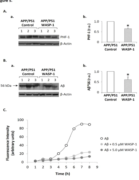
(45) 39. WASP-1 and FOXY-5 increase synaptic protein levels. Previous studies have shown that activation of Wnt signaling promotes the assembly of pre- and postsynaptic sites in developing and mature synapses (Ciani and Salinas, 2005; Farias et al., 2010). Indeed, canonical Wnt ligands have been implicated in the assembling of presynaptic proteins (Inestrosa and Arenas, 2010). Thus, Wnt-7a ligand induces the clustering of the α7-nicotinic acetylcholine receptor (α7nAChR), SV2, Synaptotagmin (Syt) (Farias et al., 2007), Synapsin-1 (Syn-1) (Hall et al., 2000) and Synaptophysin (Syp) (Cerpa et al., 2008), while Wnt-7b increases the number of clusters of Bassoon, VAMP2 and Syt (Ahmad-Annuar et al., 2006). Conversely, non-canonical Wnt ligand, Wnt-5a, promotes the clustering of postsynaptic proteins as the GABA-A receptor (Cuitino et al., 2010) or postsynaptic density protein-95 (PSD-95) (Farias et al., 2009). Here, we studied whether chronic treatment with WASP-1 and FOXY-5, also has an effect on hippocampal synaptic proteins levels of adult mice. As shown in Fig. 5A, WASP-1 treatment significantly enhanced the levels of the presynaptic proteins Syp and Syn-1, as well as the postsynaptic protein N-Methyl-D-aspartate (NMDA) receptor subunit 2B (NR2B) without affecting the levels of PSD-95. Instead, FOXY-5 treatment increased the levels of NR2B protein but did not induce changes on the levels of PSD-95 or any assayed presynaptic protein (Fig. 5B). These results indicate that chronic activation of Wnt signaling might modulate synaptic structure of pre- and postsynaptic sites in vivo. Even though, FOXY-5 treatment only affected the levels of the postsynaptic protein NR2B, the activation of canonical Wnt signaling by WASP-1 (Fig. 1A) raised the levels not only of presynaptic but also postsynaptic proteins. This dual effect on the synaptic structure could be responsible for the functional changes observed in the hippocampal CA3-CA1 synapses (Fig. 2, 3)..
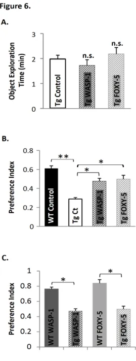
(46) 40. WASP-1 and FOXY-5 rescue cognitive impairment in adult APP/PS1 mice. Several studies suggest that Wnt signaling is involved in AD pathology (Caricasole et al., 2004; De Ferrari et al., 2007; Toledo and Inestrosa, 2010; Zhang et al., 1998). Moreover, numerous in vitro studies have shown that activation of Wnt signaling has a neuroprotective effect against Aβ-induced synaptic damage (Alvarez et al., 2004; Cerpa et al., 2010; De Ferrari et al., 2003; Shruster et al., 2011). To evaluate the effect of in vivo Wnt signaling activation on cognitive impairment associated to AD, we used here APP/PS1 double transgenic mice that coexpress mutant APPswe (K595N/M596L) and PSEN1 (exon 9 deletion of the presenilin-1 gene). The episodic memory performance in APP/PS1 mice begins to be affected as early as three-months-old, while spatial memory is impaired only after six-months-old (Trinchese et al., 2004). Therefore, to address whether WASP-1 and FOXY-5 treatments can rescue non-spatial episodic memory decline, we examined behavioral performance of seven-months-old APP/PS1 mice in the novel object recognition task. Exploration time during familiarization of the mice to the objects did not differ significantly among control, WASP-1 and FOXY-5 treated APP/PS1 mice (Fig. 6A). Upon introduction of a novel object, WASP-1 and FOXY-5 treated APP/PS1 mice exhibited stronger preference for the novel object than control APP/PS1 mice (Fig. 6B). Remarkably, performance of WASP-1 and FOXY-5 treated mice is similar to that of WT animals (Fig. 6B), indicating that chronic activation of Wnt signaling successfully overcome memory impairment in adult APP/PS1 mice. Although, APP/PS1 mice treated with WASP-1 and FOXY-5 almost reached control WT performance, still exhibited lower preference index than treated WT mice (Fig. 6C). Defective learning and memory in APP/PS1 mice has been associated with synaptic plasticity deficits (Trinchese et al., 2004). Indeed, APP/PS1 mice show decreasing on basal synaptic transmission at late ages of five to six-months-old, whereas hippocampal LTP.
Figure


+7


Documento similar