TítuloMineralogical study of silica from opaline levels (Salamanca, Spain) by scanning electron microscope with energy dispersive X RAY attachment
9
0
0
Texto completo
(2) also extended into various areas of neighbouring provinces. SANCHEZ CAMAZANO and SAAVEDRA (op. cit.) describe several outcrops and indicate the mineralogical composition. As well as opal-A (JONES et aL, 1971), opal-CT may be found. Quartz, kaolinite, iron oxides, are frequent but no constant. At times, alunite is extremely abundante Montmorillonite does not appear. A genetic mechanism for these materials has been proposed by SAAVEDRA and SANCHEZ CAMAZANO (in press). These authors underline the evolved nature of the detritic grains (angular and rounded quartzs), the presence of fragments of opaline material itself, the filling of a paleorelief and the association to shield zones in which the relict meteorization evidences a loss of silica in an arid or semi-arid climate. The evaporation of silica-bearing water, during the dry period, originated from the aforesaid meteorization, together with the contribution of more acidic percolating solutions, leads to the deposition of opal. The work described in this paper involves the use of the technique of scanning electron microscopy for the study of the siliceous materials of the above mentioned meteorization; the mineralogy has been checked by dispersive energy of X-ray.. PREPARATION OF SAMPlES Inmediately after fracturing, the samples were mounted on aluminium stubs and coated with an evaporate gold film, before examination with a scanning electron microscope Phillips 500 model at 25 kv. Al figures are scanning electron micrographs.. DESCRIPTION For the purposes of this study, 14 samples of opal which cemented and filled cracks were selected with different external characteristics (colour, beded or massive structure, etc.). They correspond to three different areas: in the first, the area inmediately around the city of Salamanca, it appears in sand and very mature cong,lomerates. In the second, Aldearodrígo, in granite which was initially meteorized and then compacted, and in the deepest zones of a lateritic type of meteorization. In all the localities, similar phenomena are observable with very variable amounts of opal. The mineralogical composition corresponds to that showed by SANCHEZ CAMAZANO and SAAVEDRA (op. cit.). The opal with massive shape seems to invade some previous minerals (fig. 1) and to set in others (fig. 2). Different types of morphology of opal-CT is founded; laminar aggregates give an aspect of compact nodules on large surfaces (figs. 3, 4 and 5). According to our news these forms were not described by others workers. The opal-CT platelets, with serrated edges, which are either subparallel (fig. 6), or penetrating each other, appear in layer alternating with other constitued for Fe minerals (figs. 7 and 8) or cOating the interior of walls of open cavities (fig. 9). Finally, spherical aggregates forms, «Iepispheres» (WISE et aL, 1972), and coalescent lepispheres appear in holes (figs. 10, 11, 12, and 13). The gradual development of these structure, from isolated blades to lepispheres and coalescente aggregates, have described (FLORKE et al., 1976). On these opal-CT shapes, sorne minerals are developed, figs. 14 (detail of fig. 11) and 15, which will be matter of a posterior publication. Similar forms to these have been reported in world literature in the study of opals of varying origin, above all in marine sediments (SEGNIT et aL, 1970, 1973; POLLARD et aL, 1973; MURATA et aL, 1975; FLORKE et aL, 1975, and many more). The descriptive studies have shown that the morphology of opal-CT varies with the conditions of crystalization.. 254.
(3) GENESIS The hypothesis of SAAVEDRA and SANCHEZ CAMAZANO (op. cit.) for the origin of opal is based on an evaporation and, at times, mixture of siliceous waters with other, more acidic and superficial, both generated in the same meteorización process. Alunite, which according to the thermodynamic data (LOPEZ AGUAYO et al., 1977), precipitates in a rather acid medium, at pH somewhat lower than 4 in the presence of kaolinite and an excess of silica, would already appears at the moment of the mixture, as may be seen from, fig. 2. The observation of the proceding micrographs shows that opal-CT only appears in the form of blades and spherical aggregates when its growth is not inhibited: compare fig. 3 with those which evidence the existence of holes, figs. 10-13. This underline the importance of the porosity of the material and of the surface phenomena. The solubility of silica is classically considered to be constants in the pH range 2-8.5. Amorphous silica is more soluble than the crystaline polymorphsj the solubility of these latter increases parallel to the increase in the degree of their disorder (FOURNIER, 1973).On the 0ther hand, the data reported by SOSMAN (1965) shows that the density of silica gel is much less than the disordered silica polymorphs cristobalite-trydimite; the transit of the amorphous variety to the crystaline ones, ,therefore, implies a decrease in volume. The use of oxygen isotopes (MURATA et aL, 1977) has made possible to show that the change from amorphous silica to disordered cristobalite must imply a solution which is followed by a deposition. Within the cristobalite, however, the change from disordered forms to more ordered ones, measured by a descrease in the d(001) spacing, seems to correspond to a zero-order reaction (il') solid state), with no solution being required. Thus, the dispositions observed in the micrographs are explained by a deposition of opalCT from solutions of amorphous silica, implying a decrease in volume and a possible creation of gaps in the places where the said mineral appears. The structural reorganizations in solid state (an increase in ordered forms) imply a parallel appearance of more empty spaces: note the morphological heterogeneity of the crystals and holes in figs. 7, 6, 9, 11, etc. ILER (1955) reports that the silica surfaces are covered with a layer of hydroxyl ions, chemically bonded to the silicium atoms, and, therefore, there are negatively charged particles (silanol groups); a strong H + concentración can annul this charge, in the case of silica with a pH less than 2. If this situation does not arise (remember that in this case the association alunite-opal CT-kaolinite and/or other, several, have a pH around 4), the surface silanol groups could react with cations to forms complexes; the thermodynamic calculation of DUGGER et al. (1964) imply that every positive ion which is hydrolyzed at certain pH forms complexes with the silanol groups of similar stability. That is to say that at pH of more than 2, reasonable for geological media, it is probable that the presence of some ions will occurs: the existence, precisely, of elements which generate cations, with easy hydrolysis at acid or weakly acid pH. This has been corroborated in practice by detection in silica of noteworthy amounts of AI+3, Fe+ 3, Mg+3 and other, capable of forming silicated compound (HURD, 1973). This fact can explain the presence of alternative layers of opal-CT and silicates of metaHc elements, fig. 7. The presence of small spheres of silica in opals was demostrated in the laboratory (JONES et aL, 1966). These authors attaked biotite with strong acid and noted the appearence of rounded masses of silica. ILER (1965) and op. cit.) had already reported that the precipitation of silica from aqueous solutions was begun by spheres. If, as is normal, metallic ions were present in the solution, the electric charge of the said spheres (which spould impede their regrouping by repulsion is neutralized by the formation of silicated complexes and these spheres, of reduced size, may also accumulate by flushing out in a liquid medium. In this case, it can nobe said that there existed an important cluster of these espheres; of course, the ILER (ops. cit.) observation with respect to siliceous is likely. With the increase in diameter of the spheres, without any evidence that they are displayed, it is necessary to point out that the normal repulsion (negatively charged surfaces) is less than its overlapping, conditions which may arise in crystalization situations particulary free and contain no cations impurities which might stop the process in its early stages, such as may be seen in figs. 10, 11 and. 255.
(4) 13. In sorne cases were ions in sufficient quantities to interrup the process of the mechanism, a fact manifested by the presence of later autormorphic minerals, in the gaps, fig. 15.. CONCLUSION The silifications in the Prelutetien continental sediments of Salamanca show morphological features similar to those reported for opals from other parts of the world, and they are interpreted as weathering products, generated by precipitation and later evolution (diagenesis) of silica from supersaturated solutions; the presence of other ions conditions the morphology of the silica polymorphs.. ACKNOWLEDGMENT The authors are greateful to Mistress Millán for their practical assistence at laboratory and for useful discussions.. 256.
(5) REFERENCES DUGGER, D. L.; STANTON, J. H.; IRBY, B. N.; MACCONNELL, B. L.; CUMMINGS, W. W. and MAATMAN, R. W. (1964): The exchange of twenty metal ions with the weakly acidic silanol group of silica gel. J. Phys. Chem. 68, 656-760. FICRKE, O. W.; JONES, J. B.; SEGNIT, E. R. (1975): Opal-CT crystals. Neues Jahrb. Mineral. Monatsh. 1975, 369-377. FLORKE, O. W.; HOLLMANN, R.; RAD, U. von and ROSCH, H. (1976): Intergrowth and twinning in Opal-CT lepispheres. Contrib. Mineral. Petrol. 58, 235-242. FOURNIER, R. O. -(1973): Silica in thermal Waters: Laboratory and field investigations, in Internat. Symposium on hydrogeochemistry and biogeochemistry, Tokyo, 1970; Washington, D. C., J. W. Clarke, Proc., 1, 122-139. HURD, D. C. (1973): Interactions of biogenic opal, sedimentand seawater in the Central Equatorial Pacific. Geochim. et Cosmochim. Acta, 37, 2257-2282. ILER, R. K. (1955): The colloid chemistry of silica and the silicates. Cornell University Press, Ithaca. ILER, R. K. (1965): Formation of precious opa!. Nature, 207, 472. JONES, J. B.; BIDDLE, J. and SEGNIT, E. R. (1966): Opal genesis. Nature, 210, 1353-1354. JONES, J. B. and SEGNIT, E. R. (1971): The nature of opal. 1. Nomenclature and constituent phases. J. Geol. Soco Aust., 18, 57-68. LOPEZ AGUAYO, F.; LA IGLESIA, A.; DOVAL, M. and MENENDEZ, F. (1977): New data on stability of alunite and jarosite. Proc. 8 th. Int. Kaolin Symposium and Meeting on Alunite, Madrid-Rome, A-4, 1-13. MURATA, K. J. and LARSON, R. R. (1975): Diagenesis of Miocene siliceous shales, Temblor range, California. Jour. Research. U. S. Geol. Survey, 3, 553-566. MURATA, K. J.; FRIEDMAN, 1. and GLEASON, J. D. (1977): Oxygen isotope relations between diagenetic silica minerals in Monterey shale, Temblor range, California. Am. Jour. Sci., 227, 259-272. POLLARD, C. O. and WEAVER, R. C. (1973): Opaline spheres: loosely-packed aggregates from silica nodule in diatomaceous miocene Fuller's Earth. Jour. Sed. Petrology, 43, 1072-1076. SAAVEDRA, J. and SANCHEZ CAMAZANO, M. (in press): Origen de niveles continentales silicificados con alunita en el Preluteciense de Salamanca. España. SANCHEZ CAMAZANO, M. and SAAVEDRA, J. (1979): Sobre la presencia de alunita en rocas postpaleozoicas de la provincia de Salamanca. IV Reunión Bienal Real Soco Esp. Hist. Nat., Como G-11, Valencia. SEGNIT, E. R.; ANDERSON, C. A. and JONES, J. B. (1970): A scanning microscope study of the morphology of opal. Search. 1, 349-351. SEGNIT, E. R.; JONES, J. B. and ANDERSON, C. A. (1973): Opaline silicas from the Murray river region West of Wentworth, N. S. W., Australia. Mem. Nat. Mus. Vic., 34, 187-194. SOSMAN, R. B. (1965): The phases of silica. Rutgers University Press, New Brunswick. WISE, S. W. and KELTS, K. M. (1972): Inferred diagenetic history of a weakly silicified deep sea chalk. Trans. Gulf Coast Ass. Geol. Soco 22, 127.. 257.
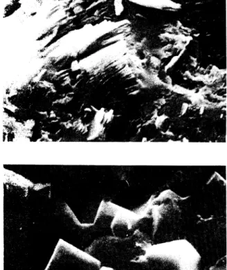
(6) . .• . . . . . . '. ,(0 ....... .'. -. -. -. :?.:,•. • ,'.,,;-. .... ~ .,.Jí1I. Fig. 1. - Kaolinite crystals (in packets of leaves) coverld by opal. Carrascalino, 2.500 x.. Fig. 2. - Dispersed alu nite crystals in an opaline mass. The holes correspond to crystals, which have disappeard, from this mineral. Salamanca, 5.000 x, scale length 1 pm.. Fig. 3. -Compact nodules of opa!. Aldearodrigo, 2.500 x.. Fig. 4. - Enlargement of part of fig. 3, 5.000 x..
(7) Fig. 5. - Enlargement of part of fig. 3, 5.000 x.. Fig. 6. -Subparallel opal-CT blades. Aldearodrigo, 5.000 x.. Fig. 7. - Opal-CT layer alternating with other constituted for Fe minerals. Carrascalino, 1.250 x.. Fig. 8. - Opal-CT blades schowing dispositions of the individ~al crystals from different orientations. Carrascalino, 20.000 x..
(8) Fig. 9. - Opal-CT crystallites coating interior of wall of a hole. Aldearodrigo, 5.000 x, scale lengt 1 }1m.. 10. -Spherical aggregates of thin platelets of opalCT. Carrascalino, 10.000 x, scale length 1 pm.. Fig. 11.-0pal-CT lepispheres in a cavity in the compact nodules opaline mass (fig. 3). Aldearodrigo, 1.250 x, scale length 10 pm.. Fig. 12. - Aggregates of lepispheres. Aldearodrigo,. 2.500 x..
(9) Fig. 13. - Lephispheres of opal-CT in a cavity in the silica mass, which enveloping kaolinite crystals. Carrascalino, 2.500 x.. Fig. 14. - Enlargement of part of fig. 11. Iron mine- . rals on Opal-CT, 10.000 x, scale length 1 pm.. Fig-. 15. -Crystals of other minerals appear on opalCT. Aldearrodrigo, 10.000 x..
(10)
Figure


Documento similar
No obstante, como esta enfermedad afecta a cada persona de manera diferente, no todas las opciones de cuidado y tratamiento pueden ser apropiadas para cada individuo.. La forma
Energy-dispersive X-ray microanalysis showed the cell walls to be the main area of Cu binding in the inner and outer cortex and infected zone of white lupin
As a first step, we verify the analysis procedure using simu- lated point-like sources with the computed spectra correspond- ing to (1) the inner part of the lunar disk (the total
In addition to two learning modes that are articulated in the literature (learning through incident handling practices, and post-incident reflection), the chapter
• Analysis of long-term variability of high-energy sources in the optical and X-ray bands, using INTEGRAL observations from IBIS (the gamma-ray imager), JEM-X (the X-ray monitor)
To do that, we have introduced, for both the case of trivial and non-trivial ’t Hooft non-abelian flux, the back- ground symmetric gauge: the gauge in which the stable SU (N )
On the other hand, sampling stations located in the northern part (SS9 and SS6) showed exogenous sediments resulting from the dredging of the seabed in other parts of the lagoon
El acceso a la versión del editor puede requerir la suscripción del recurso Access to the published version may
