Morphological and anatomical studies of the seeds and seedlings of Eucalyptus pilularis and E umbra
11
0
0
Texto completo
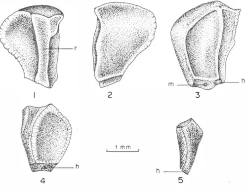
(2) REVISTA DE BIOLOGIA TROPICAL. 18 6. Anatomic studies of the seeds were done by means of microscopical observations of cross, longitudinal and paradermal hand-made sections of soaked seeds (Comer , 1976). The sections were mounted in glycerine (10%) as semi-permanent preparations (Sass, 1951). A 1 % ferríc chloride solution with 0,1 N hydrochloric acid was used to determine the ocurrence of phenolic compounds and tests with chloriódide of zinc were made for the identification of celulose and lignin (Jensen, 1962). Lignified walls were also localized by the phloroglucin test (Sass, 1951). The cuticle, the cutinized walls and lipid reserves became evident with Sudan IV (Johansen, 1940). Calose was localized with anilin blue and the proteinic reserves were identified by the Millon reagent (Jensen, 1962). The aleurone grains were stained by 1 % alcoholic eosine solution after fixation with 20% solution of mercuric chloride in absolute alcohol (Accorsi, 1941). Liquid amrnonia was used for the identification of anthocyanin in the seedlings (J ohansen, 194O). The drawings and diagrams of the structures were made with a camera lucida. For gerrnination studies, the seeds were placed on moist filter paper in covered Petri dishes at room temperature (mean 26 C) and light. RESULTS External morphology of fertile and sterile seeds: In the seeds of Eucalyptus pilularis and E. umbra there are three shapes: apparently fertile seeds, provided with embryo; and shape "A" and shape "B" embryoless sterile seeds. E. pilularis. 2. 3. 1 mm. E�_h 4. Figs. 1-5.. E. pilularis. h. 5. - Fcrtile and sterile secds. External Morphology. Figs. 1·3 Fig. 4 . shapc A sterilc secd; Fig.5 - shapc B stcrilc sccd.. -. fcrtilc seed;.
(3) BELTRAT/: Morphology and anatomy ofEucalyptus seeds. 1 87. ee. pq 10. ee m. en. Figs.6-12.. 12. E. pilularis - Fertile seed coat anato my. Figs. 6 and 7 - respectively, cross and paradermal sections of the fertile seed coat (antiraphe face); Fig. 8 vascularization pattern; Fig. 9 - transversal section through the edge; Fig. 10 basal seed face diagram showing the hilum an d micropyle positions; Fig. 11 transversal raphe section. Fig. 12 - Paradermal section at the micropylar regíon.. presented a mean of 6 ± 3% (by weight) of fertile seeds which represented an average of 1 2 ± 5 fertile seeds per gramo The mean weight of 100 fertile seeds was 155.6 ± 6.2 mg . In E. umbra the values were : a mean of 7± 3% (by weight) of fertile see ds which represented an average of 95± 7 fertile seeds per gramo The mean weight of 100 E. pilularis fertile seeds was 65.2 ± 4.1 mg. E. pilularis fertile see ds (Figs. 1-3) are red-brown; polyhedral or trapezoidal ; moderately lustrous surface with fine pitting; entire prominent edges ; hilum (h) basal, ciÍcular, conspicuous by its light color ; chalaza region not externally distinct ; long raphe (r) as a longitudinal protuberance ; antiraphe convex; exostome (m) visible as a little gap beneath the hilum ; 2.20 ± 0.14 mm long x 1.53 ± 0.21 mm broad . E. umbra: fertile seeds (Figs. 24-26) are light red-brown or light brown; polyhedral, trapezoidal or sickle-shaped ; other features similar to E. pilularis; 1.62 ± 0.29 mm long x 1.31 ± 0.24 mm broad . E. pilularis shape A sterile seeds (Fig. 4) are re d-brown ; lateral1 y flattened , trapezoidal; sickle -shape d; hilum basal; surface features similar to those of fertile seeds; 2.35 ± 0.10 mm long x 1.0 1 ± 0.10 mm broad..
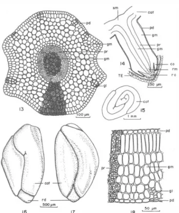
(4) 188. REVISTA DE bIOLOGIA TROPICAL. Shape. B. (Fig.. 5). are red-brown, elongated; many conical or cylindrical;. surface features similar to those of fertile seeds;. 1.95. ±. 0.70. mm long x. 1.09 ± 0. 1 5. mm broad.. E. umbra Shape A sterile seeds (Fig. 27) are light red-brown to brown; lateral1y flattened; trapezoidal; sickle-shaped; hilum basal; surface features similar. 1.5 5 :!: 0. 1 6 mm long x 0.60 ± 0.08 mm broad. � B (Fig. 28), color, shape and other surface features are similar those of shape A sterile seeds; 1 .09 ± 0. 18 mm long x 0.82 ± 0.24 mm broad.. to those of fertile seeds; In shape. to. I nte rnal morphology of fe rtil e seeds: In the two species the ripe seeds are basically constituted by the seed coat and the embryo. The seed coat is formed by the two integuments (TE and TI); also it is possible to fmd sorne remnants of the nucellar tissue (nu) and a single layer of endosperm (en). The. embryo. (Figs.. 1 6, 1 7,39. 40). and. consists. of. the. cylindrical. hypocotyl-root axis and two thick cotyledons (cot), bent down along the embryo axis (Figs.. 14, 1 5,37. and. 38).. The root meristem (rm) and root cap (rc), are almost. surrounded by the still incipient clinging disc or cupuliform organ (co). This organ is a bulge of the hypocotyl (hy) cortex (Figs.. 14. and. 38).. gm. co. �� col. 15. �. pr. 16. 17. 18. �. Figs. 13-18. E. pilularis - Embryo . Fig. 13 - middlc cross section through cmbryo axis;.Fig. 1 4Diagram of longitudinal section through embryo axis; Fig. 15 Diagram of cross section through the cotylcdons; Fig. 16· surfacc vicw of thc c mbryo (raphc facc); Fig. 17 - surfacc view of the embryo (antiraphc facc); Fig. 18 - cotyledon cross section. -.
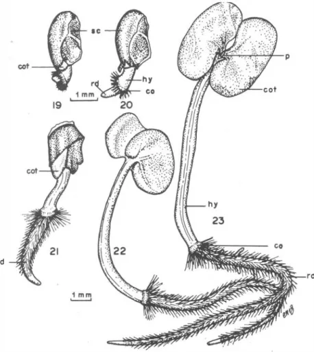
(5) BEL TRA TI: Morphology and anatorny of Eucalyptus seeds. 189. Fertile seed coat anatomy : Eucalyptus pilularis: testa (TE) multilayered; exotesta (ee) as a salid layer of sc1erotic cells with heavy lamellated thickenings on the outer and radial walls, the latter transversed by simple pit canals. The thickened walls are lignified and leave a very small lumen filled with amorphous dark-brown tannic material. The inner epidermis (ei) is made up of small thin-walled ce lIs, rectangular in cross section (Fig. 6) and poligonal in paradermal section (Fig. 7). Crystals of calcium oxalate occur dispersed in these cells, being most plentiful in the raphe parenchyma, just over the hilum and at the ribs (Figs. 6 and 11). Between the two epidermal layers there are several layers of parenchyma (tp) which frequently change in number according to their position in the seed (Fig. 6, 9 and 11). These cells have pitted walls and are filled with amorphous dark-brown tannic material. E. umbra (Fig. 29 and 30): testa generally two-Iayered. The parenchyma cells occur just over the hilum, along the raphe and at the edges (Figs. 34 and 35). The other anatomic features are similar to those of E. pilularis.. cot---'...I'!!. rd d. Fig. 19-23.. E. pi/ularis - scqucntial stagcs of dcvcloprnent of seedlings. Fig. 19 - seedlings at 4 days; Fig. 20 - at 5 days; Fig. 2 1 - at 7 days; Fig. 22 - at 10 days; Fig. 23 - at 18 days..
(6) 190. REVISTA DE BIOLOGIA TROPICAL. r. 25. 26. 28. 1. mm. Figs . 24-28. E umbra - F ertile and sterile seeds. Externa! morphology. Figs. 24-26 - fertile seed. Fig. 24 - shape A sterile seed; Fig. 28 - shape B sterile sced.. In E. pilularis, from the hilum an amphicribal vascular bundle (vb) with helically thickened tracheids extends in the raphe parenchyma right up to the chalaza where it spreads out (Figs. 8 and 11) . In E. umbra the vascular bun dle gives rise to sorne branchlets before reaching the chalaza region (Figs. 3 1, 32 and 35) The chalaza (ch) is a parenchymatous tissue made up of thin -walled suberized cells f illed with a dense reddish-brown tannic material. In E pilularis the inner integument (TI) is formed by two epidermal layers of suberized tabular cells (Fig . 6). In E. umbra the two epidermal layers of the inner integument are distinct only along the raphe. At the inner limit there is a rather evident cuticle which forms short rib-like projections of cutine between the nucellus epidermal cells. The formation of this cuticle is supressed in the chalazal region . Remnants of the nucellar tissue (nu) with empty and largely obliterated cells are present, especially in the chalaza region, where the celular structure is evident. The endosperm (en) is almost completely absorbed, remaining only a layer of cells filled with proteins and oil droplets (Figs. 6, 9, 11, 29 , 34). In both spe cie s, the hilum (h) is a scar of thin-walled non-suberized cells surrounded by a rim of sclereids (Fig . 35) In E. pilularis the micropylar gap (m) is bordered at its upper and lateral edges by small thin-walled suberized cells, each containing a calcium oxalate crystal, and at its lower edge, by sclereids (F igs. 10 and 12). In E umbra surrounding the micropylar gap there is a complete rim of small sclereids ( Fig. 3 3). .. .. .. Anatomy of the e m b ryo : In E. pilularis the hypocotyl-root axis is about 1.2 mm long and 0 .4 mm in diamete r and in E. umbra it is about 0.8 mm long and 0.3 mm in diameter. In both species the embryo axis is covered by the protoderm (pd, Figs. 1 3 and 16). Inside to the protoderm there are 7 or 8 layers of ground meristem (gm) , precursor of cortical ground tissue, of roun d thin-walled cells. Below this tissue there is a cylinder of narrow procambial cells (pr), 6 or 7 layers thick in E. pilularis and 4 or 5 layers in E. umbra..
(7) BELTRA TI: Morphology and anatomy of Eucalyptus seeds. 191. F igs. 29 -35. E. umbra - Fertile seed coat anatomy. Figs. 29 and 3 0 - respectively cross and paraderrnal sections of the fertile seed coat (antiraphe face); Fig. 3 1 - Seed diagram showing thc hllum, chalaza and micropyle positions; Fig. 32 vascularization pattern; Fig. 33 - paradermal section at the micropylar region. Fig. 34 - raphe transversal section; Fig. 35 - longitudinal section through the hllum region.. Arising from the protoderm of the axis, as well as from that of the cotyledons, and deeper in the ground meristem there are small oil glands (gl) scattered along the embryo axis, and in both faces of the cotyledons , mainly at the abaxial surface. The cotyledons (Figs. 18 and 41) are covered by thin-walled protodermal cells, rectangular in cross section and irregular in paradermal sectiori. The ground meristem below the protoderm, on the adaxial face consists of a row of palisade cells, and on the abaxial side there are 5 layers of round cells which present small intercelular spaces..
(8) 192. REVISTA DE BIOLOGIA TROPICAL. Most of the embryo tissues except procambial and glandular cells, are fiHed with aleuron grains and oil droplets. Each one of the aleuron grains contains one or more srnaH druses of ca1cium oxalate (F igs.. 18 and 41).. Germi nation and seedling morphology: Germination and morpholog y of the seedlings in the two species are similar. Three days after sowing (in pe.tri dishes) the rupture of the seed coat occurs at the micropylar region and the radicular primordium (rd) emerges, partially surrounded by the cupuliform organ (co). This. sm. � pd gm. +---+t-pr 1+-+--tT- gm. 38. ,100 �m,. 40. Figs.3641. E. umbra. Embryo, Fig. 36 - middle cross section through the embryo axis; Fig. 37 - diagram of cross section through the cotyledons; Fig. 38 - Diagram of longitudinal section through the embryo axis. Fig. 39 - surface view of the embryo (raphe face); Fig. 40 - surface view of the embryo (antiraphe face ); Fig. 41 - cotyle don cross section..
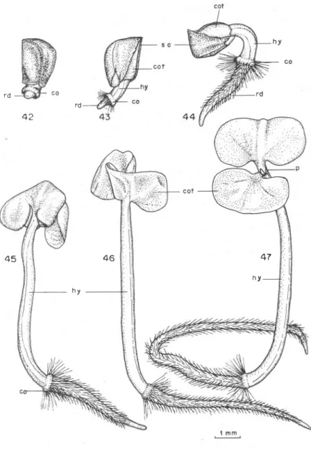
(9) 193. BELTRATI: Morphology and anatomy of Eucalyp t us seed s. organ, that is very narrow (0,3 mm in E. pilularis and 0,2 mm in E. umbra), soon develops long absorbing hairs (Figs. 20 and 43) and remains funetional for about 30 days when the hairs begin to dry. The radiele and the hypoeotyl (hy) grow simultaneously and after 7 or 8 days the seedling is fIxed to the substratum (Figs. 2 1 and 44) when the seed eoat (se) is released. The hypoeotyl is pink, due to the presenee of anthoeyanin s in the epidermal ceUs. cot. rd. rd. 42. 46. 45. hy. \ \ \¡. . .. 47. I. I. hy. l\'. F igs. 4247. E. umbra - sequential st age s of see dling deve lopment. Fig. 42 - see d ling at 4 days; Fig. 43 - at 6 d ays; Fig. 44 - at 8 days; Fig. 45 - at 10 days; Fig. 46 - at 1 1 d ays; Fig. 47 - at 1 8 days..
(10) 194. REVISTA DE BIOLOGIA TROPICAL. After about 12 days the cotyledons (cot) unfold (Figs. 22, 23 and 4547). They are bilobed, dark-green adaxially and reddish-purple abaxially, due to anthocyanins. The epicotyl (p) initiates its growth in about 20 days in E pi/ularis and in about 25 days in E umbra. Comparison of seed length averages: As additional information to the morphological comparisons, the variation of the fertile seed length Was studied in greater detail. The Kruskal-Walles technic (Sokal & Rohlf, 1969) was used to verify whether the existing difference in the average length of the fertile seeds was significant or not. According to the test, E. pi/ularis seeds differ significantly in length from those of E. umbra.. DISCUSSION The present results suggest that these two species have many common morphological and anatomical features but they can be distinguished mainly by the average length of fertile see ds and by the seed coat anatomy. In E. pi/ularis, but not in E. umbra, the testa is multilayered in all its extent and according to Comer (1976) "multiplicative seeds occur in families acknowledged to be primitive". Another important observation is that in other previously studied species (Beltrati, 1977a; 1977b; 1978a; 1978b; 1979; 1980) but not in E pilularis and in E. umbra, the exotesta is unlignified and the endotesta is composed of closely packed cells, each containing a heavy cellulosic thickening of its inner periclinal wall and one or more calcium oxalate crystals each. According to Gauba & Pryor (1958) the discontinuous presence or complete absence of calcium oxalate crystals in generally unmodified cells of the inner epidermis of the testa among the Renantherae-Normales is likely to have taxonomic significance since in the Angiosperms the ocurren ce of a "crystal epithelium" is widely distributed, especially in the more primitive families. RESUMEN Se estudiaron en detalle aspectos morfológicos y anatómicos de semillas y plántulas jóvenes de Eucalyptus pi/ularis Sm. y E. umbra R.T. Baker. Se encontró que eran similares a excepción de algunos aspectos anatómicos de la testa y del promedio de longitud de las semillas fértiles, que es significativamente diferente en estas dos especies. LITERA TU RE CITED A ccorsi, W.R. Contribution to the comparative anatomical study o f Eucalyptus tereticor nis 1 94 1 . Smith and E u ca lyptus citr íodora Hooker. Thesis. Esc. Supo Agr. "Luiz de Quciroz". Piracicaba, Brasil.. Beltrati, C. M. 1 97 7a.. Comparacio morfológica entre sementes procedentes do Brasil e da Austrália, de. Eucalyptus alba R einw. Rev. Brasil. Biol., 37 : 463-4 7 1 .. Beltrati, CM. 1 97 7b.. Eucalyptus ¡;randís (Hill) Maiden : morfologia das sementes e de sua gcrmina�ao.. Phyton, 35 : 93-10 1 ..
(11) BELTRA TI: Morphology and anatomy of Eucalyptus seeds. 1 95. Beltrati, C.M. 1 97 8a.. Morphological and anatomical studies of the seeds and seedlings of Euca[yptus citriodora and E maculata. Rey. Biol. Trop., 26 : 21 3-225.. Beltrati, C.M. 1 978b.. Morfología e anatonúa das sementes e Turrialba, 2 8 : 209-2 1 4.. phintulas de Eucalyptus maidenii.. Beltrati, C.M. 1 9 79.. Beltrati, C. M. 1 98 0 .. Morfología e anatomia das sementes de Eucalyptus punctata O C. Anais da Sociedadc Botanica do Brasil, XXX Congresso Nacional de Botánica: 1 7 -21 .. Morfología e anatomia das sementes e plantulas d e Eucalyptus saligna S m . Rey. Brasil. Biol., 4 0 : 441 -446.. Blakely, W. F. 1 95 5 .. A key t o the eucalyptus Australia.. (2d ed.). Comm. Austr. For. Timber Bur. Canberra,. Corner, E.J.lL 1 97 6 .. The Sceds o f dicotyledons. Vol. 1 . Cambridge Uniycrsity Press. Cambridge.. Gauba, E., & LD. Pryor 1 95 8 .. Seed coat anatomy and taxonomy in Eucalyptus. 1 . Proc. Linn. Soco N.S.W., 8 3 : 20-32.. Jensen, W.A. 1 96 2.. Botanical histochcmistry. Principies and pratice. Freeman, San Francisco .. Johansen, O.A. 1 94 0 .. Plant microtechnique. M (.'(}raw-Hill. New York.. Sass, J.E. 1 95 1 .. Botanical microtechnique (2d ed.). Ames, 10wa State Collegc Prcss.. Sokal, R.R., & F J . Rohlf 1 969.. B iometry. Frecman. San Francisco.. Vaughan, J.G. 1 96 8.. Sccd anatomy and Taxonomy. Proc. Linn . Soco Lond., 79 : 25 1 -255..
(12)
Figure

+2
Documento similar
In this respect, a comparison with The Shadow of the Glen is very useful, since the text finished by Synge in 1904 can be considered a complex development of the opposition
Safety and performance of the drug-eluting absorbable metal scaffold (DREAMS) in patients with de-novo coronary lesions: 12 month results of the prospective, multicentre,
1. S., III, 52, 1-3: Examinadas estas cosas por nosotros, sería apropiado a los lugares antes citados tratar lo contado en la historia sobre las Amazonas que había antiguamente
Since such powers frequently exist outside the institutional framework, and/or exercise their influence through channels exempt (or simply out of reach) from any political
Of special concern for this work are outbreaks formed by the benthic dinoflagellate Ostreopsis (Schmidt), including several species producers of palytoxin (PLTX)-like compounds,
50 The goal is to help people to reach an optimum level in the dimensions of psychological well- being: environmental mastery, personal growth, purpose in life,
In the previous sections we have shown how astronomical alignments and solar hierophanies – with a common interest in the solstices − were substantiated in the
While Russian nostalgia for the late-socialism of the Brezhnev era began only after the clear-cut rupture of 1991, nostalgia for the 1970s seems to have emerged in Algeria
