Changes in myocardial iron content following administration of intravenous iron (Myocardial‐IRON): Study design
29
0
0
Texto completo
(2) 8. Servicio de Cardiología y Unidad de Insuficiencia Cardiaca, Hospital Universitari. Germans Trias i Pujol, Badalona, Spain. Universitat Autonoma de Barcelona,. Accepted Article. Barcelona, Spain.. Short title: Changes in myocardial iron after iron administration. Address for correspondence*: Julio Núñez, MD Servicio de Cardiología. Hospital Clínico Universitario Avda. Blasco Ibáñez 17. 46010 Valencia-España Tel: +34617551562; Fax: +34963862658 e-mail: [email protected]. Funding. This work was supported in part by an unrestricted grant from Vifor Pharma and Proyectos de Investigación de la Sección de Insuficiencia Cardiaca 2017 from Sociedad Española de Cardiología. Conflict of interest: none. This article is protected by copyright. All rights reserved..
(3) BRIEF TRIAL SUMMARY Background. Treatment with intravenous ferric carboxymaltose (FCM) showed to. Accepted Article. improve symptoms, functional capacity, and quality of life in patients with heart failure (HF) and iron deficiency (ID). However, the underlying mechanisms for these beneficial effects remain undetermined. The aim of this study is to quantify cardiac magnetic resonance (CMR) changes in myocardial iron content after administration of intravenous FCM in patients with HF and ID and contrast them with parameters of HF severity. Methods. This is a multicenter, double-blind, randomized study. Fifty patients with stable symptomatic HF, left ventricular ejection fraction (LVEF) <50%, and ID will be randomly assigned 1:1 to receive intravenous FCM or placebo. Intramyocardial iron will be evaluated by T2* and T1 mapping CMR sequences before, 7 and 30 days after FCM. After 30 days, patients assigned to placebo will receive intravenous FCM in case of persistent ID. The main endpoint will be changes from baseline in myocardial iron content at 7 and 30 days. Secondary endpoints will include the correlation of these changes with LVEF, functional capacity, quality of life, and cardiac biomarkers. Results. The results of this study will add important knowledge about the effects of intravenous FCM on myocardial tissue and cardiac function. Conclusions. We hypothesize that short-term (7 and 30 days) myocardial iron content changes -evaluated by CMR- after intravenous FCM would correlate with simultaneous changes in parameters of HF severity. The study is registered at http://clinicaltrials.gov (NCT03398681).. Key words: Heart failure; Iron deficiency; Cardiac magnetic resonance; Ferric carboxymaltose; Myocardial iron.. This article is protected by copyright. All rights reserved..
(4) ABBREVIATIONS. Accepted Article. ACEI: angiotensin converting enzyme inhibitors ARB: angiotensin receptor blockers CA125: carbohydrate antigen 125 CMR: cardiac magnetic resonance eGFR: estimated glomerular filtration rate FCM: ferric carboxymaltose HF: heart failure HFrEF: heart failure with reduced ejection fraction hsTnT: high-sensitivity troponin T ID: iron deficiency KCCQ: Kansas City Cardiomyopathy Questionnaire LVEF: left ventricle ejection fraction NGAL: neutrophil gelatinase-associated lipocalin NT-proBNP: amino-terminal pro-brain natriuretic peptide sTfR: soluble transferrin receptor TSAT: transferrin saturation 6MWT: 6-minutes walking test. This article is protected by copyright. All rights reserved..
(5) INTRODUCTION Iron deficiency (ID) is a common finding in patients with heart failure (HF). It is. Accepted Article. usually associated, even in the absence of anemia, with decrease in functional capacity, quality of life, and with increased risk of mortality and readmission.1-7 Treatment with intravenous ferric carboxymaltose (FCM) in patients with HF and ID has shown improving symptoms, functional capacity, quality of life, and decrease hospitalizations under an acceptable safety profile.8,9 Such benefit has been consistent in patients with and without anemia,10,11 suggesting additional pathophysiological pathways beside anemia resolution. In addition, recent studies suggested that myocardial ID could play a direct role in the pathogenesis and progression of HF.12-14 Despite the fact that T2* cardiac magnetic resonance (CMR) sequence has shown to provide a reliable assessment of myocardial iron content15-18 newer techniques, such as T1 mapping, have emerged as an alternative tool for myocardial iron assessment.19 Changes in myocardial T1 mapping are more linear and have fewer artifacts than with T2* sequence, which translated into a more reproducible and sensitive technique.20 Thus, in this work we aim to evaluate the utility of T1 mapping for detecting myocardial iron changes after intravenous iron administration. In a recent pilot study, our group reported an association between intravenous FCM administration and myocardial iron repletion assessed by T2* CMR. Interestingly, myocardial iron changes were strongly related to an improve in left ventricular ejection fraction (LVEF).21 We sought to determine that short-term (7 and 30-d) myocardial iron content changes -evaluated by T1* CMR- after intravenous FCM would correlate with simultaneous changes in parameters of HF severity.. This article is protected by copyright. All rights reserved..
(6) METHODS Overall study design. Accepted Article. This is a multicenter, double-blind, randomized, and placebo-controlled study aimed to test the effect of intravenous FCM [Ferinject®, Vifor Pharma (Glattbrugg, Switzerland)] on myocardial iron content assessed by CMR in five academic centers in Spain (Hospital Clínico Universitario de Valencia, Hospital de Manises, Hospital General Universitari de Castelló, Hospital Universitario y Politécnico La Fe, and Consorci Hospital General Universitari de Valencia). After signing the informed consent, patients will be randomized 1:1 to receive FCM or placebo. Intramyocardial iron will be evaluated before its administration, at 7 and 30 days. At 30-day, patients assigned to placebo will receive FCM if ID persists (Figure 1). The study will be carried out in accordance with the principles of the Declaration of Helsinki (1996) and the Good Clinical Practice of the International Conference on Harmonization. The study protocol was approved by Agencia Española del Medicamento y Productos sanitarios (AEMPS) on 6th December 2016 and by Comité Ético de Investigación Clínica (CEIC) del Hospital Clínico Universitario de Valencia on 26th January 2017, with an amendment on 22nd June 2017. CMR studies will be performed by ERESA (Valencia), and laboratory parameters will be analyzed in local labs. The study is registered at http://clinicaltrials.gov (NCT03398681).. Study population Eligible patients are those with stable chronic HF (NYHA II-III), LVEF <50%, and ID, the latter defined as serum ferritin <100μg/L or 100-299μg/L with transferrin saturation (TSAT) <20% and hemoglobin <15g/dL. All patients must meet all inclusion criteria and none exclusion criteria (Table 1).. This article is protected by copyright. All rights reserved..
(7) Randomization. Accepted Article. Patients will be randomly allocated into 1:1 ratio to receive FCM or placebo by means of a web-based computer-generated block sequence. Investigators and patients will be blinded to treatment allocations.. Study procedures Summary of study procedures are detailed in Table 2.. Cardiac magnetic resonance CMR data will be blindly acquired and quantified offline by two experienced cardiologists (M.P.L.L. and J.V.M., both with 15 years’ experience in CMR imaging) on a 1.5 Tesla MR scanner (Essenza y Avanto, Siemens, Erlangen, Germany). The three consecutive CMR studies of each patient will be analyzed by the same operator. No contrast media is used. All images are obtained with electrocardiographic gating and breath-holding. Cine images are acquired at rest in short axis views every 1 cm with steady-state free precession imaging sequences (time resolution: 37ms; voxel size: 1.7x1.7x7mm). Right and left ventricle (LV) ejection fraction (%), LV end-diastolic an end-systolic volume index (mL/m2), and LV mass (g/m2) are calculated by semiautomatic planimetry of endocardial and epicardial borders in short-axis views cine images. The basic T2* pulse sequence is a breath hold, multiecho gradient echo T2* sequence (voxel size: 1.6x1.6x8 mm) with 8 echo times from 2.65 to 21ms, in midventricular short axis. For T2* analysis, a region of interest (ROI) is chosen in the midleft ventricular septum. The mean signal intensities of ROI are measured in the series of. This article is protected by copyright. All rights reserved..
(8) increasing TE images to give an exponential decay curve. The monoexponential decay model and the nonlinear curve fitting algorithm are used to fit the curve to obtain T2*. Accepted Article. measurement. T1 mapping is performed with MOLLI sequences with motion correction (voxel size: 1.5x1.5x7mm) in three short axes (basal, medial and apical). After T1-maps are generated, a ROI is chosen in the mid-left ventricular septum in the three-short axis and the average T1 values are calculated. Details of the CMR sequences used are described in Appendix 3. All measurements were made on the platform Syngo MR C15, Siemens. The same protocol will be repeated at 7 and 30 days.. Six-minutes walking test (6MWT) The 6MWT is performed in a place well equipped for cardiopulmonary resuscitation. Subjects are advised not to have undertaken vigorous exercise within the previous two hours and instructed to cover the maximum distance possible in six minutes, at a self-graded walking speed. Pausing to rest will be allowed when needed.. Kansas City Cardiomyopathy Questionnaire (KCCQ) The KCCQ it is a self-administered instrument designed to evaluate healthrelated quality of life in chronic HF patients. It is composed of 23 items (15 questions) that form seven domains: physical limitations, symptoms (frequency, severity and change over time), self-efficacy and knowledge, social interference and quality of life. It is scored by assigning each response 1 to 5, 6 or 7 points, being 1 de lowest punctuation. The sum of these items is subsequently transformed to a scale of 0 to 100. This article is protected by copyright. All rights reserved..
(9) points (Appendix 2).22 The Spanish version of the KCCQ23 will be filled-in by patients. Accepted Article. with the support of trained nurses.. Biomarkers The results from lab data will be reviewed and signed by the investigator who will record in the case report form whether they are normal, abnormal, and clinically significant. The following parameters will be assessed at baseline, 7, and 30 days: a) hematology: hemoglobin, hematocrit, red cell distribution width, mean corpuscular volume, and mean corpuscular hemoglobin; b) serum electrolytes: sodium, potassium and chloride; c) ID parameters: ferritin, TSAT, soluble transferrin receptor (sTfR), and hepcidin; d) renal function parameters: cystatin C, serum creatinine, blood urea nitrogen, and estimated glomerular filtration rate (eGFR); e) liver function parameters: alanine amino-transferase, and aspartate amino-transferase; and, f) HF-biomarkers: carbohydrate antigen 125 (CA125), amino-terminal pro-brain natriuretic peptide (NTproBNP), galectin-3, ST-2, and high-sensitivity troponin T (hsTnT).. Clinical visits Summary of study procedures performed at each visit are detailed in Table 2. Screening and eligibility assessment (visit 0) After signing and dating the informed consent, the study procedures will be initiated. Follow-up Visits Scheduled follow-up visits will be performed at 24 hours, 7 and 30 days after randomization. Patients will be censored if they withdraw from the informed consent or die.. This article is protected by copyright. All rights reserved..
(10) Additional Visits Optional visits are permitted. The main reason for each optional visit, and for. Accepted Article. any laboratory or procedure additionally performed must be recorded on the case report form. Information on concomitant medications and clinical adverse events will be recorded.. Trial Intervention Eligible patients will be randomized to receive FCM or placebo.. Intravenous ferric carboxymaltose (FCM) FCM solution [Ferinject® (FCM), Vifor Pharma (Glattbrugg, Switzerland)] will be given as a perfusion of 20 mL (equivalent to 1000 mg of iron) diluted in a sterile saline solution [0.9% NaCl] and administered over at least 15 min.. Placebo Normal saline [0.9% NaCl] will be administered as per the instructions in the placebo-group. Because FCM is a dark-brown solution easily distinguishable, the personnel responsible for its preparation and administration will not be involved in any study assessments. To ensure that patients will be unaware of the study drug, the materials used in drug administration will be covered with aluminum foil or other opaque material and the injection site shield from the patient view.. Concomitant drugs The indication of other HF-drugs will be done according to the current recommendations for clinical practice.. This article is protected by copyright. All rights reserved..
(11) Endpoints. Accepted Article. Primary endpoint The main endpoint will be the changes from baseline at 7 and 30 days in myocardial iron content assessed by T2* and T1 mapping CMR sequences. The statistical comparisons for the primary efficacy objective will test the null hypotheses of no differences in changes from baseline in myocardial iron content –as assessed by T2* and T1 mapping CMR; the alternative hypotheses will indicate differences in either direction. Strictly speaking, the primary objective will be the 30-day evaluation; the 7day evaluation will be considered as a co-primary endpoint.. Secondary endpoints - On the entire sample, to correlate these changes with the following clinical markers of disease severity a) LVEF, b) functional capacity (6MWT and NYHA class), c) quality of life (KCCQ), and d) cardiac biomarkers. - On the sample stratified into three pre-specified subgroups: a) age >70 years vs. ≤70 years, b) anemia vs. no anemia (according to WHO criteria), and c) ischemic vs. non-ischemic etiology. - On the entire sample, to correlate these changes with blood markers specific to iron biology/deficiency (ferritin, TSAT, sTfR, and hepcidin).. Safety endpoints Based on previous studies,8,9 a safety surveillance will be specifically focused on: a) general disorders and administration site conditions, b) skin and subcutaneous tissue disorders, c) nervous system disorders, d) gastro-intestinal disorders, e) vascular. This article is protected by copyright. All rights reserved..
(12) disorders, f) ear and labyrinth disorders, g) injury, poisoning and procedural. Accepted Article. complications, and, h) cardiac disorders.. Sample size calculation The sample size was calculated based on the expected changes in T2*, according to the following parameters: 1) two treatment arms; 2) statistical power of the primary endpoint of 80%; and 3) alpha error of 0.05. We used repeated measures ANOVA using the Lawley-Hotelling test to evaluate the effect of treatment. Based on studies from our group,21 we predict a mean difference of 9.25±8.69 in T2* at 30 days after treatment, and a correlation of 0.38 between T2* measurements at baseline and 1 month later. The correlation of T2* at baseline and 7 days would be 0.40, since we expect the correlation to decrease with time. For a desired power of 0.80 and a Type I error of 0.025, we need to include 42 participants to detect a mean difference of 9.25 on T2* at 30 days, assuming no differences with placebo. Assuming a loss of 10% of patients, we increased the sample size to 50 patients (25 patients per arm).. Statistical analysis All statistical comparisons will be made under an intention-to-treat principle. Continuous variables will be presented as mean (standard deviation) for normallydistributed variables, and as median (interquartile range) otherwise. Discrete data will be expressed as percentages. The primary and secondary endpoints will be tested using an ANCOVA-design within a framework of linear mixed model. The analysis will include a between (FMC vs. placebo) and within comparison (changes at 7 and 30 days). The interaction term Tx*visit will be included if the omnibus p-value ≤0.05. The ANCOVA model for the. This article is protected by copyright. All rights reserved..
(13) primary analysis will include as dependent variable the myocardial T2* CMR values; the contrast among treatment groups at 30-d and 7-d will test the primary and co-. Accepted Article. primary endpoint respectively. As a pre-specified analysis, no adjustment for multiple comparisons will be made. Baseline value of myocardial T2* CMR will be included as an obligated covariate. The use of other covariates will be dictated if important differences among treatment groups were observed after randomization. Based on the normality of residuals a decision about transforming the outcome variable will be made. A similar approach will be taken for the secondary endpoints where left and right ventricular systolic function, KCCQ, NYHA class, and serum biomarkers will be the outcome variables. A two-sided p-value of 0.05 will be considered statistically significant for all analyses. Statistical package STATA 15.1 (STATA Statistical Software: Release 15.1, College Station, TX, USA) will be used for the analysis.. Current status Patient enrollment started in May 2017. On December 31st, 2017, 25 patients have been enrolled in the study (50% of the target). Baseline characteristics of these patients are described in table 3.. Planned substudies Some substudies are planned: a) the correlation of basal T1 mapping and T2* with basal ferritin and TSAT; b) correlations of changes in T1 mapping and T2* with changes in ferritin and TSAT; c) the effect of FCM on right ventricular function; and, d) the effect of FCM on left ventricular tissue Doppler.. This article is protected by copyright. All rights reserved..
(14) RESULTS Once the study is finished the changes in T2* and T1 mapping after FCM. Accepted Article. administration at 7 and 30 days will be documented. Furthermore, we will describe changes in laboratory data, functional capacity (6MWT, NYHA class), quality of life (KCCQ), LVEF, and ventricular diameters and volumes in echocardiography and CMR. Finally, we will relate the changes in T2* and T1 mapping with secondary endpoints mentioned above. We expect the results available around October 2018.. This article is protected by copyright. All rights reserved..
(15) DISCUSSION The prevalence of ID in chronic HF is approximately 50%, and commonly. Accepted Article. associated with decreased functional capacity and quality of life, and increased risk of mortality and readmission, even in the absence of anemia.1-7 Indeed, the administration of FCM has shown to reverse these changes within an acceptable safety profile.8,9 Several studies have demonstrated clinical improvement after intravenous iron administration in patients with and without anemia, suggesting that its beneficial effect includes additional mechanisms independent of the erythropoietic pathway.10,11. Iron and myocardial function Iron plays a crucial role in: 1) oxygen transport, through the production of hemoglobin, 2) oxygen storage, through myoglobin, and 3) as a component of the mitochondrial respiratory chain involved in energy production.1 An experimental studies have shown that anemia-deficient iron rats developed LV hypertrophy and dilation due to mitochondrial ultrastructural damage.12 Another study in non-anemic iron-deficient mice showed that iron content in cardiomyocytes and mitochondrial function were restored by iron repletion.13 In humans, a small study showed a reduction in the iron content of cardiomyocytes in patients with HF and reduced ejection fraction (HFrEF) as compared to controls.14 More recently, Toblli et al, in a small randomized trial including 60 patients with HFrEF, ID, and chronic kidney disease, showed that iron sucrose administration translated into a significant 6-month improvement in LVEF.24 More recently, findings from a cohort of 232 patients undergoing renal transplantation showed an increase of LVEF which was particularly notorious in those with systolic dysfunction.25. This article is protected by copyright. All rights reserved..
(16) This preliminary evidence has led us to postulate that myocardial ID may play a direct role in the pathogenesis and progression of HF. However, the clinical impact of. Accepted Article. myocardial ID on HF has not been thoroughly evaluated, mainly because of the lack of reliable and widely available noninvasive techniques for myocardial iron quantification.. CMR and myocardial iron assessment CMR has emerged as a non-invasive accurate technique for evaluation of cardiac anatomy, function, and risk stratification.26,27 More recently, this technique has been used to assess myocardial iron content.15 T2* CMR sequence has been considered a reliable tool for myocardial iron overload assessment.16,17 Nagao et al, in a small casecontrol study, found a significant decrease in myocardial iron concentration, assessed by T2* CMR, particularly in non-ischemic HF patients.18 Later, these authors also reported that T2* CMR was related to an increased risk of adverse outcomes.18 In a pilot study of 8 patients with HFrEF, our group found that treatment with FCM was associated with significant 30-day changes in T2* CMR, and they were associated to marked improvement in LVEF.21 Some new CMR techniques, such as T1 mapping, have emerged as a potential alternative for myocardial iron quantification.19 We postulate that T1 mapping CMR sequence, a more sensitive and reproducible technique,20 could also identify myocardial ID and quantify changes in myocardial iron content after FCM administration. In summary, preliminary evidence suggests that myocardial iron content plays a key pathophysiological role in HF. We speculate that with the new CMR sequences we will be able to reliably assess changes in myocardial iron content after intravenous iron administration, and, thereby, opening new modality of treatments for HF patient’s care. In addition, these results will add new insights about the role of iron in the. This article is protected by copyright. All rights reserved..
(17) physiopathology of the disease. A randomized clinical trial is a necessary step forward. Accepted Article. to advance the knowledge in this area.. Limitations There is a possibility that large areas of fibrosis may modify T2* and T1 measurement irrespective of iron status. As CMR are only performed on 1.5T machines, and T1 mapping is performed with MOLLI sequence, the extrapolation of the findings to 3T machines or other T1 mapping protocols is unknown. Several factors inherent to the study design, such a lower dose (and one time) administration of FCM, short trial duration (endpoint assessment at 30 days), the broad inclusion criteria (LVEF up to 50%, anemia not required), might reduce the expected response to therapy. In addition, the small number of patients leading to inadequate statistical power may become a potential limitation to reliably assess the clinical response.. Conclusions We hypothesize that T2* and T1 mapping CMR sequences would be sensitive enough to detect changes in myocardial iron content following administration of FCM, and that those changes would correlate with surrogates of HF severity.. This article is protected by copyright. All rights reserved..
(18) REFERENCES 1. Cohen-Solal A, Leclercq C, Deray G, et al. Iron deficiency: an emerging therapeutic. Accepted Article. target in heart failure. Heart. 2014;100:1414-1420. 2. Comín-Colet J, Enjuanes C, González G, et al. Iron deficiency is a key determinant of health-related quality of life in patients with chronic heart failure regardless of anaemia status. Eur J Heart Fail. 2013;15:1164-1172. 3. Núñez J, Domínguez E, Ramón JM, et al. Iron deficiency and functional capacity in patients with advanced heart failure with preserved ejection fraction. Int J Cardiol. 2016;207:365-367. 4. Jankowska EA, Rozentryt P, Witkowska A, et al. Iron deficiency predicts impaired exercise capacity in patients with systolic chronic heart failure. J Card Fail. 2011;17:899-906. 5. Jankowska EA, Rozentryt P, Witkowska A, et al. Iron deficiency: an ominous sign in patients with systolic chronic heart failure. Eur Heart J. 2010;31:1872-1880. 6. Klip IT, Comin-Colet J, Voors AA, et al. Iron deficiency in chronic heart failure: an international pooled analysis. Am Heart J. 2013;165:575-582.e3. 7. Núñez J, Comín-Colet J, Miñana G, et al. Iron deficiency and risk of early readmission following a hospitalization for acute heart failure. Eur J Heart Fail. 2016;18:798-802. 8. Anker SD, Comin Colet J, Filippatos G, et al; FAIR-HF Trial Investigators. Ferric carboxymaltose in patients with heart failure and iron deficiency. N Engl J Med. 2009;361:2436-2448.. This article is protected by copyright. All rights reserved..
(19) 9. Ponikowski P, van Veldhuisen DJ, Comin-Colet J, et al; CONFIRM-HF Investigators. Beneficial effects of long-term intravenous iron therapy with ferric. Accepted Article. carboxymaltose in patients with symptomatic heart failure and iron deficiency. Eur Heart J. 2015;36:657-668. 10. Filippatos G, Farmakis D, Colet JC, et al. Intravenous ferric carboxymaltose in irondeficient chronic heart failure patients with and without anaemia: a subanalysis of the FAIR-HF trial. Eur J Heart Fail. 2013;15:1267-1276. 11. Silverberg DS, Iaina A, Schwartz D, Wexler D. Intravenous iron in heart failure: beyond targeting anemia. Curr Heart Fail Rep. 2011;8:14-21. 12. Dong F, Zhang X, Culver B, et al. Dietary iron deficiency induces ventricular dilation, mitochondrial ultrastructural aberrations and cytochrome c release: involvement of nitric oxide synthase and protein tyrosine nitration. Clin Sci (Lond). 2005;109:277-286. 13. Haddad S, Wang Y, Galy B, et al. Iron-regulatory proteins secure iron availability in cardiomyocytes to prevent heart failure. Eur Heart J. 2017;38:362-372. 14. Maeder MT, Khammy O, dos Remedios C, Kaye DM. Myocardial and systemic iron depletion in heart failure implications for anemia accompanying heart failure. J Am Coll Cardiol. 2011;58:474-480. 15. Anderson LJ, Holden S, Davis B, et al. Cardiovascular T2-star (T2*) magnetic resonance for the early diagnosis of myocardial iron overload. Eur Heart J. 2001;22:2171-2179.. This article is protected by copyright. All rights reserved..
(20) 16. Kirk P, He T, Anderson LJ, et al. International reproducibility of single breathhold T2* MR for cardiac and liver iron assessment among five thalassemia centers. J Magn. Accepted Article. Reson Imaging. 2010;32:315-319. 17. He T. Cardiovascular magnetic resonance T2* for tissue iron assessment in the heart. Quant Imaging Med Surg. 2014;4:407-412. 18. Nagao M, Matsuo Y, Kamitani T, et al. Quantification of myocardial iron deficiency in nonischemic heart failure by cardiac T2* magnetic resonance imaging. Am J Cardiol. 2014;113:1024-1030. 19. Schelbert EB, Messroghli DR. State of the Art: Clinical Applications of Cardiac T1 Mapping. Radiology. 2016;278:658-676. 20. Kellman P, Hansen MS. T1-mapping in the heart: accuracy and precision.J Cardiovasc Magn Reson. 2014;16:2. 21. Núñez J, Monmeneu JV, Mollar A, et al. Left ventricular ejection fraction recovery in patients with heart failure treated with intravenous iron: a pilot study. ESC Heart Fail. 2016;3:293-298. 22. Green CP, Porter CB, Bresnahan DR, Spertus JA. Development and evaluation of the Kansas City Cardiomyopathy Questionnaire: a new health status measure for heart failure. J Am Coll Cardiol. 2000;35:1245–55. 23. Comin-Colet J, Garin O, Lupón J, et al. Validation of the Spanish Version of the Kansas City Cardiomyopathy Questionnaire. Rev Esp Cardiol. 2011;64:51-58.. This article is protected by copyright. All rights reserved..
(21) 24. Toblli JE, Di Gennaro F, Rivas C. Changes in echocardiographic parameters in iron deficiency patients with heart failure and chronic kidney disease treated with. Accepted Article. intravenous iron. Heart Lung Circ. 2015;24:686–695. 25. Hawwa N, Shrestha K, Hammadah M, et al. Reverse remodeling and prognosis following kidney transplantation in contemporary patients with cardiac dysfunction. J Am Coll Cardiol. 2015;66:1779–1787. 26. Pilz G, Heer T, Harrer E, et al. Clinical applications of cardiac magnetic resonance imaging. Minerva Cardioangiol. 2009;57:299-313. 27. Husser O, Monmeneu JV, Bonanad C, et al. Prognostic value of myocardial ischemia and necrosis in depressed left ventricular function: a multicenter stress cardiac magnetic resonance registry. Rev Esp Cardiol (Engl Ed). 2014;67:693-700.. This article is protected by copyright. All rights reserved..
(22) FIGURE LEGENDS. Accepted Article. Figure 1. Study design. V: visit; HF: heart failure; R: randomization; FCM: ferric carboxymaltose.. This article is protected by copyright. All rights reserved..
(23) Accepted Article This article is protected by copyright. All rights reserved.. Figure 1.
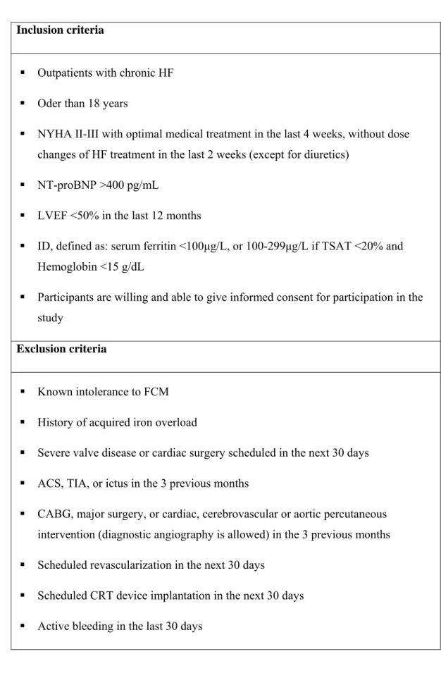
(24) Table 1. Inclusion and exclusion criteria. Accepted Article. Inclusion criteria . Outpatients with chronic HF. . Oder than 18 years. . NYHA II-III with optimal medical treatment in the last 4 weeks, without dose changes of HF treatment in the last 2 weeks (except for diuretics). . NT-proBNP >400 pg/mL. . LVEF <50% in the last 12 months. . ID, defined as: serum ferritin <100μg/L, or 100-299μg/L if TSAT <20% and Hemoglobin <15 g/dL. . Participants are willing and able to give informed consent for participation in the study. Exclusion criteria . Known intolerance to FCM. . History of acquired iron overload. . Severe valve disease or cardiac surgery scheduled in the next 30 days. . ACS, TIA, or ictus in the 3 previous months. . CABG, major surgery, or cardiac, cerebrovascular or aortic percutaneous intervention (diagnostic angiography is allowed) in the 3 previous months. . Scheduled revascularization in the next 30 days. . Scheduled CRT device implantation in the next 30 days. . Active bleeding in the last 30 days. This article is protected by copyright. All rights reserved..
(25) Accepted Article. . Active infection or malignancy. . Immediate need for transfusion or hemoglobin ≥15g/dL. . Anemia for reasons other than ID. . Immunosuppressive therapy or dialysis. . History of treatment with erythropoietin, intravenous iron, or transfusion in the previous 12 weeks. . Treatment with oral iron at doses >100mg/day in the previous week. . Contraindications to CMR, including non-compatible pacemakers or defibrillators, cochlear implants, cerebral aneurysm clips, claustrophobia, or large body size that does not allow the performance of the test.. . Pregnant or lactating women. . Subject of childbearing age who is unwilling to use adequate contraceptive measures during the study and up to 5 half-lives after the administration of study treatment. . Participation in another trial at the time of inclusion or in the previous 30 days. . Any disorder that compromises the ability to sign informed consent and/or comply with study procedures. HF: heart failure; NYHA: New York Heart Association; NT-proBNP: amino-terminal pro-brain natriuretic peptide; LVEF: left ventricle ejection fraction; ID: iron deficiency; TSAT: transferrin saturation; FCM: ferric carboxymaltose, ACS: acute coronary síndrome; TIA: transient ischemic attack, CABG: coronary artery by-pass surgery, CRT: cardiac resincronization therapy, CMR: cardiac magnetic resonance.. This article is protected by copyright. All rights reserved..
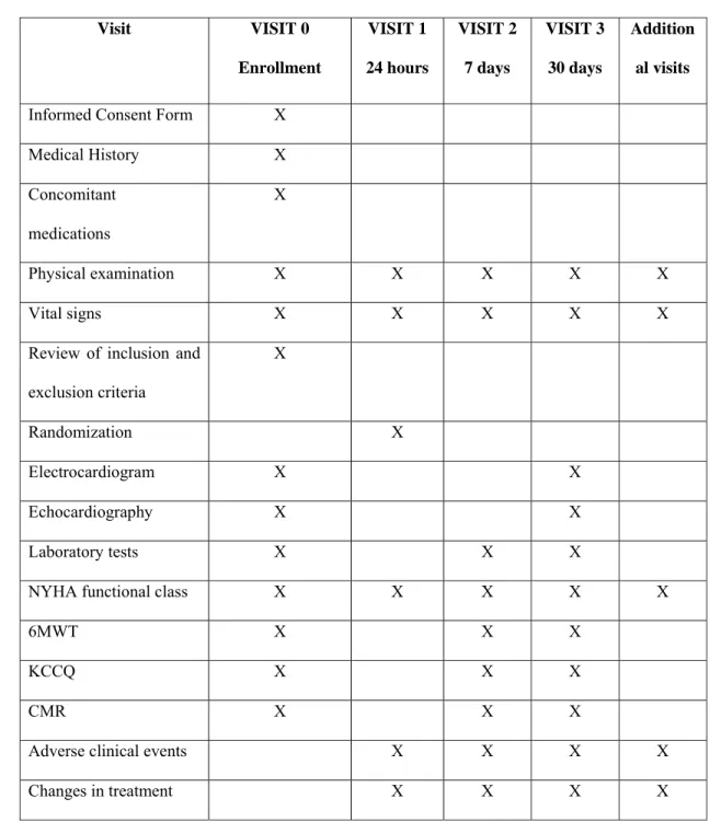
(26) Table 2. Study procedures. Accepted Article. Visit. VISIT 0. VISIT 1. VISIT 2. VISIT 3. Addition. Enrollment. 24 hours. 7 days. 30 days. al visits. Informed Consent Form. X. Medical History. X. Concomitant. X. medications Physical examination. X. X. X. X. X. Vital signs. X. X. X. X. X. Review of inclusion and. X. exclusion criteria Randomization. X. Electrocardiogram. X. X. Echocardiography. X. X. Laboratory tests. X. NYHA functional class. X. 6MWT. X. X. X. X. X. X. X. KCCQ. X. X. X. CMR. X. X. X. X. X. Adverse clinical events. X. X. X. X. Changes in treatment. X. X. X. X. NYHA: New York Heart Association, MWT: 6-minute walking test; KCCQ: Kansas City Cardiomyopathy Questionnaire, CMR: cardiac magnetic resonance.. This article is protected by copyright. All rights reserved..
(27) Table 3. Baseline characteristics. Accepted Article. Variables. n=25 Demographics and medical history. Age, years. 72.5 (67-78.5). Male, n (%). 17 (68.0). Hypertension, n (%). 16 (64). Dyslipidemia, n (%). 15 (60). Diabetes Mellitus, n (%). 12 (48). Smoker, n (%). 3 (12.0). Former smoker, n (%). 13 (52.0). Coronary artery disease, n (%). 9 (36.0). Hospital admission for AHF in the last. 14 (56.0). year, n (%) COPD, n (%). 6 (24.0). CKD, n (%). 8 (32.0). Stroke, n (%). 5 (20.0). NYHA functional class, n (%) II. 23 (92.0). III. 2 (8.0) Vital signs. Heart rate, bpm SBP, mmHg. 70 (60-79) 118 (106-130). Electrocardiogram and echocardiography Atrial fibrillation, n (%). This article is protected by copyright. All rights reserved.. 9 (36.0).
(28) LVEF, %. 40 (34-44) Laboratory. Accepted Article. Hemoglobin, g/dL. 12 (12.1-13.3). Anemia (WHO criteria), n (%) Transferrin saturation, % Ferritin, ng/mL. 8 (32.0) 14.9 (11-18.9) 78 (42-148). Absolute iron deficiency, n (%). 14 (56). Relative iron deficiency, n (%). 11 (44.0). Lymphocyte count, x103 cells/ml Sodium, mEq/L. 1720 (1210-2130) 140 (139-142). Potassium, mEq/L. 4.6 (4.3-4.9). Urea, mEq/L. 62 (50-82). Serum creatinine, mg/dl eGFR <60 mg/dL/1.73 m2, n (%) NT-proBNP, pg/ml,. 1.17 (.94-1.57) 62 (44-83) 1690 (1117-2836). Medical treatment Diuretics, n (%). 23 (92.0). Beta-blockers, n (%). 22 (80.0). ACEI, n (%). 6 (24.0). ARB, n (%). 6 (24.0). Sacubitril/Valsartan, n (%). 6 (24.0). MRI, n (%). 13 (52.0). ACEI: angiotensin converting enzyme inhibitors; AHF: acute decompensate heart failure; ARB: angiotensin II receptor blockers; CKD: chronic kidney disease; COPD:. This article is protected by copyright. All rights reserved..
(29) chronic pulmonary obstructive disease; eGFR: estimated glomerular filtration rate; LOS: length of stay; LVEF: left ventricular ejection fraction; MI: myocardial infarction;. Accepted Article. MRI: mineralocorticoid receptor inhibitors; NT-proBNP: amino-terminal pro-brain natriuretic peptide; NYHA: New York Heart Association; SBP: systolic blood pressure; WHO: World Heart Organization. WHO criteria for anemia: adult male, hemoglobin 13 g/dL, adult, non-pregnant female, hemoglobin 12 g/dL, adult pregnant female, hemoglobin 11 g/dL. Absolute iron deficiency: ferritin <100 ng/mL Relative iron deficiency: ferritin 100-299 ng/mL and transferrin saturation <20%. Values expressed as median (interquartile range); categorical variables are presented as percentages.. This article is protected by copyright. All rights reserved.. View publication stats.
(30)
Figure

Documento similar
In conclusion, supported iron oxide nanocatalysts were synthesized under continuous flow conditions and investigated as efficient catalysts in the microwave-assisted conversion of
Beta-blocker improves survival, left ventricular function, and myocardial remodeling in hypertensive rats with diastolic heart failure. Induction of cardiac fibrosis by β- blocker
In addition, the size distribution of carbon coated iron nanoparticles is broad by using this method as shown in TEM image (a) and size distribution histogram (b) of Figure
The content of this dissertation comprises the design of two arc-discharge plasma (ADP) reactors (a conventional and a modified one); the experimental study of the different
From this point of view, this thesis has a very broad scope focused on each and every one of the stages required to fabricate the high quality nanoelements studied: (i) the growth
Tie2-expressing macrophages (TEMs) are recruited into the myocardium in response to myocardial infarction and represent a cell population distinct from the Ly6C lo
Restless legs syndrome in patients with high serum ferritin and normal iron
The experimental and clinical work contained in this dissertation shows that the degree and extent of post-myocardial infarction tissue composition changes (mainly
