&
Chemical Biology
Binding Studies of Metal–Salphen and Metal–Bipyridine
Complexes towards G-Quadruplex DNA
Anna Łe˛czkowska,
[a]Jorge Gonzalez-Garcia,
[a]Cristina Perez-Arnaiz,
[a, b]BegoÇa Garcia,
[b]Andrew J. P. White,
[a]and Ramon Vilar*
[a]Abstract:The proposed in vivo formation of G-quadruplex DNA (G4 DNA) in promoter regions of oncogenes and in te-lomeres has prompted the development of small molecules with high affinity and selectivity for these structures. Herein we report the synthesis of a new di-substituted bipyridine ligand and the corresponding complexes with Ni2+ and VO2+. Both these new complexes have been characterized spectroscopically and by X-ray crystallography. Detailed DNA binding studies of these two complexes, together with three previously reported metal salphen complexes, are presented.
Using FRET melting assays, the binding affinity and selectivi-ty of the five metal complexes against six different G4 DNA structures as well as a duplex DNA have been determined. In addition, we present detailed ITC and UV/Vis studies to characterize the interaction of the complexes with human telomeric G4 DNA. Finally, we show via a polymerase stop assay that these complexes are able to stabilize a G4 DNA structure (from the c-Myconcogene promoter) and halt the activity ofTaqpolymerase.
Introduction
Besides its canonical double-stranded helix, DNA can fold into a range of different structures such as hairpins, triplexes, cruciform junctions, i-motifs and G-quadruplexes (G4).[1] The latter forms
from stacks of two or more guanine tetrads that in turn arise from hydrogen bonding network of four guanines (see Figure 1). A large number of putative G-quadruplex forming sequences (ca. 700,000) have been identified in vitro in the human genome[2]and there is increasing evidence they are involved in
essential biological processes.[3, 4]G-quadruplex-forming
sequen-ces in the human genome are found in telomeres[5]and
promot-er regions of various oncogenes.[6]Consequently, these G4
struc-tures have been proposed as potential targets for therapeutic in-tervention and therefore significant research has been carried out to develop small molecules with high affinity for G4 DNA.[7–9]
One of the biggest challenges when developing small mole-cules as quadruplex binders is to make them selective for a given G4 structure over others as well as over duplex DNA. While all G4 structures share some structural features—for
ex-ample, the stacks of guanine tetrads stabilized by K+ ions— they show wide structural diversity caused by the strand orien-tation, base composition, nature of pentose ring, conformation of glycosidic bonds of guanine bases in the G-quartets, length and sequence composition of loops/grooves formed.[10, 11]
Initially, most G-quadruplex binding molecules were based on organic heteroaromatic compounds.[12, 13] Subsequently, it
Figure 1.(a) Schematic representation of a G4 DNA structure highlighting the guanine tetrad. (b) Chemical structures of metal-salphen (1–3) and metal-bipyridine (4,5) complexes studied in this work.
[a]Dr. A. Łe˛czkowska, Dr. J. Gonzalez-Garcia, C. Perez-Arnaiz, Dr. A. J. P. White, Prof. R. Vilar
Department of Chemistry Imperial College London London SW7 2AZ (UK) E-mail: r.vilar@imperial.ac.uk
[b]C. Perez-Arnaiz, Prof. B. Garcia Universidad de Burgos Departamento de Qumica 09001 Burgos (Spain)
Supporting information and the ORCID identification number(s) for the au-thor(s) of this article can be found under:
https://doi.org/10.1002/chem.201802248.
was shown that metal complexes can also be excellent G4 DNA binders thanks to their unique electronic and structural features, as has been discussed in detail elsewhere.[14, 15]An
ex-ample of this, are metal salphen and salen complexes, which have been extensively studied over the past 10 years (see Figure 1).[16, 17]A wide range of derivatives with different metals
(and hence geometries),[18–20] salphen/salen core structure[21, 22]
and nature/number/position of substituents[19, 23–27] have been
reported as excellent G4 DNA binders. In spite of this, there is still little information regarding the selectivity of metal-sal-phens against several different G4 DNA structures.[26]
With the aim of increasing the aromatic planar surface of metal salphens and hence their G4 DNA binding affinity, herein we report the synthesis and structural characterisation of two new metal complexes where Ni2+ (4) and [VO]+ (5) are coordi-nated to a new 6,6’-diphenyl-2,2’-bipyridine ligand (Figure 1). The affinities of these complexes (and of the previously report-ed metal-salphens1–3) towards six different G-quadruplex DNA structures have been determined by fluorescence reso-nance energy transfer (FRET) melting assays. We have also es-tablished by detailed isothermal titration calorimetry (ITC) and UV/Vis spectroscopic studies, the affinities and thermodynamic binding parameters for these complexes towards human telo-meric G-quadruplex DNA. Finally, using polymerase stop assay (PSA), we report on the ability of these complexes to stabilise the G-quadruplex structure in the promoter region of c-Myc
and in doing so halt the activity of a polymerase.
Results and Discussion
Design and synthesis of metal complexes
We previously reported the structural characterization of a nickel(II) and a copper(II)-salphen complexes bound to human telomeric G-quadruplex DNA.[17] These structures confirmed
that the square planar metal complexes bind to the external G-tetrads viap–p stacking interactions. From these structures, we hypothesized that a system based on a di-substituted bi-pyridine with four aromatic rings (Figure 1) would have similar dimensions to the salphen ligand but possess an extra phenyl ring which could increase the ability of the resulting metal complexes to interact with the G-tetrad. Besides the polyaro-matic tetradentate core of the ligand, we designed the system with two ethyl piperidine substituents analogous to those present in the successful metal-salphen complexes we have previously developed.[16, 18]The presence of such substituents is
particularly important to increase the compounds’ water solu-bility as well as the affinity towards DNA thanks to the positive charge on the piperidine groups, which are protonated at physiological pH.
Complexes1–3 were synthesized as we previously report-ed[18] while the new complexes4 and 5 were prepared as
shown in Scheme 1. Several complexes with 6,6’-diphenyl-2,2’ -bipyridine ligand derivatives have been reported.[28, 29] We
therefore adapted some of the previous protocols to prepare our new ligand (7) and corresponding metal complexes (4and 5). A Suzuki cross-coupling reaction using 6,6’-dibromo-2,2’ -bi-pyridine and 2,4-dimethoxyphenyl boronic acid was carried out, followed by demethylation with boron tribromide to yield compound7. The phenolic scaffold was metalated by using Ni2+ or VO2+ salts to yield the corresponding metal-bipyridine precursor complexes8 and 9, which were then reacted with chloroethyl piperidine to yield the new complexes4 and5. It should be pointed out that no vanadyl complexes with 6,6’ -di-phenyl-2,2’-bipyridine ligands have been previously reported. All compounds were fully characterized by1H NMR
spectrosco-py (except for the vanadyl complexes5and9which are para-magnetic), mass spectrometry and their purity confirmed by el-emental analyses (see Experimental Details). As described in the following section, the X-ray crystal structures of
pounds4 and 5 were also determined confirming their pro-posed square planar and square-based pyramidal geometries. To investigate the ability of the nickel(II) complex4 to dis-playp–pinteractions in solution (and hence its potential to do so with G4 DNA), variable concentration1H NMR spectroscopic
studies were carried out. As has been previously reported,p–p stacking interactions results in shielding of protons and hence in lower 1H NMR chemical shifts in comparison to the
non-ag-gregated species. As can be seen in Figure S12, upon increas-ing the concentration of the complex all the aromatic signals shifted upfield consistent with the expected increase inp–p in-teractions.
Similarly, we carried out a variable temperature 1H NMR
spectroscopic study of complex4 (at constant concentration see Figure S13). Upon heating the sample up from 20 to 708C, all the aromatic protons shifted. However, in this case two dif-ferent trends were observed: the resonances between 7.70 and 8.30 ppm all shifted downfield as the temperature in-creased which is consistent with a decrease ofp–pinteractions at higher temperature. On the other hand, the resonances be-tween 6.20 and 6.40 ppm (which correspond to the protons next to the O atoms in the di-phenolic ring) shifted upfield. The opposite shift of these signals in comparison to the other aromatic protons reflects a more complex set of intermolecular interactions for the protons in the phenolic ring. It is likely that these two protons display stronger hydrogen bonding interac-tions with the solvent which decrease as the temperature in-creases and therefore an upfield shift is observed.
Structural characterisation of 4 and 5
Single crystals of both complexes4 and 5 suitable for X-ray crystallography were obtained. The four aryl rings in the struc-ture of 4 adopt a distorted conformation (Figure 2); though the bipyridyl unit is relatively coplanar (the torsion angle be-tween ringsAandBbeing only ca. 2.58), ringsCandDare no-ticeably twisted with respect to their adjacent pyridyl rings, theA–CandB–Dtorsion angles being around 11.9 and 8.68 re-spectively. For rings A and C there is also a significant fold component such that O19 lies ca. 1.00 out of the plane of ringA. This fold distorts the square planar coordination at the nickel atom such that O14 lies around 0.22 out of the (Ni,N1,N7,O29) plane (the atoms of which are coplanar to better than 0.01 ). Similar distortions from a square planar ge-ometry have been previously observed for other bipyridine bis-phenol nickel(II) complexes.[28]
The p-ring systems of adjacent molecules pack across two independent centres of symmetry to form a loosely-linked ex-tended stack along the crystallographic a axis direction (Figure 3). The closest approaches are between ringA in one
molecule and ring B in the “below” counterpart (centroid···-centroid and mean interplanar separations of ca. 3.90 and 3.44 , rings inclined by around 38, interaction a in Figure 3), between ring B in one molecule and ring C in the “below” counterpart (centroid···centroid and mean interplanar separa-tions of ca. 4.00 and 3.33 , rings inclined by ca. 218, interac-tionb in Figure 5), and between ring Bin one molecule and ring D in the “above” counterpart (centroid···centroid and mean interplanar separations of around 4.05 and 3.49 , rings inclined by around 98, interactioncin Figure 3).
The four aryl rings in the structure of 5 (Figure 4) adopt a similar distorted conformation to that seen in the related nickel species4. The bipyridyl unit is again relatively coplanar with anA–B torsion angle of around 3.08, and rings CandD are twisted with respect to their adjacent pyridyl rings by ap-proximately 12.38 and 8.38 respectively. For rings A and C there is again a significant fold component such that O19 here
Figure 2.The crystal structure of4. Selected bond lengths () and angles (8); NiN1 1.877(2), NiN7 1.876(2), NiO14 1.8317(18), NiO29 1.8348(17), N1-Ni-N7 86.75(9), N1-Ni-O14 94.25(8), N1-Ni-O29 178.16(9), N7-Ni-O14 173.46(9), N7-Ni-O29 94.96(8), O14-Ni-O29 84.12(7).
Figure 3.Part of one of the looselyp–plinked stacks of molecules along the aaxis direction present in the crystal of4. Interactionsa,b, andchave cen-troid···centroid and mean interplanar separations () of ca.a) 3.90 and 3.44, b) 4.00 and 3.33, andc) 4.05 and 3.49, the ring pairs being inclined by ca. 3, 21 and 98respectively.
Figure 4.The crystal structure of5. Selected bond lengths () and angles (8); V1O1 1.6012(11), V1N1 2.0730(12), V1N7 2.0775(12), V1O14 1.9095(10), V1O29 1.9001(11), V1-N1 103.58(5), V1-N7 105.00(5), O1-V1-O14 112.72(5), O1-V1-O29 110.32(6), N1-V1-N7 79.75(5), N1-O1-V1-O14 86.12(5), N1-V1-O29 145.74(5), N7-V1-O14 141.87(5), N7-V1-O29 86.83(5), O14-V1-O29 85.33(4).
lies approximately 0.84 out of the plane of ringA. Unlike the case with3, however, where this fold affects the square planar coordination to the nickel atom, here the square-based pyra-midal coordination geometry of the vanadium centre is unaf-fected; the four basal atoms are coplanar to within 0.03 with the metal lying around 0.61 out of the basal plane in the di-rection of the apical atom O1.
Thepring systems of adjacent molecules pack across three independent centres of symmetry to form a sheet of molecules in the crystallographic ab plane. Figure S4 in the Supporting Information gives a detailed view of the six p–p interactions around one molecule, whilst Figure 5 here shows part of one
of the resulting sheets. The closest approaches are between ringAin one molecule and ring Bin aCi-related counterpart (centroid···centroid and mean interplanar separations of ca. 3.61 and 3.32 , rings inclined by ca. 48, interaction a in Figure 5), between ring Ain one molecule and ring C across an independent centre of symmetry (centroid···centroid and mean interplanar separations of ca. 3.83 and 3.43 , rings in-clined by ca. 168, interaction bin Figure 5), and between ring Bin one molecule and ringDin the next across another inver-sion centre (centroid···centroid and mean interplanar separa-tions of ca. 3.93 and 3.39 , rings inclined by ca. 88, interaction cin Figure 5).
DNA Affinity: Fluorescence resonance energy transfer (FRET) melting assays
Previously, we have reported that metal-salphen complexes1– 3have high affinity (in particular1and2) towards human telo-meric G-quadruplex DNA and good selectivity over duplex DNA,[16, 18]but their binding to a wider range of G-quadruplex
structures and therefore their G4-selectivity has not been pre-viously studied. Thus, with the aim of comparing the G4
affini-ty of1–3with that of the new complexes4and5as well as to establish preferences for a given G4 topology, we carried out FRET meting assays with a range of G4 DNA and RNA struc-tures. We studied G-quadruplexes with antiparallel (i.e. 22CTA), parallel (i.e. 25CEB, c-kit2, c-Myc) as well as mixed/hybrid (i.e. HTelo21 and Bcl-2) conformations. Structures HTelo21, 25CEB and 22CTA are derived from telomeric regions whilec-Myc,
c-kit2andBcl-2are sequences from oncogene promoter regions.
A duplex DNA (ds26) was also studied to investigate the selec-tivity for G4 vs. duplex DNA (see Experimental Details for de-tails of all sequences).
All metal complexes showed higher affinity for G4 over duplex DNA, with the new complex4 exhibiting the highest thermal stabilization effect of all complexes under investigation (see Figure 6). Amongst the metal-salphens, complex1
dis-played the highest stabilization for all G4s confirming our pre-vious observations for HTelo21 andc-MycG4 DNA. In the case of HTelo21, two different transitions in the melting curves were clearly observed for the square planar metal complexes1, 2 and4(see Figures S18, S19 and S21). This suggests more than one binding mode for the square planar complexes with the hybrid HTelo21 structure, which is also consistent with the ITC data discussed in the next section. With regards to the most potent stabilizer4, it showed a moderate affinity towards duplex DNA suggesting a possible intercalation of the bipyri-dine moiety between base-pairs of the double-stranded DNA. Complex5showed a similar stabilizing effect on the G-quadru-plexes than the most potent salphen complex (1) in spite of having a square based pyramidal geometry. This is likely to be due to the more extended p-system in the bipyridine bis-phenol ring as compared to salphen which allows the square-based pyramidal complex5to display a better G4 DNA affinity than the analogous V-salphen complex3.
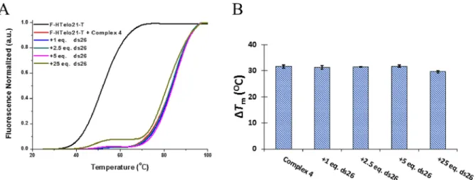
The selectivity of4 for G4 structures was studied by using a competition FRET experiment with non-labeled duplex DNA (ds26). The addition of duplex competitor did not yield any de-crease in the melting transition up to 25 equiv. of ds26 (i.e. Figure 5.Part of one of the sheets ofp–plinked molecules in theabplane
present in the crystal of5. Interactionsa,b, andchave centroid···centroid and mean interplanar separations () of ca.a) 3.61 and 3.32,b) 3.83 and 3.43, andc) 3.93 and 3.39, the ring pairs being inclined by ca. 4, 16 and 88
respectively.
Figure 6.Plot ofDTm(radial plot between 0 and 408C) of different
over 250 equiv. of base pairs) suggesting high selectivity for G-quadruplexes over duplex DNA as expected from the previous-ly studied metal-salphen complexes (Figure 7).
DNA binding affinity determined by isothermal titration cal-orimetry and UV/Vis spectroscopy
The binding affinities (Ka) along with the thermodynamic pa-rameters of the Gibb’s equation of complexes1–4 towards human telomeric G-quadruplex DNA were determined using isothermal titration calorimetry (ITC) experiments. The binding isotherms showed one binding event for complex3, while for 1 and 2 two binding processes were observed (see Figure 8 and S23 in the Supporting Information). Interestingly, com-plex4 displayed three independent binding processes (Figure 10). All the salphen complexes showed binding iso-therms consistent with exothermic reactions while for the bi-pyiridine complex4, the first binding event is governed by an endothermic process, while the 2ndand 3rdare exothermic (see
Figure 8).
Single site, two-site or three-site binding models[30] were
used to obtain the thermodynamic parameters for the interac-tion between the complexes and human telomeric G4 DNA (Table 1). For compounds1 and 2, the first G4 DNA affinity Figure 7.A) Melting curves for F-HTelo21-T with increasing concentrations of ds26 in the presence of 1mmof complex4. B) Variation of the F-HTelo21-T
melt-ing temperature with increasmelt-ing concentrations of unlabelled duplex DNA.
Figure 8.ITC titrations of human telomeric G4 DNA (20mm) with complexes1(A) and4(B). The concentration of the respective metal complexes was 1 mm.
Table 1.Thermodynamic parameters obtained for the interaction be-tween the metal complexes and human telomeric G4 DNA by ITC at 298 K.
1 2 3 4
K1 1015, M1 27060 495 0.90.1 3600900
DH1, kJ mol1 1353 991 7.10.2 1.20.3
TDS1, kJ mol1 93.8 60.4 21.1 50
n1 3.70.8 3.170.08 3.330.06 1.00.3
K2 10 –5
, M1 5.70.9 1.80.9 – 1100700
DH2, kJ mol1 893 1612 – 7010
TDS2, kJ mol1 56.3 131.2 – 23.5
n2 8.50.6 9.30.1 3.80.4
K3 10
–5, M1 – – – 5010
DH3, kJ mol1 – – – 111
TDS3, kJ mol1 – – – 12.70.9
n3 8.00.7
constant (K1) is higher (47 and 27-fold, respectively) than the second one (K2) (see Table 1) indicating that the first G4 bind-ing site is less hindered than the second. It could be hypothe-sized that the two binding events correspond to the com-pounds binding with different affinity to each of the two exter-nal G-tetrads in the G4 structure. However, this model is com-plicated by the observed stoichiometry for the first binding event (see Table 1) which suggests that ca. 3 to 4 molecules of the compounds are needed per G4 DNA structure (see below for further comments on stoichiometry). Both complexes ex-hibited a higher value ofDHthanTDSindicating that enthalpy is the main driver for the interaction between these complexes and telomeric G4 DNA.
Consistent with the FRET-melting assays and our previous work,[18] the affinity of 3 for G4 DNA is lower than that of1
and2, which can be attributed to the geometries of the com-plexes : square-based pyramidal for 3 and square planar for1 and 2. Interestingly, the binding process for 3 is entropically driven (see Table 1) in contrast to the observations with1and 2. This suggests the possibility of a conformational change of complex3, the G4 DNA structure or both upon binding. Taking into account the number of binding processes and the affinity constants obtained, the affinity of the complexes for telomeric G4 DNA follow the trend3!2<1<4which is consistent with the FRET melting assays.
There have been relatively few previous reports where ITC was used to determine the affinities of metal complexes to-wards G4 DNA.[31–33]One of these studies with metalated
por-phyrin (TMPyP4), found that NiII-TMPyP4 and CuII-TMPyP4
dis-play high affinities to G4 DNA and four independent binding sites,[31]which is similar to our observations. In different study,
ITC showed that the binding process (i.e., enthalpy, entropy and number of binding sites) between a NiII
-bis(phenanthro-line) complex and telomeric DNA can be affected by the exact choice of telomeric sequence.[32]With HTelo22 (i.e. the same
se-quence we have used for our studies) it was found that this NiII complex displayed higher binding affinities than towards
the structurally constrained 26-mer sequence.
As indicated above, for the first binding event of com-plexes1–3, ca. 3 to 4 equivalents of the respective compound per G4 molecule are needed (see Table 1). A similar behavior has been recently reported for other metal complexes target-ing G-quadruplex structures.[34, 35]In contrast, complex4shows
a one-to-one stoichiometry (n=1) for the first binding event although, the subsequent two binding events also show higher ratios. Interestingly, an analogous three-event model withn=1 for the first binding event, has been recently report-ed for a terpyridine-CuII complex interacting with G4 DNA.[36]
While the number of binding events can be correlated to dif-ferent sites in the G4 structure (e.g. the two tetrads) it is not clear why the observed stoichiometries are so high for the complexes.
We also studied the possibility that different protonation species could lead to the two-binding process for the interac-tion of 1 and 2 with HTelo22. Therefore, the basicity of the complexes was examined by means of UV/Vis spectroscopy at different pH values. The overlapping of the absorption spectra
showed two distinct isosbestic points at different pH ranges for1–4(see Figure 9 for compound4), indicating the presence of three different protonated species (see Figures S24–27 in Supporting Information). The pKa values were obtained by plotting the absorbance versus pH and ranged between 7.6– 8.4 (see Supporting Information for all the values) suggesting that the protonation steps occur in the amino group of piperi-dine rings in good agreement with values in the literature.[37]
Thus, these values pointed out that the di-protonated species is the only one present under our experimental conditions and therefore this species should be involved in both binding events with telomeric G4 DNA.
Following on from the results described above, the affinities and binding mode of all metal complexes were studied by using UV/Vis titration experiments. In agreement with the ITC studies, the UV/Vis binding isotherms showed one binding mode for 3 and 5, two for 1 and 2 and three for 4 (see Figure 10 and Figures S28–S30). It is worth mentioning that the titrations of metal-salphen complexes were already
report-ed but reaching lower molar ratios (r=[L]/[G4 DNA] and thus, a second binding process wasn’t previously monitored.
The first binding process of the three salphens complexes displayed hypochromism. In the case of1and2, this was also accompanied by a red-shift of the bands which is consistent with our previous observations, and can be attributed to the p–p stacking interactions of the metal complexes with the guanine tetrad. Interestingly, the second binding event for 1 and2 showed hyperchromism (and no red shift) which is usu-ally attributed to non-specific interactions such as electrostatic or van der Waals forces.
Figure 9.Absorbance spectra of4recorded with increasing pH (arrow sense) from 6.0 to 7.8 (A) and from 7.8 to 8.4 (B).
Figure 10.Plots of the UV/Vis titrations of HTelo22 and complexes1(A) and 4(B) ([complex]=20mm). Insets are the absorbance binding isotherms
Complex4 showed three binding processes : the first and third events resulted in hyperchromism in contrast with the second one that showed hypochromism (Figure S31). More-over, there is a general red-shift of the spectrum suggesting the stacking between this nickel(II) complex and the guanine tetrad. This data is summarized in Table S3.
Due to the different independent DNA binding sites for the square-planar metal complexes (i.e.1,2and4), it was not pos-sible to fit the UV/Vis titration data to a model from which we could derive the affinity constants for these three complexes. In the case of the square-based pyramidal V=O complexes3 and5, only one binding process was determined by UV/Vis ti-trations (in agreement with the ITC data previously discussed). Therefore, it was possible to fit the titration data to a 1:1 model and determine the affinity constants for3(logKa=4.97) and5 (logKa=5.11). The former is consistent with the affinity determined by ITC (i.e. logK=4.95, see Table 1)
Polymerase stop assay
To test the ability of the complexes to stabilise G-quadruplexes in a functional manner, we performed a Polymerase Stop Assay (PSA) adapted from previously reported protocols.[38, 39] A
60 bases-long template oligonucleotide containing a G4-form-ing sequence derived from c-Myc promoter was used as a model for the quadruplex-forming sequences. This was select-ed since the metal complexes under investigation have an in-termediate affinity for this sequence as compared to for exam-ple, HTelo G4 and ds26 DNA. This sequence was mixed with a short (22 bases long) labelled oligonucleotide with a sequence complementary to the 3’overhang of the template. As previ-ously reported, the ability of a polymerase to elongate the la-belled-template under different conditions (namely in the pres-ence/absence of K+ ions or DNA binders) was then studied.
Two control experiments were performed before addition of the compounds to be tested. In the first of these, no K+ (or compounds) were added to the mixture which led to the full elongation of the primer by the polymerase. In contrast, addi-tion of high concentraaddi-tion of K+ ions to the mixture and hence formation of G4 DNA in the template sequence led to the appearance of a new band of intermediate size between
that of the initial (labelled) primer and the fully elongated one. This indicates that the polymerase is able to extend the primer but only up to the point where the template forms a G4 DNA structure that is, roughly a 40-base long oligonucleotide. Having established this, we then investigated the effect that increasing amounts of the different compounds had on the polymerase activity. As can be seen in Figure 11 and Fig-ure S32–S35, a dose response was observed with the com-pounds under study. The amount of truncated oligo (i.e. the 40 bases long that stops at the point where the G4 forms on the template strand) could be quantified by analyzing the in-tensity of the corresponding band in the gel. This clearly became more intense as the corresponding G4 binder concen-tration increased (see Figure 11 for complex4 as an example). The intensity of this band was then plotted vs. the concentra-tion of the compound to obtain EC50 values (see Figure 11 B). The lowest EC50 value (0.920.03mm)—that is, the highest
ability to inhibit polymerase mediated by G4 stabilisation— was obtained for complex4. This is consistent with the data discussed in previous sections which showed that this com-pound has the highest affinity for G4 DNA. On the other hand, the highest EC50value (3.80.4mm), and hence lowest activity,
was obtained for the square-based pyramidal complex3— which is again consistent with the affinity of this compound for G4 DNA as determined by FRET, ITC and UV/Vis studies). Complexes1and2displayed EC50values of 1.30.1 and 1.5
0.2mmrespectively.
Conclusions
The new metal complexes4and5display high affinity for G4 DNA and selectivity over duplex DNA. As compared to the cor-responding metal salphen complexes (i.e.4cf. 1and2; and5
cf. 3), both these complexes are better G4 DNA binders—al-though in the case of complex4 a modest affinity to duplex DNA is also observed. The selectivity of these five complexes against six different G4 DNA structures of different topology has been studied via FRET melting assays. All of them show a preference towards antiparallel and hybrid conformations over parallel ones.
Figure 11.Polymerase Stop Assay (PSA) with a template strand containing the G4-forming sequence from thec-Mycgene promoter. (A) Image of the poly-acrylamide gel electrophoresis showing the control lanes (1, 2 and 8) as well as the effect of increasing concentration of compound4(0.3, 1, 5, 10 and 20mm) on the product distribution (lanes 3–7). The band corresponding to the pausing product is indicated with a box. (B) Fitting of the experimental data
obtained from the PSA with metal complexes1–4.
To characterise in more detail the interaction between these complexes and G4 DNA, detailed ITC and UV/Vis studies were carried out with HTelo DNA as a representative example of a G-quadruplex structure. The data obtained from these studies is consistent with the FRET melting assays showing that the new complex4 has the highest affinity for G4 HTelo DNA. On the other hand, as expected, the square-base pyramidal com-plex3 has the lowest affinity for G4 DNA. Interestingly, the analogous vanadyl-bipyridine complex5with an extra aromat-ic ring—has signifaromat-icantly higher affinity for G4 than 3 and of roughly the same magnitude than the square planar salphen complexes1 and 2. This highlights the importance of p–p stacking in binding to the guanine tetrads. ITC studies have also showed differences in the detailed binding profile of the complexes, with distinct number binding events for the square planar complexes than for the square-based pyramidal ones. Finally, we show that these complexes have the ability to stop the Taq polymerase from elongating a primer when a G4 struc-ture forms in the template strand. The EC50values determined
by PSA follow the same trend than the affinity of the metal complexes for G4 DNA.
Experimental Details
Chemicals were purchased from Sigma Aldrich, Alfa Aesar or Acros Organics and used as received. 1
H and 13
C NMR spectra were re-corded by using a 400 or 500 MHz Bruker Avance Ultrashield NMR spectrometer at 296 K, respectively. Chemical shifts are referenced to residual deuterated solvent. Mass spectra were obtained by using electrospray ionisation (ESI) by Mrs. L. Haigh (Imperial Col-lege London) on a LCT Premier Mass Spectrophotometer. IR spec-tra were recorded on PerkinElmer Spectrum 100 FT-IR Spectrome-ter. Compounds microanalysis were performed by Mr. A. Dickerson (University of Cambridge). Absorption measurements were made on a PerkinElmer UV/Vis spectrometer. The synthesis of metal-sal-phen complexes (1–3) was achieved following a previously report-ed protocol. 6,6’-dibromo-2,2’-bipyridine was prepared by previ-ously reported procedure.[40]
Synthesis of 6.6,6’-dibromo-2,2’-bipyridine (0.63 g, 2.0 mmol),
2,4-dimethoxyphenylboronic acid (0.90 g, 5.0 mmol), 2m aq. K2CO3
(10 mL) in THF (50 mL) were stirred under N2 atmosphere for
30 min. [Pd(PPh3)4] was then added and the reaction mixture was
left stirring at 708C for 48 h. After cooling to room temperature, the solution was filtered off and activated carbon was added to the filtrate. The mixture was stirred for further 20 min and then fil-tered through celite. The solvent was removed under reduced pressure and the compound was taken up with 2 mL of CHCl3.
Ad-dition of MeOH (30 mL) afforded a white precipitate. The solid was filtered off and dried under reduced pressure. Yield (0.72 g, 84 %).
1
154.6, 136.4, 132.3, 124.5, 122.3, 118.5, 105.3, 98.9, 55.6, 55.5 ppm. MS (ES+, CH
3Cl)m/z=429 [M+H+]. HRMS (ES+); found: 429.1817,
elemental analysis calcd (%) for C26H25N2O4: 429.1814. EA: found: C
72.69, H 5.59, N 6.76, calcd for C26H24N2O4: C 72.88, H 5.65, N 6.54;
mp. 218–2208C
Synthesis of 8. Compound6 (0.11 g, 0.27 mmol) in dry DCM
(15 mL) was stirred under nitrogen at798C for 30 min and BBr3
(0.30 g, 1.2 mmol) was added and stirred overnight at room tem-perature. The solution was combined with H2O (20 mL) and the
or-ganic phase extracted with CH2Cl2(3 20 mL). The organic phase
was evaporated, dried under reduced pressure to afford 7 and used in the following step without further purification. Yield (0.1 g, 98 %). MS (ES+) m/z
=373 [M+]. Crude compound7 (0.1 g,
0.27 mmol) and Ni(OAc)2·4 H2O (0.07 g, 0.27 mmol) in EtOH (30 mL)
were stirred at 608C for 3 h during which time a dark red/brown precipitate formed. The solution was then cooled down to room temperature and the precipitate was filtered off, washed with wa-ter:EtOH (1:1, 3 15 mL) and dried under reduced pressure. Yield (0.11 g, 92 %). 1
(0.45 g, 1.8 mmol) was added and stirred overnight at room tem-perature. The solution was combined with H2O (20 mL) and the
or-ganic phase extracted with CH2Cl2(3 20 mL). The organic phase
was evaporated, dried under reduced pressure to afford 6 and used in the following step without further purification. Yield (0.17 g, 98 %). MS (ES+) m/z=373 [M+]. Crude compound6
(0.17 g, 0.46 mmol) and VOSO4·0.5H2O (0.08 g, 0.46 mmol) in MeOH
(50 mL) were stirred at 508C for 15 h while a dark green precipitate formed. The solution was then cooled down to room temperature, filtered off and the precipitate washed with water (10 mL), MeOH (10 mL) and EtOH (10 mL) and dried under reduced pressure. Yield (0.09 g, 44 %). MS (ES+
mosphere for 10 min. 1-(2-chloroethyl)piperidine hydrochloride (0.085 g, 0.46 mmol) was then added and the reaction mixture was stirred at room temperature for 72 h. The solution was concentrat-ed under rconcentrat-educconcentrat-ed pressure to ca. 2 mL and water addconcentrat-ed (50 mL). The red precipitate that formed was filtered off, washed with water (40 mL) and diethyl ether (20 mL) and dried under reduced pres-sure to give an orange/red powder. Yield: (0.092 g, 62 %). Crystals of4suitable for X-ray analysis were obtained by slow diffusion of Et2O into a CH2Cl2 solution of the complex.
0.46 mmol) and 1-(2-chloroethyl)piperidine hydrochloride (0.08 g, 0.46 mmol) in dry DMF (20 mL) were stirred at room temperature under N2atmosphere for 48 h. The solution was filtered, the filtrate
concentrated to ca. 2 mL and water added (50 mL). The precipitate that formed was filtered off, washed with water (30 mL) and dieth-yl ether (20 mL) and dried under reduced pressure to give a light green powder. The residue was then dissolved in CH2Cl2 (3 mL)
and the complex was precipitated withn-pentane. Yield: (0.064 g, 43 %). MS (ES+, CHCl
3) m/z=660.3 [M+H+]; EA (%): Found: C
62.26, H 5.98, N 8.04; calcd for C36H40N4O5V·2 H2O: C 62.15, H 6.37,
1397 (m), 1353 (m), 1242 (m), 1179 (s), 1116 (m), 1033 (m), 1001 Oxford Diffraction Xcalibur PX Ultra diffractometer; 6452 independ-ent measured reflections (Rint=0.0287), F2
refinement,[41, 42]
R1(obs)=0.0486,wR2(all)=0.1393, 5167 independent observed
ab-sorption-corrected reflections [jFoj>4s(jFoj), 2qmax=1458], 457
parameters. CCDC 1005613 contains the supplementary crystallo-graphic data for this paper. These data can be obtained free of charge from The Cambridge Crystallographic Data Centre.
Crystal data for 5: C36H40N4O5V, M=659.66, monoclinic, P21/c
,T=173 K, dark orange tabular needles, Oxford Diffrac-tion Xcalibur PX Ultra diffractometer; 6162 independent measured reflections (Rint=0.0233), F2
refinement,[41, 42]
R1(obs)=0.0335,
wR2(all)=0.0943, 5763 independent observed absorption-corrected
reflections [jFoj>4s(jFoj), 2qmax=1458], 444 parameters. CCDC
1005614.
Oligonucleotides and stock solution preparation. Non-labelled
oligonucleotides and labelled oligonucleotides of double HPLC-grade purity were purchased from Eurogentec (Belgium). The la-belled sequences used were 5’-FAM-dGGG(TTAGGG)3-TAMRA-3’for telomeric G4 DNA (F-HTelo21-T), 5’ -FAM-dTGAGGGTGGG-TAGGGTGGGTAA-TAMRA-3’ forc-Myc, 5’ -FAM-dGGGAGGGCGCTGG-GAGGAGGG-TAMRA-3’forc-kit2, 5’ -FAM-dAGGGCTAGGGCTAGGGC-TAGGG-TAMRA-3’ for 22CTA, 5’-FAM- dAAGGGTGGGTG-TAAGTGTGGGTGGGT-TAMRA-3’ for 25CEB, 5’ -FAM-dGGGCGCGGGAGGAAGGGGGCGGG-TAMRA-3’forBcl-2and 5’ -FAM-CAATCGGATCGAATTCGATCCGATTG-TAMRA-3’for ds26 (donor fluo-rophore FAM, 6-carboxyfluorescein; acceptor fluofluo-rophore TAMRA, 6-carboxy-tetramethylrhodamine). The unlabelled oligonucleotides used had the same sequence except for telomeric G4 DNA that 5’ -dAGGG(TTAGGG)3-3’was used (HTelo22).
Tested compounds were dissolved in DMSO to give 10 mmstock
solutions. All solutions were stored in 100mL aliquots at 208C, thawed, and diluted immediately before use. Oligonucleotide con-centrations were calculated using the following extinction coeffi-cients e (L cm1
mol1
) at 260 nm given by the supplier: 228500 (HTelo22), 232000 (c-Myc), 258 900 (ds26), 205 600 (c-kit2), 220 400 (22CTA), 265 100 (25CEB), 231 300 (Bcl-2) and 220 590 (F-HTelo21-T). Under our experimental conditions the absorbance values were proportional to the metal complex concentrations, thus excluding aggregate formation. Extinction coefficients were calculated in Tris buffer (50 mm) supplemented with 10 mmKCl, pH 7.0,T=258C.e
(L cm1m1): 23 200 for 1 (l
=370 nm), 22 500 for2 (l=380 nm), 23 700 for3(l=322 nm), 18 500 for4(l=290 nm).
Isothermal Titration Calorimetry (ITC).The ITC experiments were
performed at 258C with a Nano ITC (TA Instruments, Newcastle, USA). Solutions were degassed in a degassing station (TA, Waters LLC, New Castle, USA) to minimize the formation of bubbles during the titration. In the course of the titration, the compound was injected (25 injections of 2.02mL) into the calorimetric cell (187mL) containing the HTelo22 (i.e. dGGG(TTAGGG)3A) solution in
50 mmTris-HCl, 10 mmKCl at pH 7.0. The stirring speed was
main-tained constant at 250 rpm. Control experiments were carried out to subtract the contribution of the heat of dilution of the
com-plexes. The resulting thermograms (integrated area of the peak/ mole of injectant versus [complex]/[HTelo22] ratio) obtained in the titrations were treated with model equations for single, two or three independent modes of binding using the Nano Analyze Soft-ware (TA Instruments, Newcastle, USA).[30]
The thermodynamic pa-rameters were obtained from two independent experiments.
UV/Vis spectroscopy. Spectrophotometric measurements were
performed with a HP 8453A photodiode array spectrophotometer (Agilent Technologies, Palo Alto, CA) endowed with a temperature control Peltier system. Titrations were carried out by adding in-creasing amounts of HTelo22 solution (between 0 and 2 equiv) to the complex solution in 1 cm pathlength cells with black quartz sides (50 mm Tris-HCl, 10 mm KCl, pH 7.0, T=258C). The
absorb-ance data were corrected by the dilution factor. For complexes3
and5, the affinity constants were obtained according to the inde-pendent-site model by nonlinear fitting (see details in the Support-ing Information).[43]The titrations were performed in triplicate.
FRET melting assays.Doubly labelled and non-labelled
oligonucle-otides (FAM/TAMRA labelling) were prepared as 20mmstock
solu-tions in MilliQ water and stock solusolu-tions of 0.4mmwere annealed
in cacodylate buffer (10 mm, pH 7.4) supplemented with potassium
depending on the sequence (c-Myc: 1 mm KCl+99 mm LiCl;
HTelo22, 22CTA,c-kit2, ds26, 25CEB: 10 mmKCl+90 mmLiCl;Bcl-2:
100 mm KCl). For FRET melting experiments 8-well optical tube
strips were used. The final volume of each sample was 40mL, with a final DNA concentration of 0.20mmand increasing concentration
of tested compound (0–8mm). Measurements were performed on
a PCR Stratagene Mx3005P (Agilent Technologies) with FAM excita-tion at 450–495 nm and detecexcita-tion at 515–545 nm. Readings were taken from 258C to 958C every 0.58C and at 18C min1
melting rate. To obtain melting curves, normalised FAM fluorescence signal was plotted against temperature. From the non-linear fitting of the plotDTm[Tm(with ligand)—Tm(without ligand) obtained from at
least three independent measurements] vs. ligand concentration, theDTmvalue for 1mmconcentration of ligand was obtained. In
the case of the competition assays, each well was prepared with a final labelled oligo concentration of 0.20mm, 1mmcompound, and
increasing concentration of non-labelled oligonucleotide to test (0.2, 1 and 2mm).
Polymerase Stop Assay.The oligonucleotide containing a
quadru-plex-forming sequence derived from c-Myc oncogene (5’ - GCGGCTCCTGTGAGGGTGGGGAGGGTGGGGAAGATTCCCGACTTCG-TATTAAGTACTCTAGCCTT-3’) and the corresponding partially com-plementary FAM label-sequence (5’ -FAM-AAGGCTAGAGTACTTAA-TACGA-3’) were used. A mixture of each oligonucleotide (1mm
each) and increasing concentrations of the compounds (0, 0.3, 1, 5, 10 and 20mm) were annealed in Tris buffer (50 mm, pH 7.4). After
cooling to room temperature, the PSA reactions were performed in 1x Taq buffer, containing the previously annealed solution (0.2mm),
dNTPs (0.2 mm), MgCl2(1.25 mm) andTaqDNA polymerase (2.5 U)
at 378C during 30 min. The reactions were quenched by adding an equal volume of stop buffer (90 % formamide, 10 mmNaOH). PSA
products were then analysed on a denaturing polyacrylamide gel (20 %) in 1x TBE and visualized in FAM dye channel. As a control experiment, the same protocol was followed using the non-form-ing quadruplex sequence (5’ -GCGGCTCCTGTGAGGGTGAA-GAGGGTGGGGA AGATTCCCGACTTCGTATTAAGTACTCTAGCCTT-3’) as a template oligonucleotide.
Acknowledgements
The UK’s Engineering and Physical Sciences Research Council (EPSRC) (grant number: EP/H005285/1) is thanked for financial support. J. G.-G. thanks the Royal Society and the British Acad-emy for a Newton Fellowship.
Conflict of interest
The authors declare no conflict of interest.
Keywords: cancer · DNA binding · heterocycles · metal complexes·polymerase
[1] M. Kaushik, M. Kaushik, S. Kaushik, K. Roy, A. Singh, S. Mahendru, M. Kumar, S. Chaudhary, S. Ahmed, S. Kukreti,Biochem. Biophys. Rep.2016, 5, 388 – 395.
[2] S. Chambers Vicki, G. Marsico, M. Boutell Jonathan, P. Smith Geoffrey, M. Di Antonio, S. Balasubramanian,Nat. Biotechnol.2015,33, 877 – 881. [3] D. Rhodes, H. J. Lipps,Nucleic Acids Res.2015,43, 8627 – 8637. [4] R. Hnsel-Hertsch, M. Di Antonio, S. Balasubramanian,Nat. Rev. Mol. Cell
Biol.2017,18, 279 – 284
[5] S. Neidle,J. Med. Chem.2016,59, 5987 – 6011.
[6] R. Hnsel-Hertsch, D. Beraldi, S. V. Lensing, G. Marsico, K. Zyner, A. Parry, M. Di Antonio, J. Pike, H. Kimura, M. Narita, D. Tannahill, S. Balasubrama-nian,Nat. Genet.2016,48, 1267 – 1272.
[7] S. A. Ohnmacht, S. Neidle, Bioorg. Med. Chem. Lett. 2014, 24, 2602 – 2612.
[8] S. Neidle,Nat. Rev. Chem.2017,1, 0041.
[9] S. Balasubramanian, L. H. Hurley, S. Neidle, Nat. Rev. Drug Discovery 2011,10, 261 – 275.
[10] G. W. Collie, G. N. Parkinson,Chem. Soc. Rev.2011,40, 5867 – 5892. [11] S. M. Haider, S. Neidle, G. N. Parkinson,Biochimie2011,93, 1239 – 1251. [12] A. R. Duarte, E. Cadoni, A. S. Ressurreicao, R. Moreira, A. Paulo,
Chem-MedChem2018,13, 869 – 893.
[13] D. Monchaud, M.-P. Teulade-Fichou,Org. Biomol. Chem.2008,6, 627 – 636.
[14] S. N. Georgiades, N. H. Abd Karim, K. Suntharalingam, R. Vilar, Angew. Chem. Int. Ed.2010, 49, 4020 – 4034;Angew. Chem.2010, 122, 4114 – 4128.
[15] Q. Cao, Y. Li, E. Freisinger, P. Z. Qin, R. K. O. Sigel, Z.-W. Mao,Inorg. Chem. Front.2017,4, 10 – 32.
[16] J. E. Reed, A. A. Arnal, S. Neidle, R. Vilar,J. Am. Chem. Soc.2006,128, 5992 – 5993.
[17] N. H. Campbell, N. H. A. Karim, G. N. Parkinson, M. Gunaratnam, V. Pet-rucci, A. K. Todd, R. Vilar, S. Neidle,J. Med. Chem.2012,55, 209 – 222. [18] A. Arola-Arnal, J. Benet-Buchholz, S. Neidle, R. Vilar,Inorg. Chem.2008,
47, 11910 – 11919.
[19] N. H. Abd Karim, O. Mendoza, A. Shivalingam, A. J. Thompson, S. Ghosh, M. K. Kuimova, R. Vilar,RSC Adv.2014,4, 3355 – 3363.
[20] S. Bandeira, J. Gonzalez-Garcia, E. Pensa, T. Albrecht, R. Vilar, Angew. Chem. Int. Ed.2018,57, 310 – 313.
[21] C.-Q. Zhou, T.-C. Liao, Z.-Q. Li, J. Gonzalez-Garcia, M. Reynolds, M. Zou, R. Vilar,Chem. Eur. J.2017,23, 4713 – 4722.
[22] A. Ali, M. Kamra, S. Roy, K. Muniyappa, S. Bhattacharya,Bioconjugate Chem.2017,28, 341 – 352.
[23] A. Terenzi, R. Bonsignore, A. Spinello, C. Gentile, A. Martorana, C. Ducani, B. Hogberg, A. M. Almerico, A. Lauria, G. Barone,RSC Adv.2014, 4, 33245 – 33256.
[24] A. Terenzi, D. Loetsch, S. van Schoonhoven, A. Roller, C. R. Kowol, W. Berger, B. K. Keppler, G. Barone,Dalton Trans.2016,45, 7758 – 7767. [25] P. Wu, D.-L. Ma, C.-H. Leung, S.-C. Yan, N. Zhu, R. Abagyan, C.-M. Che,
Chem. Eur. J.2009,15, 13008 – 13021.
[26] L. Lecarme, E. Prado, A. D. Rache, M.-L. Nicolau-Travers, R. Bonnet, A. V. D. Heyden, C. Philouze, D. Gomez, J.-L. Mergny, H. Jamet, E. De-francq, O. Jarjayes, F. Thomas,Inorg. Chem.2014,53, 12519 – 12531. [27] V. Rakers, P. Cadinu, J. B. Edel, R. Vilar,Chem. Sci.2018,9, 3459 – 3469. [28] H. Arora, C. Philouze, O. Jarjayes, F. Thomas, Dalton Trans.2010,39,
10088 – 10098.
[29] Y.-Y. Lin, S.-C. Chan, M. C. W. Chan, Y.-J. Hou, N. Zhu, C.-M. Che, Y. Liu, Y. Wang,Chem. Eur. J.2003,9, 1263 – 1272.
[30] C. A. Brautigam,Methods2015,76, 124 – 136.
[31] J. I. DuPont, K. L. Henderson, A. Metz, V. H. Le, J. P. Emerson, E. A. Lewis, Biochim. Biophys. Acta Gen. Subj.2016,1860, 902 – 909.
[32] C. Musetti, A. P. Krapcho, M. Palumbo, C. Sissi, PLoS One 2013, 8, e58529.
[33] X.-H. Zheng, Y.-F. Zhong, C.-P. Tan, L.-N. Ji, Z.-W. Mao, Dalton Trans. 2012,41, 11807 – 11812.
[34] A. Garci, K. J. Castor, J. Fakhoury, J.-L. Do, J. Di Trani, P. Chidchob, R. S. Stein, A. K. Mittermaier, T. Friscic, H. Sleiman,J. Am. Chem. Soc.2017, 139, 16913 – 16922.
[35] L. Hahn, N. J. Buurma, L. H. Gade,Chem. Eur. J.2016,22, 6314 – 6322. [36] Y. Li, M. Cheng, J. Hao, C. Wang, G. Jia, C. Li,Chem. Sci.2015,6, 5578 –
5585.
[37] A. V. Rayer, K. Z. Sumon, L. Jaffari, A. Henni,J. Chem. Eng. Data2014,59, 3805 – 3813.
[38] L. Ren, A. Zhang, J. Huang, P. Wang, X. Weng, L. Zhang, F. Liang, Z. Tan, X. Zhou,ChemBioChem2007,8, 775 – 780.
[39] H. Han, L. H. Hurley, M. Salazar,Nucleic Acids Res.1999,27, 537 – 542. [40] J. E. Parks, B. E. Wagner, R. H. Holm,J. Organomet. Chem.1973,56, 53 –
66.
[41] SHELXTL v5.1, Bruker AXS, Madison, WI,1998.
[42] SHELX-2013, G. M. Sheldrick,Acta Crystallogr. Sect. C,2015,71, 3 – 8. [43] F. H. Stootman, D. M. Fisher, A. Rodger, J. R. Aldrich-Wright, Analyst
2006,131, 1145 – 1151.
Manuscript received : May 4, 2018
Accepted manuscript online: June 13, 2018
FULL PAPER
&
Chemical BiologyA. Łe˛czkowska, J. Gonzalez-Garcia, C. Perez-Arnaiz, B. Garcia, A. J. P. White, R. Vilar*
&&–&&
Binding Studies of Metal–Salphen and Metal–Bipyridine Complexes towards G-Quadruplex DNA
G4 Binders: G-quadruplex DNA is emerging as a new target for anticancer agents, which has prompted the devel-opment of small molecules with high af-finity and selectivity for these structures. Herein a detailed study is reported of the G4 DNA binding properties of five metal complexes and shows their selec-tivity profile. It is demonstrated that these complexes stabilize G4 DNA and in doing so stop the activity of a poly-merase.


![Figure 10. Plots of the UV/Vis titrations of HTelo22 and complexes 1 (A) and 4 (B) ([complex] = 20 mm)](https://thumb-us.123doks.com/thumbv2/123dok_es/3908901.664182/6.892.465.818.683.819/figure-plots-uv-vis-titrations-htelo-complexes-complex.webp)

