Original Article
Extracorporeal membrane oxygenation improves
survival in a novel 24-hour pig model of severe
acute respiratory distress syndrome
Joaquín Araos1, Leyla Alegría1, Patricio García2, Felipe Damiani1, Pablo Tapia3, Dagoberto Soto1, Tatiana Salomon4, Felipe Rodriguez5, Macarena Amthauer5, Benjamín Erranz6, Gabriel Castro5, Pamela Carreño7, Tania Medina7, Jaime Retamal1, Pablo Cruces7,8, Guillermo Bugedo1, Alejandro Bruhn1
1Departamento de Medicina Intensiva, Facultad de Medicina, Pontificia Universidad Católica de Chile, Santiago, Chile; 2Carrera de Kinesiología, UDA Ciencias de la Salud, Facultad de Medicina, Pontificia Universidad Católica de Chile, Santiago, Chile; 3Unidad de Pacientes Críticos, Hospital La Florida, Santiago, Chile; 4Unidad de Cuidados Intensivos Pediátricos, Clinica Alemana, Santiago, Chile; 5Centro de Pacientes Críticos, Hospital Clínico de Red de Salud UC CHRISTUS, Santiago, Chile; 6Centro de Medicina Regenerativa, Facultad de Medicina, Clínica Alemana Universidad del Desarrollo, Santiago, Chile; 7Unidad de Cuidados Intensivos Pediátricos, Hospital El Carmen, Dr. Luis Valentín Ferrada, Santiago, Chile; 8Centro de Investigación de Medicina Veterinaria, Escuela de Medicina Veterinaria, Facultad de Ecología y Recursos Naturales, Universidad Andres Bello, Santiago, Chile
Received February 23, 2016; Accepted April 27, 2016; Epub June 15, 2016; Published June 30, 2016
Abstract: Extracorporeal membrane oxygenation (ECMO) is increasingly being used to treat severe acute respira-tory distress syndrome (ARDS). However, there is limited clinical evidence about how to optimize the technique.
Experimental research can provide an alternative to fill the actual knowledge gap. The purpose of the present study was to develop and validate an animal model of acute lung injury (ALI) which resembled severe ARDS, and which could be successfully supported with ECMO. Eighteen pigs were randomly allocated into three groups: sham, ALI, and ALI + ECMO. ALI was induced by a double-hit consisting in repeated saline lavage followed by a 2-hour period of injurious ventilation. All animals were followed up to 24 hours while being ventilated with conventional ventilation (tidal volume 10 ml/kg). The lung injury model resulted in severe hypoxemia, increased airway pressures, pulmo
-nary hypertension, and altered alveolar membrane barrier function, as indicated by an increased protein concen
-tration in bronchoalveolar fluid, and increased wet/dry lung weight ratio. Histologic examination revealed severe
diffuse alveolar damage, characteristic of ARDS. Veno-venous ECMO was started at the end of lung injury induction with a flow > 60 ml/kg/min resulting in rapid reversal of hypoxemia and pulmonary hypertension. Mortality was 0,
66.6 and 16.6% in the SHAM, ALI and ALI + ECMO groups, respectively (p < 0.05). This is a novel clinically relevant
animal model that can be used to optimize the approach to ECMO and foster translational research in extracorpor -eal lung support.
Keywords: ECMO, ARDS, mechanical ventilation
Introduction
Despite the increasing use of extracorporeal membrane oxygenation (ECMO) in acute res-piratory distress syndrome (ARDS), there is quite low evidence about its efficacy or how to optimize its application [1, 2]. Although the number of patients receiving ECMO is increas-ing steadily worldwide [3], it is still difficult to perform controlled studies in these patients. At this stage, the few published large clinical trials have been focused on trying to
A few animal studies with ECMO have been published during the last years exploring physi-ologic consequences of ECMO on coagulation, pharmacokinetics or organ function [8-11]. Some of these studies have used acute lung injury models. However, the reported models do not appear well suited or relevant to study optimal approaches to care during ECMO. Major limitations of current models are doubtful clini-cal relevance concerning severe ARDS, insuffi-cient duration of the models to detect more subtle modifications, or insufficient severity compared to ARDS patients treated with ECMO [10, 12].
The goal of the present study was to develop and characterize a large animal model of acute lung injury which resembled clinical severe ARDS, refractory to conventional mechanical ventilation, and which could be supported suc-cessfully with high flow veno-venous ECMO (vvECMO) for at least 24 hours.
Materials and methods
This study was conducted with the approval of the Pontificia Universidad Católica de Chile Animal Ethics Committee, approval number 12-029. All experiments were performed in accordance to the Guide for the Care and Use of Laboratory Animals, 8th Edition, from the National Academy of Sciences of the United States of America.
Animal preparation
Eighteen pigs (Sus scrofa domestica) (30±5 kg) were used in the study. Animals were housed at the research facility the day before, fasted for 12 hours before experiments, with water access ad libitum. Animals were pre medicated with ketamine (20 mg·kg-1) + xylazine (2 mg·kg-1) + Atropine (0.05 mg·kg-1) intramuscularly. Once sedated, a catheter was placed in the marginal ear vein, and anesthesia induced with a combi-nation of fentanyl (30 µg·kg-1), midazolam (0.25 mg·kg-1) and atracurium (0.5 mg·kg-1) intrave-nously. Pigs were then intubated with an endo-tracheal tube (6.0-7.0 ID), and connected to a mechanical ventilator in volume controlled ven-tilation (VCV) mode (Dräger Evita XL®, Lübeck, Germany). Initial ventilatory settings included positive end-expiratory pressure (PEEP) of 5 cmH2O, tidal volume (Vt) 10 ml·kg-1, and an I:E ratio 1:2. Respiratory rate (RR) was initially set
at 16-18·min-1, and adjusted thereafter to keep PaCO2 between 30-50 mmHg. Inspired oxygen fraction (FiO2) was kept at 1.0 throughout all the experiment. Anesthesia was maintained with a continuous intravenous infusion of a solution consisting of midazolam 2 mg·ml-1, fen-tanyl 20 μg·ml-1, and ketamine 20 mg·ml-1, set at 0.5 ml·kg-1·h-1 during invasive procedures and induction of lung injury, and at 0.25 ml·kg-1·h-1 thereafter until the end of the experiment. Depth of anesthesia was assessed regularly by checking for movements or hemodynamic response to a painful stimulus. Muscle paraly-sis was maintained with a continuous infusion of atracurium (0.5 mg·kg-1·h-1) throughout the experiment. At the time of instrumentation, 30 mg·kg-1 of cephazolin was administered intrave-nously and repeated every 8-hours thereafter. Animals received normal saline at 10 ml·kg-1·h-1 during preparation and while inducing lung inju-ry, and 2 ml·kg-1·h-1 during the 24-hour study period. The body temperature of the animals was kept at 38±1°C.
Under sterile conditions, the left carotid artery and left external jugular vein were surgically exposed for insertion of arterial and pulmonary artery catheters, respectively. A pulmonary artery catheter was placed under direct pres-sure curve guidance. A percutaneous cystosto-my was placed to measure urine output. After completing instrumentation, baseline data was collected. Electrocardiogram, arterial blood pressure, pulmonary artery pressure, cardiac output, heart rate, pulse oximetry, and core temperature were monitored periodically.
Induction of lung injury
and inspiratory pressure of 40 cmH2O, at a respiratory rate of 10·min-1, I:E of 1:1, and FiO
2 1.0. The first hour was in prone position and the second in supine position. After completing this two-hour period, ventilator settings were returned back to those used at baseline for 10 minutes and a full assessment of all variables was registered (time 0-T0), before starting the 24-hour study period.
Extracorporeal membrane oxygenation (ECMO) setup
The ECMO equipment included a magnetic Medtronic Bio-Medicus® 540 centrifuged pump (Eden Praire, MN, USA), a coagulation monitor (Hemochron® Response, ITC, USA), and a heat exchanger HU-35 (Maquet, USA). The circuit comprised a HILITE® 2400LT poly-methylpentene hollow fiber membrane oxygen-ator, 0.65 m2 (MEDOS, Stolberg, Germany), polyvinyl chloride ¼-inch lines coated with rheoparin, and a Rotaflow 32 head pump (Maquet, USA). The circuit was primed with saline. Pressure transducers were placed before and after the membrane, and a negative pressure transducer was connected to the drainage line.
In animals allocated to ECMO, cannulation was performed during the second hour of injurious ventilation, while pigs were in supine position. Under sterile conditions, the right external jugu-lar vein was surgically exposed and a 23-F bi-caval dual lumen (BCDL) catheter (AVALON ELITE®, Maquet, USA) was inserted and direct-ed towards the inferior vena cava, and securdirect-ed at 18 cm from the tip. In pilot experiments we observed that the infusion port consistently remained facing the right atrium at this depth. Anticoagulation was induced with an intrave-nous heparin bolus (100 IU·kg-1), followed by a continuous infusion targeting an activated clot-ting time (ACT) of 180-220 s. The BCDL cathe-ter was connected to the circuit afcathe-ter T0 mea-surements and extracorporeal circulation start-ed progressively. The pump was adjuststart-ed to target a blood flow > 65 ml/kg/min, but keep-ing pressure in the drainage line above -70 mmHg. Heat exchanger was set at 38°C. The initial sweep gas flow (FiO2 1.0) was set at 1:1 with blood flow, and then titrated to keep an arterial PaCO2 between 40±10 mmHg. In pilot experiments we observed that hypotension
was frequent after connection to ECMO, and that moderate doses of noradrenaline were required despite adequate fluid loading. Therefore, our protocol established that a noradrenaline infusion was started promp- tly after connection to ECMO in case mean arte-rial pressure felt below 65 mmHg. If hypoten-sion persisted despite noradrenaline (0.1 mcg·kg-1·min-1), or if pulse pressure variation was > 15%, animals received a fluid challenge with normal saline 2 ml·kg-1. Further fluid chal-lenges were decided according to fluid and vasopressor responsiveness.
Study protocol
After instrumentation and baseline measure-ments, 18 animals were randomly allocated to one of the three following study groups:
Sham: animals were ventilated with the same parameters as baseline for 3 hours (to parallel the time spent in inducing lung injury in the other groups), but without inducing lung injury. Thereafter, the 24-hour study period was start-ed with conventional mechanical ventilation: VCV, Vt 10 ml·kg-1, PEEP 5 cmH
2O, RR adjusted to PaCO2 30-50 mmHg, I:E ratio 1:2 (n = 6). Acute lung injury (ALI): lung injury was induced as described above, followed by the 24-hour study period with conventional mechanical ven-tilation: VCV, Vt 10 ml·kg-1, PEEP 5 cmH
2O, RR adjusted to PaCO2 30-50 mmHg, I:E ratio 1:2 (n = 6).
Acute lung injury + ECMO (ALI + ECMO): lung injury was induced as described above, but per-forming cannulation during the second hour of injurious ventilation. Thereafter, ECMO was started at T0, along with conventional mechani-cal ventilation for the 24-hour study period: VCV, Vt 10 ml·kg-1, PEEP 5 cmH
2O, RR as set at baseline, I:E ratio 1:2 (n = 6).
Monitoring, sampling and measurements
Blood was drawn for blood gas and biochemical analysis at baseline, T0, T3, T12 and T24. Non-bronchoscopic bronchoalveolar lavages (BAL) with 0.5 ml·kg-1 of warm saline were performed at T0, T3 and T24. Plasma and cell-free BAL fluid (BALF) samples were obtained by centrifu-gation and immediately frozen and kept at -80°C for further analysis. Changes in BALF total protein content were determined as a sur-rogate of alveolar-capillary membrane perbility. Total protein content in BALF was mea-sured using the Bradford method [13]. Plasma and BALF urea concentrations were determined in order to account for the dilution of BALF
sam-ples. Epithelial lining fluid (ELF) protein concentra-tions were calculated using the following equa-tion: [protein in ELF] = ([protein in BALF] x [plas-ma urea])/[BALF urea] [14].
At the end of the 24-hour study period, animals were euthanized by an overdose of thiopental and T-61 euthanasia so- lution (Intervet Interna- tional B.V, Netherlands). Immediately after eutha-nasia, a thoracotomy was performed in order to access the lungs. Lungs were kept inflated at an end inspiratory pressure of 20 cmH20 by clamping the endotracheal tube. The left bronchus was tightly sutured in order to separate the left from the right lung. Samples were obtained from the depen-dent and non-dependepen-dent regions of the left superi-or, middle and inferior lobes, for total lung water estimation [15]. Briefly, all sections were weighted immediately upon extrac-tion and then dried for 24 hours at 120°C. After weighting the dry portions of the lungs, a wet-dry-weight ratio was obtained
Table 1. Hemodynamic variables
Variable SHAM ALI ALI + ECMO
HR (·min-1)
All results show median [percentile 25, 75] except for ALI group at T12, T24
(median[range]). #p < 0.05 compared to T0; *p < 0.05 compared to SHAM; £p < 0.05
compared to ALI. HR, Heart rate; MAP, mean arterial pressure; CO, Cardiac output;
mPAP, mean pulmonary arterial pressure; NA, Noradrenaline; NM, not measured.
and reported as an average of all sections. The right lung was filled with formaldehyde at 20 cmH2O.
Lung tissue histology
pro-teinaceous debris, and hyaline membranes in the alveolar space [15].
Statistical analysis
All data are presented as median [percentile 25, 75]. Comparison between groups was performed by Kruskal Wallis test, fol-lowed by Bonferroni post hoc test. Since several ani-mals of group ALI died before completing the study period, we analyzed changes along time by comparing all time points versus T0 using Wilcoxon signed-rank test, instead of performing a multiple comparison analysis. Sur- vival analysis was per-formed by Log-rank test (Mantel-Cox). A P-value < 0.05 was considered to be statistically significant. All analyses were performed using Statistical Package for Social Science (SPSS) injury required 8 [7, 9] lava- ges to reach a PaO2/FiO2 < 250. After the 2-hour period of injurious ventila-tion, animals developed severe hypoxemia, increas- ed airway pressures and moderate pulmonary hy- pertension (Tables 1 and
2). Due to persistent hypoxemia 4 of 6 animals died in the ALI group before completing the study period (Figure 1). The 2 animals that sur-vived evolved with tachy-cardia and increased
car-Table 2. Respiratory variables
Variable SHAM ALI ALI + ECMO
PaO2 (mmHg)
All results show median [percentile 25, 75] except for ALI group at T12, T24 (median
[range]). #p < 0.05 compared to T0; *p < 0.05 compared to SHAM; £p < 0.05 compared
to ALI. MV, minute ventilation; Pplat, plateau pressure; Ppeak, peak pressure; Pmean,
diac output during the first hours but their oxy-genation slowly improved regaining a PaO2 > 60 mmHg by the end of the study period. In con-trast, airway pressures remained high until the end of the experiment. Protein concentration in the ELF was markedly increased in both ALI and ALI + ECMO groups, and remained high through-out the study period (Figure 3A).
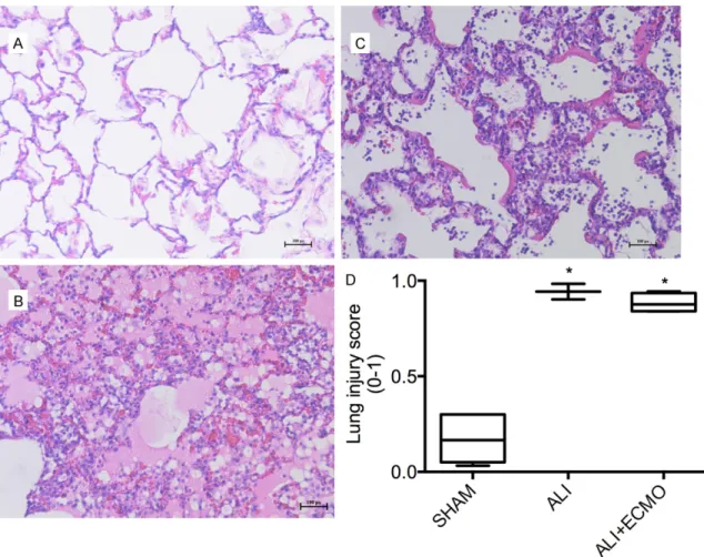
Only 2 animals from the ALI group and 5 from the ALI + ECMO group completed the study period and had lung tissue samples retrieved for analysis. Pulmonary wet/dry weight ratios were increased, indicating significant lung edema in all the animals subjected to ALI (Figure 3B). Histology revealed a severe pat-tern of diffuse alveolar damage, the pathologic hallmark of ARDS, with presence of hyaline membranes, alveolar wall thickening and exten-sive infiltration with inflammatory cells, as shown in Figure 2.
Concerning biochemical data, induction of ALI was associated to a non-significant trend towards increased lactate at T0, and increased serum glutamic oxaloacetic transaminase at T12 and T24.
Performance of extracorporeal membrane oxygenation
Cannulation could be performed successfully in all animals. Connection to ECMO was fol-lowed by rapid reversal of hypoxemia and pul-monary hypertension (Tables 1 and 2). However, hemodynamic instability was common during the first hour, with variable degrees of hypoten-sion, which exhibited no response to fluids and
therefore required moderate doses of noradren-aline. Thereafter, most animals regained hemo-dynamic stability, but remained dependent on noradrenaline until the end of the study period. One animal evolved with progressive hypoten-sion non-responsive to vasopressor and fluids, and died before T2 (Figure 1). The other five animals completed the study period. Total fluid loading was 2472 ml [2188, 2933], 3532 ml [3470, 3549], and 3015 ml [2683, 3234], in the SHAM, ALI and ALI + ECMO groups, respec-tively (p < 0.05 for SHAM vs ALI). Urine output was 1500 ml [945,2225], 2495 ml [2100, 2890], 1405 ml [888, 1566], in the SHAM, ALI and ALI + ECMO groups, respectively (no signifi-cant differences). When analysing biochemistry data in the ECMO group the only relevant obser-vation was a trend towards increased creati-nine concentrations, with no other side effects (Table 3). Oxygenation continued to improve throughout the 24-hour study period. At T24 PaO2 was 255 mmHg [111, 314] while mixed venous PO2 was 77 mmHg [56, 100]. However, airway pressures remained high up to the end of the experiment indicating that lung compli-ance did not improve.
Extracorporeal blood flow reached 2.1 L·min-1 [2.0, 2.2] at T1, which corresponded to 68.1 % [66.3, 69.4] of the last cardiac output meas-ured at T0, just before starting ECMO. The pump speed was 2970 rpm [2305, 3000], with pressure in the drain line at -60 mmHg [-71, -43]. Pre and post membrane pressures were 160 mmHg [97, 171] and 136 mmHg [82, 148], while pre and post membrane partial pressures of oxygen were 37 mmHg [36.8, 37.8] and 143.5 mmHg [121, 183.8], respectively. As ventilator settings were not modified in the ALI + ECMO group after starting ECMO, sweep gas flow had to be decreased to 0.7 L·min-1 [0.5, 1.3] to avoid hypocapnia. However, in 2 sepa-rate pilot experiments not included in the pre-sent analysis, near-apneic ventilation (minute ventilation < 1 L·min-1) was applied during the 24-hour study period. By increasing sweep gas flow, PaO2 and PaCO2 could be kept at rather normal levels (Supplementary Figure 1).
Discussion
We have developed a reproducible porcine 24-hour model of severe acute respiratory fail-ure supported with ECMO, which resembles Figure 1. Survival curves. Survival curves obtained
from SHAM, ALI and ALI + ECMO groups, with six ani
several features of clinical severe ARDS. Acute lung injury was induced by repeated saline lav-age, followed by a 2-hour period of injurious mechanical ventilation, with both hits applied alternating supine and prone position to pro-mote a more diffuse injury. The model is char-acterized by severe hypoxemia refractory to mechanical ventilation with PEEP 5 cmH2O and FiO2 1.0, exhibiting a high lethality when not supported with ECMO. A high flow vvECMO could be provided with minimal recirculation, rapidly correcting hypoxemia and improving survival.
Our goal was to develop a model of ARDS char-acterized histologically by diffuse alveolar dam-age, and functionally by severe hypoxemia and
decreased compliance, to reproduce the con-text in which ECMO is applied clinically, but without significant hemodynamic instability, so that vvECMO could be well tolerated. A few models of ALI supported with ECMO have been reported, including surfactant depletion [12, 16], endotoxin [17], oleic acid infusion [18], and smoke inhalation [10], among others. Concerning optimal species, sheep and pigs are the preferred options. Although sheep offer advantages in terms of an easier cannulation, pigs have some physiologic advantages for translation to humans concerning coagulation [19]. It is clear that all animal models have sig-nificant limitations and the optimal choice will depend on the particular goals and the research context.
Figure 2. Histologic evaluation of lung sections. Representative histologic examinations of lung samples obtained from the dependent region of the right middle lobe of animals from the SHAM (A), ALI (B) and ALI + ECMO (C) groups. A normal lung architecture is observed in (A), whereas diffuse alveolar damage, as represented by the presence of inflammatory cells, both in the interstitial and alveolar space, capillary congestion, alveolar flooding, and the presence of hyaline membranes, are observed in (B and C). Lung injury was quantified by a validated score which includes alveolar wall thickening, the presence of inflammatory cells, proteinaceous debris, and hyaline membranes in the alveolar space, with a range from 0 to 1 [15] (D). Data corresponds to median [percentile 25, 75]. *p < 0.05
After some preliminary experiments and ana-lysing the published data we decided that sur-factant depletion was the best option for our goal. However, lung inflammation is usually low in surfactant depletion models. Therefore we decided to add a second hit: injurious ventila-tion. With this double hit model significant lung inflammation has been described [20]. In pilot experiments we observed that this model led to marked diffuse alveolar damage and severe hypoxemia, however, injury had an excessive gravitational distribution, and hypoxemia was easily reversible even with low PEEP levels. For this reason we decided to perform the double hit injury alternating supine and prone position, so that dorsal and ventral regions were equally affected, and promoting a more widespread development of atelectasis. This approach resulted in severe hypoxemia and a high mor-tality as observed in the ALI group. In contrast, hypoxemia was rapidly corrected in the ALI + ECMO group, and 5/6 animals could complete the 24-hour study period. Furthermore, at the end of the 24-hour study period animals were in good condition and the model could have easily been extended. Interestingly, although gas exchange in the native lungs had partially recovered, as reflected by the large difference between PaO2 and mixed venous PO2 at T24, compliance remained low. We speculate that improvement in oxygenation may have not been
due to reopening of atelectatic lung, but instead to a better ventilation-perfusion relationship.
Since one of our goals was to develop a model of ECMO, with the ability to provide full lung support if necessary, one of the major challeng-es was cannulation. Cervical veins in the pig are rather small compared to other species, while femoral veins are difficult to cannulate because of their deep location. The largest vein is the jugular external vein, which measures 5 to 8 mm diameter for 30 kg pigs. We aimed to have a high blood flow extracorporeal setting with low recirculation, so that full support could be provided. Therefore we decided to use a BCDL catheter. After trying different sizes we found that the largest cannula we could place was a 23 French. In the first experiments we used transthoracic ultrasound to make sure that the cannula was directed towards the infe-rior vena cava and not into the right ventricle. However, thereafter we were able to introduce it correctly without ultrasound, just by advanc-ing blindly the wire guide at least 40 cm without observing arrhythmias. The cannula was advanced with the introducer over the wire guide and secured at 18 cm from the tip. In
post mortem analysis we observed that by
using this approach the cannulas were correct-ly placed towards the inferior vena cava and the return port remained at the right atrium level in Figure 3. Surrogates of alveolar membrane barrier integrity (A). Epithelial lining fluid protein concentration calculat
all cases. Although we could not perform reliable calculations of recircula-tion, because of the impossibility of obtaining representative pure ve- nous blood samples from the vena cava, the very low pre-membrane PO2 indicates that recircula-tion was probably very low [21].
In the present study we did not modify ventilator settings after connecting the animals to ECMO, as usually done in the clinical scenario, because our goal at this stage was to assess the isolated effect of extracorporeal circula-tion and gas exchange in our ALI model, without the additional influence of protective ventilation. However, in two separate pilot experiments we were able to induce near-apneic ventilation keeping nor-mal arterial blood gases, as shown in Supple- mentary Figure 1, indicat-ing that the ECMO settindicat-ing applied is able to provide full lung support. In fact, the following studies will compare different strate-gies of lung protection during ECMO in this model. In addition, we decided to keep FiO2 at 1.0 during all the experi-ment and in all the groups, even after ECMO connec-tion, to avoid the potential influence of applying dif-ferent inspired oxygen concentrations on gas exchange and lung injury. Although this may deviate from clinical practice and certainly is not an ideal setting to protect the
Table 3. Biochemistry data
Variable SHAM ALI ALI + ECMO
Creatinine (mg·dL-1)
Baseline 1.18 [1.17, 1.97] 0.81 [0.73, 0.88]* 1.03 [0.90, 1.04]
All results show median [percentile 25, 75] except for ALI group at T12, T24 (median
[range]). #p < 0.05 compared to T0; *p < 0.05 compared to SHAM; £p < 0.05 compared
to ALI. BUN, blood urea nitrogen; SGOT, Serum glutamic oxaloacetic transaminase; BE,
lungs, we thought for the purpose of research validity it was important to minimize the poten-tial covariates.
The major strengths of our model are first, its severity, which is important concerning poten-tial translations to patients with severe respira-tory failure treated with ECMO. Animals subject-ed to ALI without ECMO developsubject-ed severe hypoxemia and most of them died before com-pleting the study period. A second attribute is hemodynamic stability, which otherwise could preclude using vvECMO. Although we had to use moderate doses of Noradrenaline after starting ECMO, most animals regained stability. A third strength of our model is the use of a single BCDL catheter. Cannulation could be performed without advanced imaging, which frequently is not available in most research labs. In addition, we could avoid using the fem-oral veins, in which due to their deep location, it is cumbersome to place large cannulas in large animals [10, 22]. Fourth, histologic findings in the injured lungs indicate diffuse alveolar dam-age, as expected in ARDS, which is one of the main indications for vvECMO in adults. Finally, with the ECMO configuration applied we obtained minimal recirculation and a relatively high extracorporeal blood flow (> 60 ml·kg -1·min-1), which allows full extracorporeal lung support. This is important to explore more extreme forms of lung protection as near-apneic ventilation.
However, the model also has limitations. It is rather expensive, highly demanding and labor-intensive, therefore requiring a large research team. Experimental preparation takes 5 to 6 hours. Changing repeatedly between supine and prone position during induction of lung inju-ry is laborious. Insertion of a 23 F cannula in the external jugular vein is challenging and requires dissecting the vein very distally towards the sternal notch, where it has a larger diameter. Once vvECMO is started measure-ments of cardiac output by thermodilution are no longer reliable due to indicator loss into the extracorporeal circulation. In addition, connec-tion to ECMO while the animals are hypoxemic and hypercapnic is followed by a short period of hemodynamic instability, which requires vaso-pressor support. Almost all animals regained stability with moderate vasopressor support but one of them developed progressive hypo-tension and finally died early during the model.
However, this is a price worth to pay in order to have a clinically relevant scenario.
Conclusions
The porcine model of ALI induced by repeated saline lavage, followed by injurious ventilation, exhibits severe hypoxemia that is refractory to conventional mechanical ventilation, and has a high lethality, accurately resembling severe ARDS in humans. The present model can be successfully rescued by vvECMO, and support-ed for 24 hours, providing an adequate model for translational research in extracorporeal lung support.
Acknowledgements
AB, PC, JA, PG and GB acknowledge support from Fondo Nacional de Desarrollo Científico y Tecnológico (FONDECYT) through grant 1130428. JA acknowledges support from Pro- grama Nacional de Becas de Posgrado, from Comisión Nacional de Investigación Científica y Tecnológica (CONICYT).
Disclosure of conflict of interest
None.
Authors’ contribution
JA, PC and AB participated in all aspects of the study and wrote the manuscript. LA organized the experiments, and performed animal experi-ments. FD performed statistical analysis and assisted in the animal experiments. PG, PT, TS, BE, GC, TM and PC assisted and per-formed animal experiments. DS contributed to biochemical analysis. FR and MA managed the ECMO circuit. JR contributed to study design and animal experiments. GB contributed to study design and placed catheters and cannu-las. All authors read and approved the final manuscript.
Address correspondence to: Dr. Alejandro Bruhn, Departamento de Medicina Intensiva, Facultad de
Medicina, Pontificia Universidad Católica de Chile,
Marcoleta 367, 3° Piso, Centro de Pacientes Críticos, Santiago, Chile. Tel: 56-223543972; E-mail: [email protected]
References
[1] Australia and New Zealand Extracorporeal
Investigators, Davies A, Jones D, Bailey M,
Beca J, Bellomo R, Blackwell N, Forrest P, Gattas D, Granger E, Herkes R, Jackson A, McGuinness S, Nair P, Pellegrino V, Pettilä V, Plunkett B, Pye R, Torzillo P, Webb S, Wilson M, Ziegenfuss M. Extracorporeal Membrane Oxygenation for 2009 Influenza A(H1N1) Acute
Respiratory Distress Syndrome. JAMA 2009; 302: 1888-1895.
[2] Brodie D and Bacchetta M. Extracorporeal
membrane oxygenation for ARDS in adults. N
Engl J Med 2011; 365: 1905-1914.
[3] Sauer CM, Yuh DD and Bonde P. Extracorporeal
membrane oxygenation use has increased by
433% in adults in the United States from 2006
to 2011. ASAIO J 2015; 61: 31-36.
[4] Peek GJ, Mugford M, Tiruvoipati R, Wilson A,
Allen E, Thalanany MM, Hibbert CL, Truesdale A, Clemens F, Cooper N, Firmin RK and
Elbourne D. Efficacy and economic assess
-ment of conventional ventilatory support ver
-sus extracorporeal membrane oxygenation for severe adult respiratory failure (CESAR): a mul -ticentre randomised controlled trial. Lancet 2009; 374: 1351-1363.
[5] Marhong JD, Munshi L, Detsky M, Telesnicki T
and Fan E. Mechanical ventilation during
ex-tracorporeal life support (ECLS): a systematic
review. Intensive Care Med 2015; 41: 994-1003.
[6] Marhong JD, Telesnicki T, Munshi L, Del Sorbo L, Detsky M and Fan E. Mechanical ventilation
during extracorporeal membrane oxygenation. An international survey. Ann Am Thorac Soc 2014; 11: 956-961.
[7] Schmidt M, Stewart C, Bailey M, Nieszkowska
A, Kelly J, Murphy L, Pilcher D, Cooper DJ,
Scheinkestel C, Pellegrino V, Forrest P, Combes
A and Hodgson C. Mechanical ventilation man-agement during extracorporeal membrane
ox-ygenation for acute respiratory distress syn -drome: a retrospective international multi-center study. Crit Care Med 2015; 43: 654-664.
[8] Hayes RA, Foley S, Shekar K, Diab S, Dunster
KR, McDonald C and Fraser JF. Ovine platelet
function is unaffected by extracorporeal mem
-brane oxygenation within the first 24 h. Blood
Coagul Fibrinolysis 2015; 26: 816-822. [9] Ni L, Chen Q, Zhu K, Shi J, Shen J, Gong J, Gao
T, Yu W, Li J and Li N. The influence of extracor -poreal membrane oxygenation therapy on
in-testinal mucosal barrier in a porcine model for
post-traumatic acute respiratory distress syn-drome. J Cardiothorac Surg 2015; 10: 20. [10] Shekar K, Fung YL, Diab S, Mullany DV,
McDonald CI, Dunster KR, Fisquet S, Platts DG, Stewart D, Wallis SC, Smith MT, Roberts JA
and Fraser JF. Development of simulated and
ovine models of extracorporeal life support to improve understanding of circuit-host interac -tions. Crit Care Resusc 2012; 14: 105-111. [11] Shekar K, Roberts JA, Barnett AG, Diab S,
Wallis SC, Fung YL and Fraser JF. Can physico
-chemical properties of antimicrobials be used to predict their pharmacokinetics during extra -corporeal membrane oxygenation? Illustrative
data from ovine models. Crit Care 2015; 19:
437.
[12] Iglesias M, Jungebluth P, Petit C, Matute MP, Rovira I, Martinez E, Catalan M, Ramirez J and Macchiarini P. Extracorporeal lung membrane provides better lung protection than
conven-tional treatment for severe postpneumonecto -my noncardiogenic acute respiratory distress syndrome. J Thorac Cardiovasc Surg 2008; 135: 1362-1371.
[13] Bradford MM. A rapid and sensitive method for the quantitation of microgram quantities of protein utilizing the principle of protein-dye
binding. Anal Biochem 1976; 72: 248-254. [14] Rennard SI, Basset G, Lecossier D, O’Donnell
KM, Pinkston P, Martin PG and Crystal RG. Estimation of volume of epithelial lining fluid recovered by lavage using urea as marker of
dilution. J Appl Physiol (1985) 1986; 60: 532-538.
[15] Matute-Bello G, Downey G, Moore BB,
Groshong SD, Matthay MA, Slutsky AS and Kuebler WM. An official American Thoracic Society workshop report: features and mea
-surements of experimental acute lung injury in
animals. Am J Respir Cell Mol Biol 2011; 44: 725-738.
[16] Sanchez-Lorente D, Go T, Jungebluth P, Rovira I, Mata M, Ayats MC and Macchiarini P. Single double-lumen venous-venous pump-driven ex-tracorporeal lung membrane support. J Thorac Cardiovasc Surg 2010; 140: 558-563, 563 e551-552.
[17] Song J, Palmer K and Sun B. Effects of inhaled nitric oxide and surfactant with extracorporeal life support in recovery phase of septic acute
lung injury in piglets. Pulm Pharmacol Ther 2010; 23: 78-87.
[18] Langer T, Vecchi V, Belenkiy SM, Cannon JW,
Chung KK, Cancio LC, Gattinoni L and
Batchinsky AI. Extracorporeal gas exchange and spontaneous breathing for the treatment of acute respiratory distress syndrome: an al -ternative to mechanical ventilation?*. Crit Care Med 2014; 42: e211-220.
[19] Peek GJ, Scott R, Killer HM, Jarvis MA, Kolvekar S, Forbes D and Firmin RK. A porcine model of
prolonged closed chest venovenous extracor-poreal membrane oxygenation. ASAIO J 1999; 45: 488-495.
[20] Ballard-Croft C, Wang D, Sumpter LR, Zhou X
of acute respiratory distress syndrome. Ann
Thorac Surg 2012; 93: 1331-1339.
[21] Abrams D, Bacchetta M and Brodie D. Recirculation in venovenous extracorporeal membrane oxygenation. ASAIO J 2015; 61: 115-121.
[22] Hayes D Jr, Yates AR, Duffy VL, Tobias JD, Mansour HM, Olshove VF Jr and Preston TJ. Rapid placement of bicaval dual-lumen cathe
-ter in a swine model of venovenous ECMO. J
Supplementary Figure 1. Pilot experiments using near-apneic ventilation. PaO2, PaCO2 (left axis) and minute ventila
-tion (right axis) along the 24-hour study period, corresponding to two pilot animal experiments performed using the